

 PDF
PDF ePub
ePub Citation
 Print
Print


Abstract
Purpose
To investigate the clinical availability of AL-Scan® (Nidek, GAMAGORI, Japan) by comparing anterior segment param-eters measured with AL-Scan® and Pentacam® (Oculus, Wetzlar, Germany).
Methods
Seventy-three patients (117 eyes) who received refractive surgery at our hospital were tested with AL-Scan® and Pentacam®. We compared measurements including anterior chamber depth, central corneal thickness, white-to-white, and cor-neal curvature.
Results
When comparing measurements obtained with AL-Scan® and Pentacam®, the anterior chamber depth (p < 0.001), cen-tral corneal thickness (p < 0.001) and 2.4 mm zone K value (p = 0.038) showed significant differences; the white-to-white (p =0.348) and 3.3 mm zone K value (p = 0.429) showed no significant differences. All AL-Scan® and Pentacam® parameters had a strong positive linear correlation (p < 0.001). The Bland-Altman plots showed a high degree of agreement between AL-Scan® and Pentacam® in all parameters except for anterior chamber depth.
Conclusions
AL-Scan® is convenient to use clinically because simultaneous measurements of ocular biometry including axial length, intraocular lens power, and topography are possible. However, because differences in some anterior segment parame-ters exist when compared with Pentacam®, measurements with AL-Scan® may require comparisons with other instruments.
Go to : 
References
1. Kim DW, Yi KY, Choi DG, Shin YJ. Corneal thickness measured by dual Scheimpflug, anterior segment optical coherence tomog-raphy, and ultrasound pachymetry. J Korean Ophthal Soc. 2012; 53:1412–8.

2. Rüfer F, Schröder A, Arvani MK, Erb C. Central and peripheral cor-neal pachymetry–standard evaluation with the Pentacam system. Klin Monbl Augenheilkd. 2005; 222:117–22.
3. Lee DM, Ahn JM, Seo KY, et al. Comparison of corneal measure-ment values between two types of topography. J Korean Ophthalmol Soc. 2012; 53:1584–90.
4. Shankar H, Taranath D, Santhirathelagan CT, Pesudovs K. Anterior segment biometry with the Pentacam: comprehensive assessment of repeatability of automated measurements. J Cataract Refract Surg. 2008; 34:103–13.
5. Kawamorita T, Nakayama N, Uozato H. Repeatability and reprodu-cibility of corneal curvature measurements using the Pentacam and Keratron topography systems. J Refract Surg. 2009; 25:539–44.
6. Menassa N, Kaufmann C, Goggin M, et al. Comparison and re-producibility of corneal thickness and curvature readings obtained by the Galilei and the Orbscan II analysis systems. J Cataract Refract Surg. 2008; 34:1742–7.
7. Li H, Leung CK, Wong L, et al. Comparative study of central cor-neal thickness measurement with slit-lamp optical coherence to-mography and visante optical coherence tomography. Ophthalmology. 2008; 115:796–801.
8. Savini G, Carbonelli M, Barboni P, Hoffer KJ. Repeatability of au-tomatic measurements performed by a dual Scheimpflug analyzer in unoperated and post-refractive surgery eyes. J Cataract Refract Surg. 2011; 37:302–9.
9. Savini G, Barboni P, Carbonelli M, Hoffer KJ. Repeatability of au-tomatic measurements by a new Scheimpflug camera combined with Placido topography. J Cataract Refract Surg. 2011; 37:1809–16.
10. Rosa N, Lanza M, Borrelli M, et al. Comparison of central corneal thickness measured with Orbscan and Pentacam. J Refract Surg. 2007; 23:895–9.
11. Kim SI, Kang SJ, Oh TH, et al. Accuracy of ocular biometry and postoperative refraction in cataract patients with AL-Scan®. J Korean Ophthalmol Soc. 2013; 54:1688–93.
12. Buehl W, Stojanac D, Sacu S, et al. Comparison of three methods of measuring corneal thickness and anterior chamber depth. Am J Ophthalmol. 2006; 141:7–12.
13. Shin YJ, Kim NH, Kim DH. Comparison of Pentacam with Orbscan. J Korean Ophthalmol Soc. 2007; 48:637–41.
14. Park JY, Kim SY, Jung MS. Comparison of corneal thickness and anterior chamber depth measured with Orbscan, Pentacam, and ultrasound pachymetry. J Korean Ophthalmol Soc. 2009; 50:664–9.
15. Ryu HW, Kim KR, Chung SK. Comparison of A-scan, Scheimpflug camera, and Orbscan for measurement of anterior chamber depth. J Korean Ophthalmol Soc. 2006; 47:1287–91.
16. Kwag JY, Choi SH. Comparison of ocular biometry measured by ultrasound and two kinds of partial coherence interferometers. J Korean Ophthalmol Soc. 2011; 52:169–74.
17. Park Y, Hwang HB, Chung SK. Comparison of anterior chamber depth obtained from applanation and optical principle devices. J Korean Ophthalmol Soc. 2013; 54:1219–26.
18. Savini G, Carbonelli M, Sbreglia A, et al. Comparison of anterior segment measurements by 3 Scheimpflug tomographers and 1 Placido corneal topographer. J Cataract Refract Surg. 2011; 37:1679–85.
19. Amano S, Honda N, Amano Y, et al. Comparison of central corneal thickness measurements by rotating Scheimpflug camera, ultra-sonic pachymetry, and scanning-slit corneal topography. Ophthalmology. 2006; 113:937–41.
20. O'Donnell C, Maldonado-Codina C. Agreement and repeatability of central thickness measurement in normal corneas using ultrasound pachymetry and the OCULUS Pentacam. Cornea. 2005; 24:920–4.
21. Lackner B, Schmidinger G, Pieh S, et al. Repeatability and reprodu-cibility of central corneal thickness measurement with Pentacam, Orbscan, and ultrasound. Optom Vis Sci. 2005; 82:892–9.
22. Kawamorita T, Uozato H, Kamiya K, et al. Repeatability, reprodu-cibility, and agreement characteristics of rotating Scheimpflug photography and scanning-slit corneal topography for corneal power measurement. J Cataract Refract Surg. 2009; 35:127–33.
23. Jain R, Dilraj G, Grewal SP. Repeatability of corneal parameters with Pentacam after laser in situ keratomileusis. Indian J Ophthalmol. 2007; 55:341–7.
Go to :
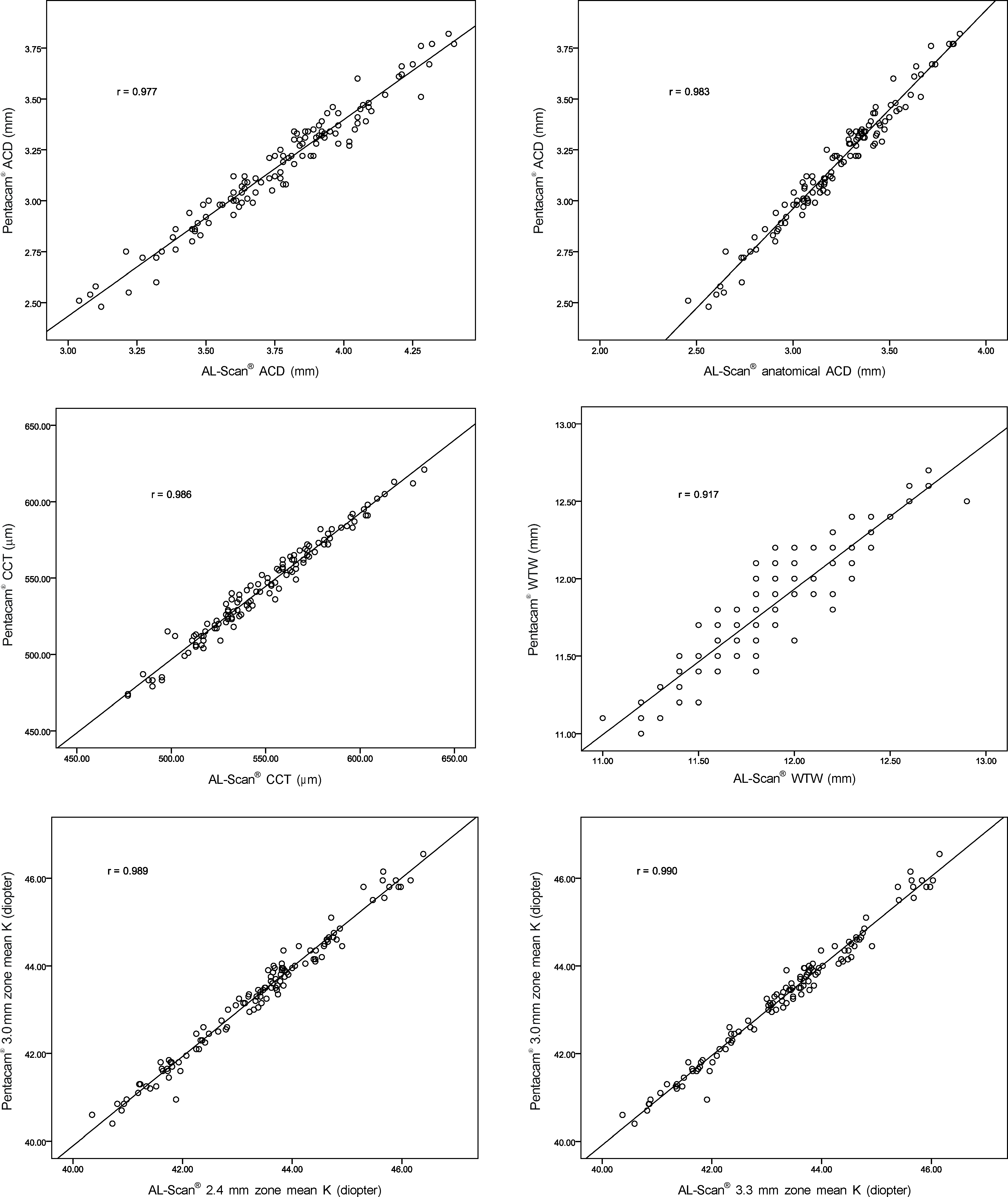 | Figure 1.Pearson correlation of AL-Scan® and Pentcam® in anterior segment parameters. ACD = anterior chamber depth; CCT = central corneal thickness; WTW = white-to-white; K = keratometric diopter. |
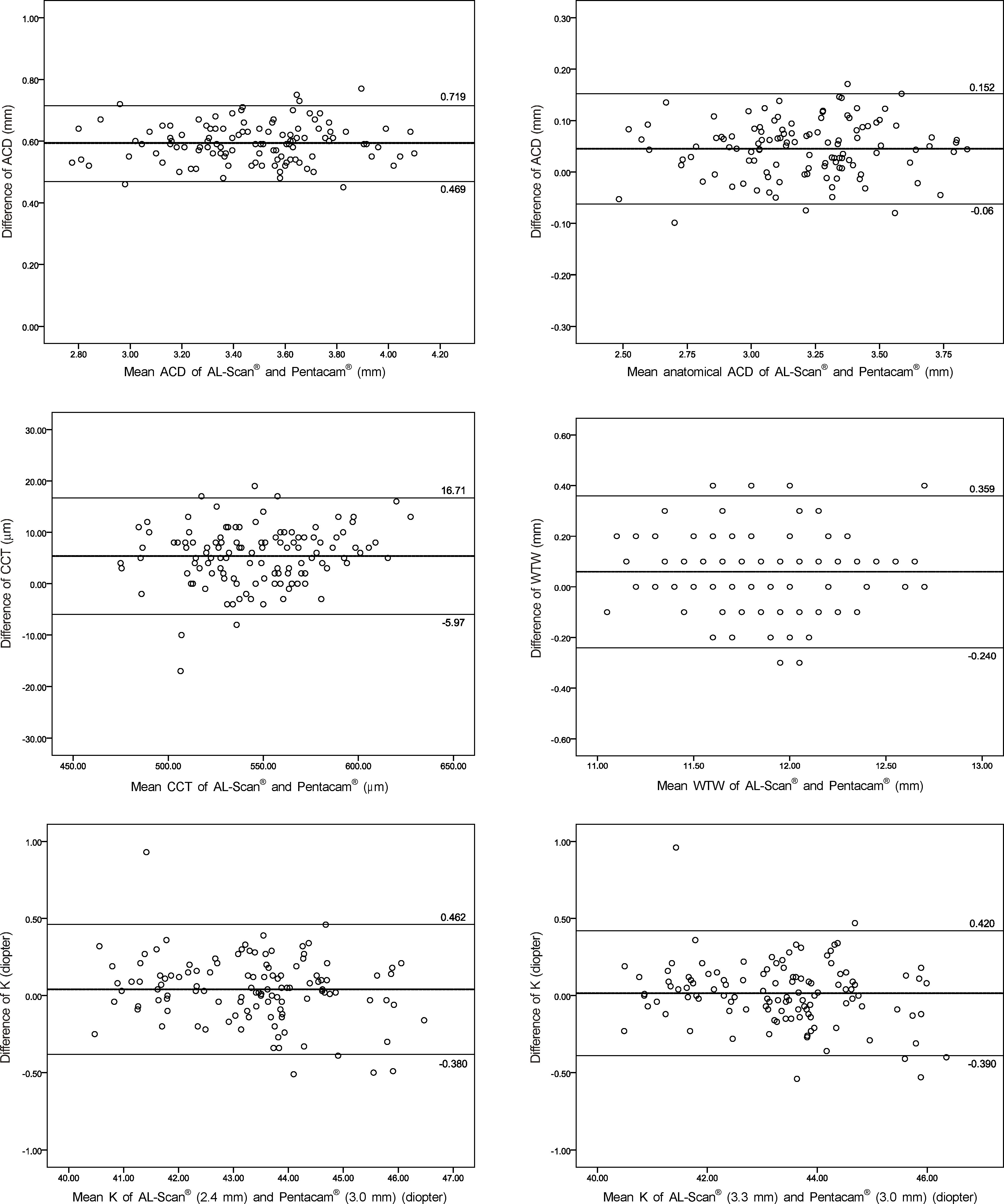 | Figure 2.Bland-Altman plots of AL-Scan® and Pentacam® in anterior segment parameters. ACD = anterior chamber depth; CCT = central corneal thickness; WTW= white-to-white; K = keratometric diopter. |
Table 1.
Comparison of AL-Scan® and Pentacam® in anterior segment parameters
| AL-Scan® | Pentacam® | p-value | |
|---|---|---|---|
| Anterior chamber depth (mm) | 3.77 ± 0.30 | 3.18 ± 0.29 | <0.001* |
| Anatomical anterior chamber depth (mm) | 3.22 ± 0.29 | 3.18 ± 0.29 | <0.001* |
| Central corneal thickness (μm) | 548.74 ± 33.41 | 543.38 ± 32.49 | <0.001* |
| White-to-white (mm) | 11.81 ± 0.32 | 11.77 ± 0.34 | 0.348* |
| K1 (2.4 mm zone) (diopter) | 42.68 ± 1.24 | 42.61 ± 1.30† | 0.004* |
| K2 (2.4 mm zone) (diopter) | 44.03 ± 1.55 | 44.03 ± 1.60† | 0.876* |
| Kmean (2.4 mm zone) (diopter) | 43.36 ± 1.35 | 43.32 ± 1.40† | 0.038* |
| K1 (3.3 mm zone) (diopter) | 42.67 ± 1.25 | 42.61 ± 1.30† | 0.005* |
| K2 (3.3 mm zone) (diopter) | 43.99 ± 1.54 | 44.03 ± 1.60† | 0.177* |
| Kmean (3.3 mm zone) (diopter) | 43.33 ± 1.35 | 43.32 ± 1.40† | 0.429* |
 XML Download