Abstract
Purpose
To report a case of melanocytoma originating from the iris observed for the first time in Korea.
Case summary
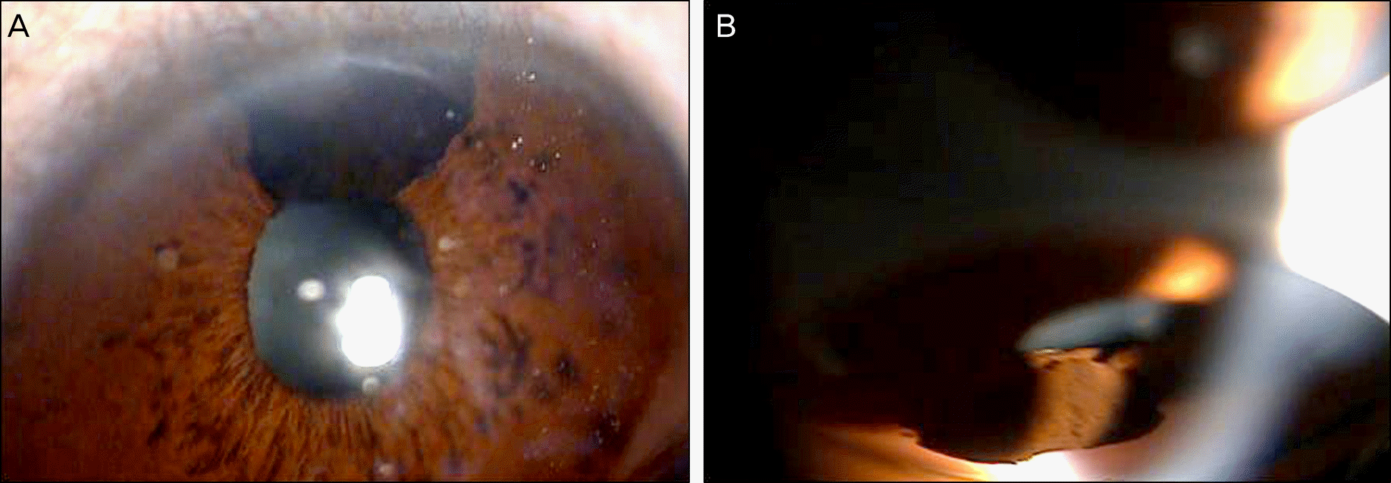
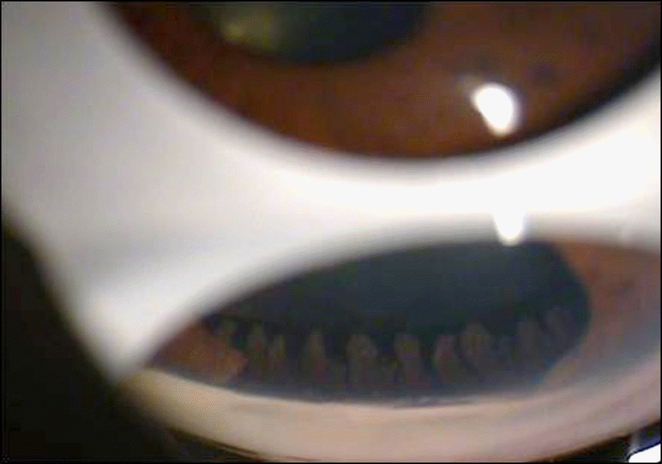
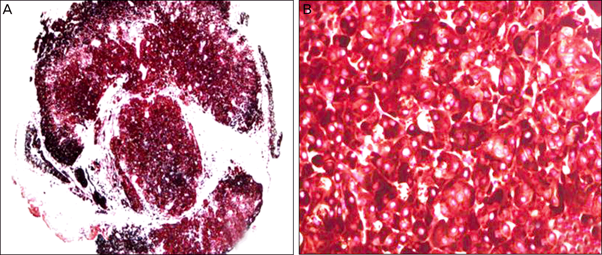
A 53-year-old female with an unexpected iris mass was referred to our clinic. A round, 2.5 mm x 3.5 mm-sized iris mass was found on slit lamp examination in the 12 o'clock area of the patient's left eye. The mass was densely pigmented and had a smooth surface. Gonioscopy showed the mass had reached the peripheral cornea frontward and the lens backward. An excisional biopsy was performed for diagnosis. After the operation, a gonioscopic examination showed an intact ciliary body behind the surgical margin of the iris. A melancytoma of the iris was observed on subsequent histopathological examination. The patient has remained symptom-free with no iris mass recurrence since the operation.
References
1. Zimmerman LE, Garrón LK. Melanocytoma of the optic disc. International Ophthalmology Clinics. 1962; 2:431–40.

2. Al-Hinai A, Edelstein C, Burnier MN Jr. Unusual case of melanocytoma. Can J Ophthalmol. 2004; 39:461–3.

3. Choi SW, Seo SG, Her J. A case of melanocytoma of the ciliary body. J Korean Ophthalmol Soc. 2009; 50:946–50.

4. Lee CS, Kim DK, Lee SC. A case of ciliary body melanocytoma pre-senting as a painful iris mass. Korean J Ophthalmol. 2010; 24:44–6.

5. Zimmerman LE. MELANOCYTES, MELANOCYTIC NEVI, AND MELANOCYTOMAS. Invest Ophthalmol. 1965; 4:11–41.
6. Shields CL, Kancherla S, Patel J, et al. Clinical survey of 3680 iris tumors based on patient age at presentation. Ophthalmology. 2012; 119:407–14.

7. Singh AD, Turell ME, Topham AK. Uveal melanoma: trends in in-cidence, treatment, and survival. Ophthalmology. 2011; 118:1881–5.

8. Esmaili DD, Mukai S, Jakobiec FA, et al. Ocular melanocytoma. Int Ophthalmol Clin. 2009; 49:165–75.

9. Shields JA, Demirci H, Mashayekhi A, Shields CL. Melanocytoma of optic disc in 115 cases: the 2004 Samuel Johnson Memorial Lecture, part 1. Ophthalmology. 2004; 111:1739–46.
10. Lee CS, Bae JH, Jeon IH, et al. Melanocytoma of the optic disk in the Korean population. Retina. 2010; 30:1714–20.

11. LoRusso FJ, Boniuk M, Font RL. Melanocytoma (magnocellular nevus) of the ciliary body: report of 10 cases and review of the literature. Ophthalmology. 2000; 107:795–800.

12. Shields JA, Shields CL, Eagle RC Jr. Melanocytoma (hyperpi-gmented magnocellular nevus) of the uveal tract: the 34th G. Victor Simpson lecture. Retina. 2007; 27:730–9.
13. Howard GM, Forrest AW. Incidence and location of melano-cytomas. Arch Ophthalmol. 1967; 77:61–6.

14. Demirci H, Mashayekhi A, Shields CL, et al. Iris melanocytoma: clinical features and natural course in 47 cases. Am J Ophthalmol. 2005; 139:468–75.

15. Cialdini AP, Sahel JA, Jalkh AE, et al. Malignant transformation of an iris melanocytoma. A case report. Graefes Arch Clin Exp Ophthalmol. 1989; 227:348–54.
Figure 1.
Preoperative image of the iris mass (A) A slit lamp exam shows a 2.5 mm ×3.5 mm sized, brownish-black mass occupying 12 o'clock area of the iris. (B) The gonioscopic view of a protruded, convex mass that is contacting the posterior surface of the cornea and the anterior surface of the lens.

Figure 2.
The postoperative image of the intact ciliary body behind the surgical margin of the iris shown through gonioscopic examination.

Figure 3.
The histopathologic images of the specimen (A) Abundant melanin pigments obscuring nuclear details in the cytoplasm (×40). (B) Microscopic appearance of the specimen with polyhedral cells heavily pigmented with prominent nucleoli (× 400).

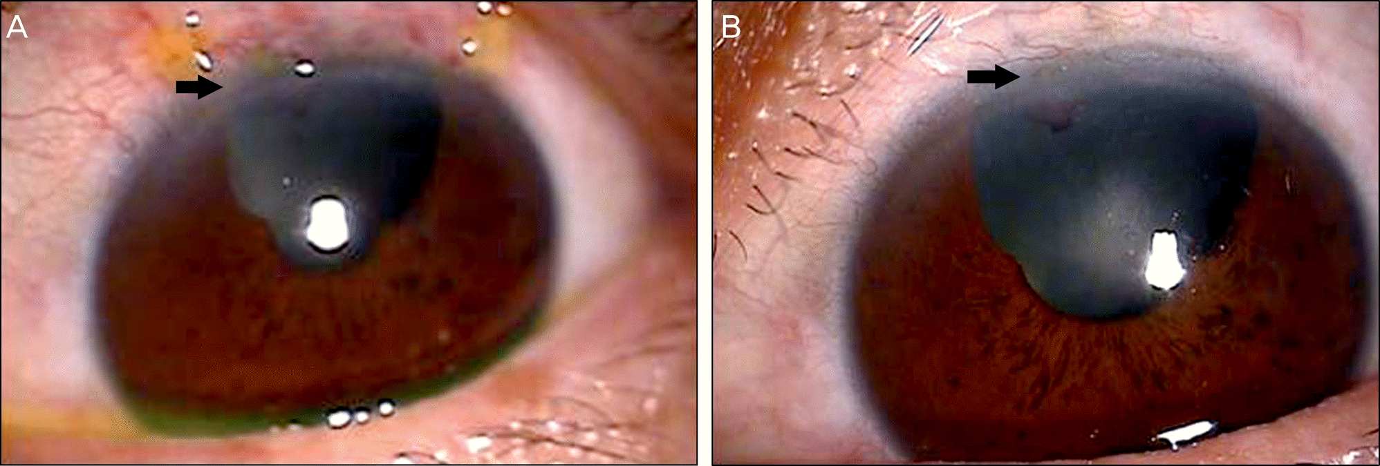
Figure 4.
(A) A slit lamp examination at postoperative day 1. (B) A slit lamp examination at postoperative 3 month. The Photograph of the resected iris taken 3 months after excisional biopsy shows no recurrence. (Arrow: tinged corneal endothelial lesion by original mass shows no change between the intervals.)


 PDF
PDF ePub
ePub Citation
 Print
Print


 XML Download