Abstract
Purpose
To report a case of delayed idiopathic macular hole closure after vitrectomy, internal limiting membrane peeling, and gas tamponade.
Case summary
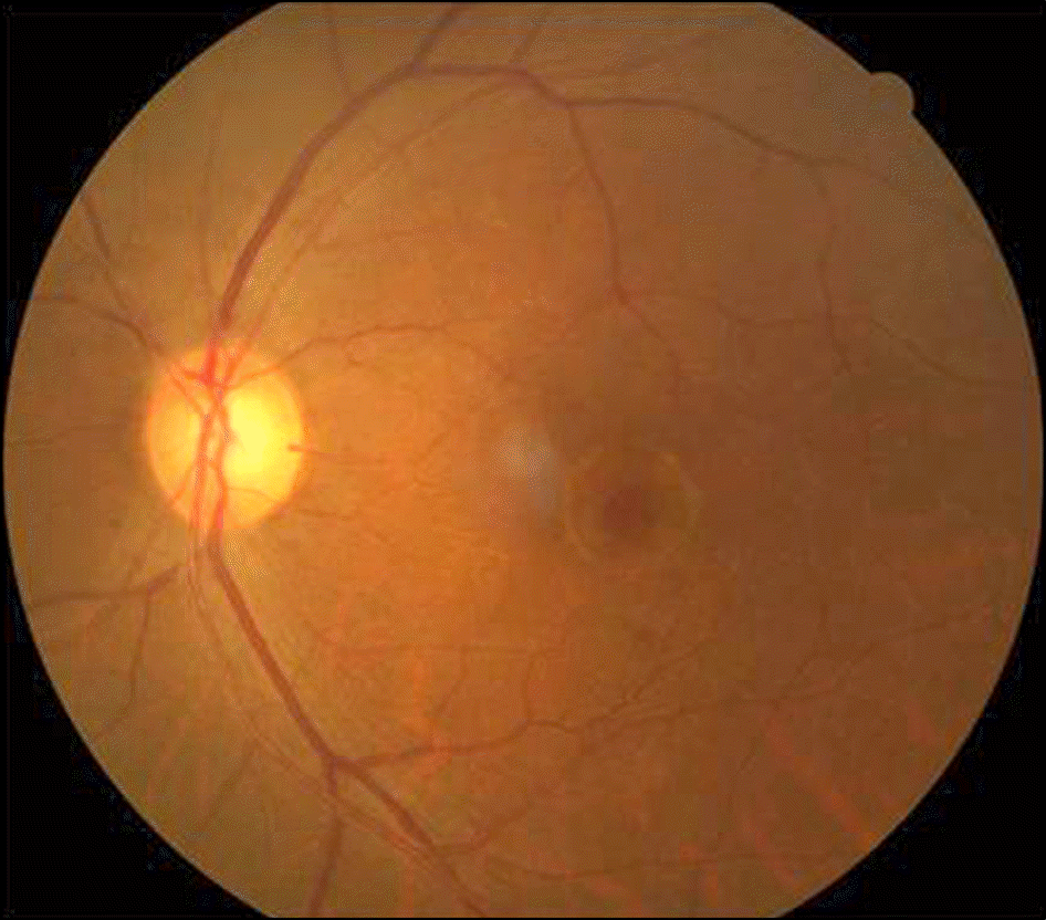
A 69-year-old female complained of visual disturbance in her left eye. At presentation, her visual acuity was 20/100 in the left eye. Fundus examination and optical coherence tomography revealed a full-thickness macular hole 489 μm in diameter as well as posterior vitreous detachment. Hence, vitrectomy, concurrent cataract surgery, internal limiting membrane peeling and gas tamponade were performed. One month postoperatively, the hole remained unclosed, although decreased in size to 378 μm. At 2 months, cystoid macular edema developed and postoperatively the hole diameter decreased gradually to 311 μm, 252 μm and 156 μm at 2, 3, and 5 months, respectively. Finally, the hole was closed upon the resolution of macular edema at 9 months. However, the visual acuity of 20/100 remained unchanged.
Go to : 
References
1. Johnson RN, Gass JD. Idiopathic macular holes. Observations, stages of formation, and implications for surgical intervention. Ophthalmology. 1988; 95:917–24.
2. Mori K, Abe T, Yoneya S. Dome-shaped detachment of premacular vitreous cortex in macular hole development. Ophthalmic Surg Lasers. 2000; 31:203–9.

3. Gross JG. Late reopening and spontaneous closure of previously repaired macular holes. Am J Ophthalmol. 2005; 140:556–8.

4. Yamashita T, Yamashita T, Kawano H, et al. Early imaging of mac-ular hole closure: a diagnostic technique and its quality for gas-fil-led eyes with spectral domain optical coherence tomography. Ophthalmologica. 2013; 229:43–9.

5. Kasuga Y, Arai J, Akimoto M, Yoshimura N. Optical coherence to-mograghy to confirm early closure of macular holes. Am J Ophthalmol. 2000; 130:675–6.

6. D'Souza MJ, Chaudhary V, Devenyi R, et al. Re-operation of idio-pathic full-thickness macular holes after initial surgery with in-ternal limiting membrane peel. Br J Ophthalmol. 2011; 95:1564–7.
7. Imai M, Gotoh T, Iijima H. Additional intravitreal gas injection in the early postoperative period for an unclosed macular hole treated with internal limiting membrane peeling. Retina. 2005; 25:158–61.

8. Cho HY, Kim MR, Kang SW. The effects of laser photocoagulation on reopened macular holes, as assessed by optical coherence tomography. Korean J Ophthalmol. 2005; 19:183–8.

9. Kelly NE, Wendel RT. Vitreous surgery for idiopathic macular holes. Results of a pilot study. Arch Ophthalmol. 1991; 109:654–9.

10. Wender J, Iida T, Del Priore LV. Morphologic analysis of stage 3 and stage 4 macular holes: implications for treatment. Am J Ophthalmol. 2005; 139:1–10.

11. Costa RA, Cardillo JA, Morales PH, et al. Optical coherence to-mography evaluation of idiopathic macular hole treatment by gas-assisted posterior vitreous detachment. Am J Ophthalmol. 2001; 132:264–6.

12. Frangieh GT, Green WR, Engel HM. A histopathologic study of macular cysts and holes. Retina. 2005; 25:311–36.

13. Smiddy WE, Feuer W, Cordahi G. Internal limiting membrane peeling in macular hole surgery. Ophthalmology. 2001; 108:1471–6.

14. Tan MH, Chen FK. “Doctor, why is my macular hole still open?”. Graefes Arch Clin Exp Ophthalmol. 2014; 252:165–7.

15. Choi YM, Oh J, Kim SW, Huh K. Delayed sealing of macular hole after vitrectomy with silicone oil tamponade. J Korean Ophthalmol Soc. 2013; 54:686–90.

16. Georgalas I, Ezra E. Delayed closure after surgery for a full-thick-ness macular hole in a highly myopic eye. Can J Ophthalmol. 2007; 42:341–2.

17. Wong SC, Neuwelt MD, Trese MT. Delayed closure of paediatric macular hole in Coats’ disease. Acta Ophthalmol. 2012; 90:e326–7.

18. Shaikh S, Garretson B. Spontaneous closure of a recurrent macular hole following vitrecomy corroborated by optical coherence tomography. Ophthalmic Surg Lasers Imaging. 2003; 34:172–4.
19. Rossetti L, Autelitano A. Cystoid macular edema following cata-ract surgery. Curr Opin Ophthalmol. 2000; 11:65–72.

20. Miyake K, Ibaraki N. Prostaglandins and cystoid macular edema. Surv Ophthalmol. 2002; 47(Suppl 1):S203–18.

21. Kalish BT, Kieran MW, Puder M, Panigrahy D. The growing role of eicosanoids in tissue regeneration, repair, and wound healing. Prostaglandins Other Lipid Mediate. 2013; 104-105:130–8.

22. Solomon LD. Efficacy of topical flurbiprofen and indomethacin in preventing pseudophakic cystoid macular edema. Flurbiprofen-CME Study Group I. J Cataract Refract Surg. 1995; 21:73–81.
Go to : 
 | Figure 2.Optical coherence tomography images before and after macular hole surgery. (A) A macular hole was detected and its diameter was 489 μm at baseline. (B) One month after surgery, the hole reduced in size to 378 μm. (C) Two months after vitrectomy, the hole decreased in size to 311 μm. Cystoid macular edema was observed. (D, E) At 3 and 5 months, the hole reduced in size to 252 and 156 μm, respectively, and the cystoid edema decreased gradually. (F) At 9 months, the hole was sealed up. (G) At 12 months after surgery, the contour of the fovea improved. Disruption of inner segment/outer segment junction of photoreceptor layer was detected at fovea (between arrows). |


 PDF
PDF ePub
ePub Citation
 Print
Print


 XML Download