

 PDF
PDF ePub
ePub Citation
 Print
Print


Abstract
Purpose
To report a case of peripheral ulcerative keratitis in a case of primary herpes simplex infection.
Case summary
A 7-year-old male complaining of pain, redness, and decreased vision in the left eye 5 days in duration visited our clinic. The patient had also vesicular lesions over the edematous left upper eyelid. Slit-lamp examination revealed peripheral ulcerative keratitis with stromal infiltration involving the superior peripheral cornea. Corneal sensation in the left eye was decreased. The patient was advised to use topical and oral antiviral agents and a topical steroid. After 5 days of follow-up, the corneal lesions were decreased and vesicles were absent. Six months later, only minor opacities remained in the cornea.
Go to : 
References
1. Binder PS. Herpes simplex keratitis. Surv Ophthalmol. 1977; 21:313–30.

2. Labetoulle M, Auquier P, Conrad H, et al. Incidence of herpes sim-plex virus keratitis in France. Ophthalmology. 2005; 112:888–95.
3. Kaye S, Choudhary A. Herpes simplex keratitis. Progress in Retinal and Eye Research. 2006; 25:355–80.
4. Herpetic Eye Disease Study Group. Acyclovir for the prevention of recurrent herpes simplex virus eye disease. N Engl J Med. 1998; 339:300–6.
5. Liesegang TJ. Herpes simplex virus epidemiology and ocular importance. Cornea. 2001; 20:1–13.
6. Hong WS, Rhee SW. Clinical observation of herpes keratitis. J Korean Ophthalmol Soc. 1977; 18:129–33.
7. Schwartz GS, Holland EJ. Oral acyclovir for the management of herpes simplex virus keratitis in children. Ophthalmology. 2000; 107:278–82.
8. Darougar S, Hunter PA, Viswalingam M, et al. Acute follicular conjunctivitis and keratoconjunctivitis due to herpes simplex virus in London. Br J Ophthalmol. 1978; 62:843–9.
9. Liu S, Pavan-Langston D, Colby KA. Pediatric herpes simplex of the anterior segment: characteristics, treatment, and outcomes. Ophthalmology. 2012; 119:2003–8.
10. Darougar S, Wishart MS, Viswalingam ND. Epidemiological and clinical features of primary herpes simplex virus ocular infection. Br J Ophthalmol. 1985; 69:2–6.
11. Liesegang TJ. Herpes simplex virus epidemiology and ocular importance. Cornea. 2001; 20:1–13.
12. Beyer CF, Hill JM, Reidy JJ, Beuerman RW. Corneal nerve dis-ruption reactivates virus in rabbits latently infected with HSV-1. Invest Ophthalmol Vis Sci. 1990; 31:925–32.
13. Remeijer L, Maertzdorf J, Buitenwerf J, et al. Corneal herpes sim-plex virus type 1 superinfection in patients with recrudescent her-petic keratitis. Invest Ophthalmol Vis Sci. 2002; 43:358–63.
14. Jeong IY, Lee KH, You IC, Yoon KC. Recurrent herpes simplex keratitis after penetrating keratoplasty. J Korean Ophthalmol Soc. 2009; 50:657–63.
15. Chong EM, Wilhelmus KR, Matoba AY, et al. Herpes simplex vi-rus keratitis in children. Am J Ophthalmol. 2004; 138:474–5.
16. Hwang JS, Wee WR, Lee JH, Kim MK. Clinical analysis of her-petic keratitis in Korea. J Korean Ophthalmol Soc. 2007; 48:1212–9.
17. Byon IS, Lee JE, Lee JS. Treatment of herpes simplex ocular dis-ease with ganciclovir ophthalmic gel. J Korean Ophthalmol Soc. 2005; 46:164–70.
18. Kuzushima K, Kudo T, Kimura H, et al. Prophylactic oral acyclovir in outbreaks of primary herpes simplex virus type 1 infection in a closed community. Pediatrics. 1992; 89:379–83.
19. Wilhelmus KR, Gee L, Hauck WW, et al. Herpetic Eye Disease Study. A controlled trial of topical corticosteroids for herpes sim-plex stromal keratitis. Ophthalmology. 1994; 101:1883–95.
20. Messmer EM, Foster CS. Vasculitic peripheral ulcerative keratitis. Surv Ophthalmol. 1999; 43:379–96.
Go to :
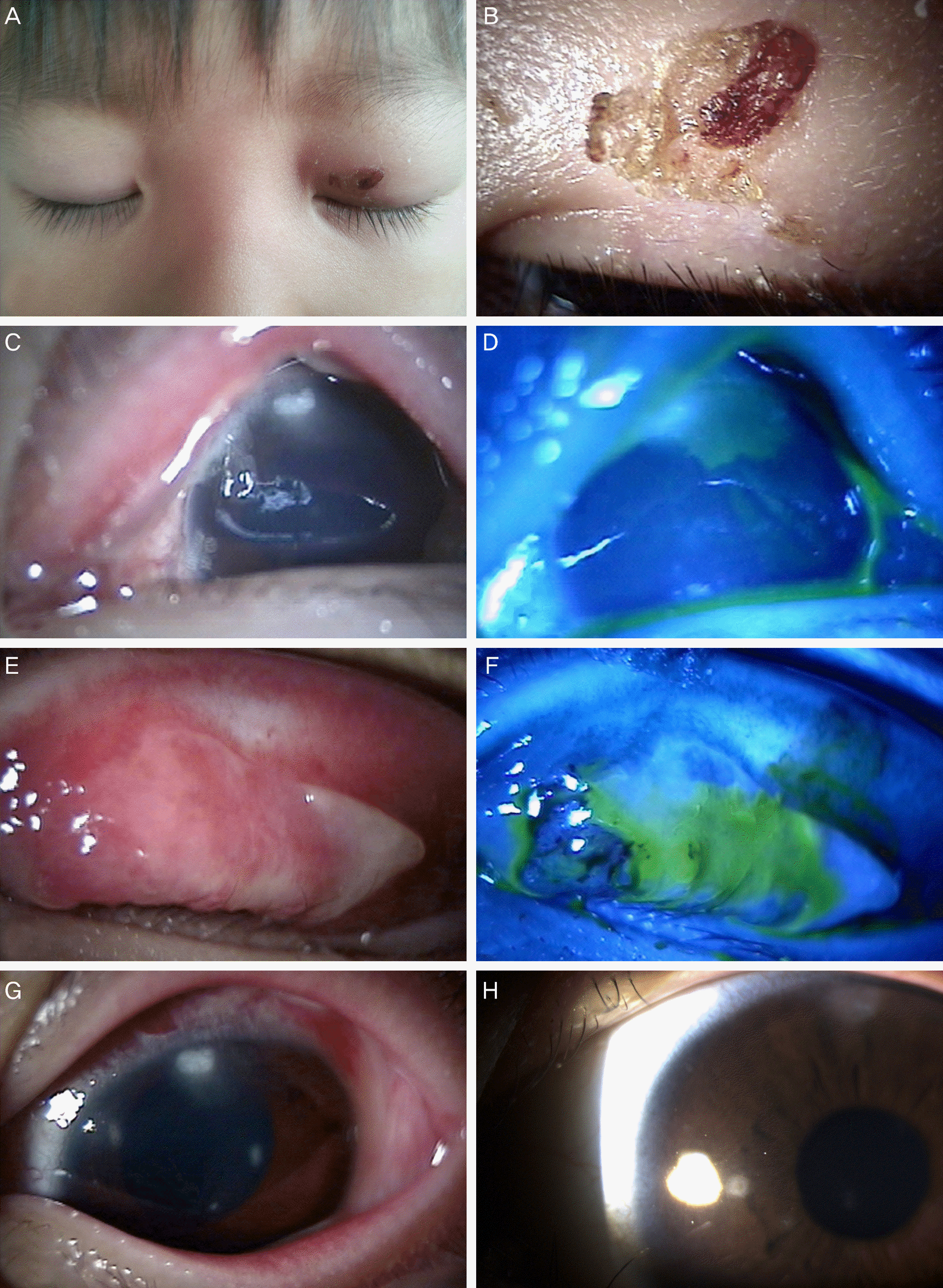 | Figure 1.(A) Vesicular lesions over the left upper eyelid with mild erythema and edema. (B) Yellow and brown scabs over the left upper eyelid after 5 days of treatment. (C) Corneal epithelial defects with stromal infiltration were detected. (D) The fluorescence staining of Fig. 1C. (E) Upper bulbar conjunctiva revealed follicles with pseudomembrane. (F) The fluorescence staining of Fig. 1E. (G) After 1 week, corneal epithelial defects with stromal infiltration were improved. (H) After 1 month, epithelial lesions were healed, but the corneal opacity remained. |
 XML Download