

 PDF
PDF ePub
ePub Citation
 Print
Print


Abstract
Purpose
To report 2 cases of Actinomyces infection in a hydroxyapatite orbital implant with a motility peg.
Case summary
A 44-year-old male and a 55-year-old male who underwent evisceration and implantation of a hydroxyapatite implant in the left eye 17 and 15 years prior, respectively, presented with a conjunctival sac granuloma with discharge and bleeding of 1 year duration. Both patients had a history of motility peg implantation. A large-area of the hydroxyapatite implant was exposed after removal of the granuloma. The previous orbital implant was removed, and the exposed area was covered with a dermis fat graft in both patients. On histopathological examination, Actinomyces infection in the orbital implant was observed in both patients.
Conclusions
To the best of our knowledge, this is the first case report of actinomycosis of hydroxyapatite orbital implant in Korea. In a patient with a porous orbital implant, the possibility of Actinomyces infection of the orbital implant should be considered after a long-duration and large-area exposure of the implant.
Go to : 
References
1. Smego RA Jr. Actinomycosis of the central nervous system. Clin Infect Dis. 1987; 9:855–65.

2. Hussain I, Bonshek RE, Loudon K, et al. Canalicular infection caused by Actinomyces. Eye. 1993; 7:542–4.
3. Weese WC, Smith IM. A study of 57 cases of actinomycosis over a 36-year period. A diagnostic “failure” with good prognosis after treatment. Arch Intern Med. 1975; 135:1562–8.
4. Sudhakar SS, Ross JJ. Short-term treatment of actinomycosis: two cases and a review. Clin Infect Dis. 2004; 38:444–7.
5. Bennhoff DF. Actinomycosis: diagnostic and therapeutic consid-erations and a review of 32 cases. Laryngoscope. 1984; 94:1198–217.
6. Schaal KP, Lee HJ. Actinomycete infections in humans - a review. Gene. 1992; 115:201–11.
7. Karcioglu ZA. Actinomyces infection in porous polyethylene orbi-tal implant. Graefes Arch Clin Exp Ophthalmol. 1997; 235:448–51.
8. Brown JR. Human actinomycosis. A study of 181 subjects. Hum Pathol. 1973; 4:319–30.
9. Brook I. Actinomycosis: diagnosis and management. South Med J. 2008; 101:1019–23.
10. Moghimi M, Salentijn E, Debets-Ossenkop Y, et al. Treatment of Cervicofacial Actinomycosis: A report of 19 cases and review of literature. Med Oral Patol Oral Cir Bucal. 2013; 18:e627–32.
11. Park JW, Choi WC, Sires BS, La TY. Orbital implant infection after drilling procedure. J Korean Ophthalmol Soc. 2007; 48:1449–58.
12. Goldberg RA, Holds JB, Ebrahimpour J. Exposed hydroxyapatite orbital implants: Report of 6 Cases. Ophthalmology. 1992; 99:831–6.
13. You SJ, Yang HW, Lee HC, Kim SJ. 5 Case of infected Hydroxya-patite Orbital Implant. J Korean Ophthalmol Soc. 2002; 43:1553–7.
14. Yang YH, Ahn M. Outcomes of autogenous dermis fat grafting with different donor sites in exposed porous orbital implants. J Korean Ophthalmol Soc. 2013; 54:545–51.
Go to :
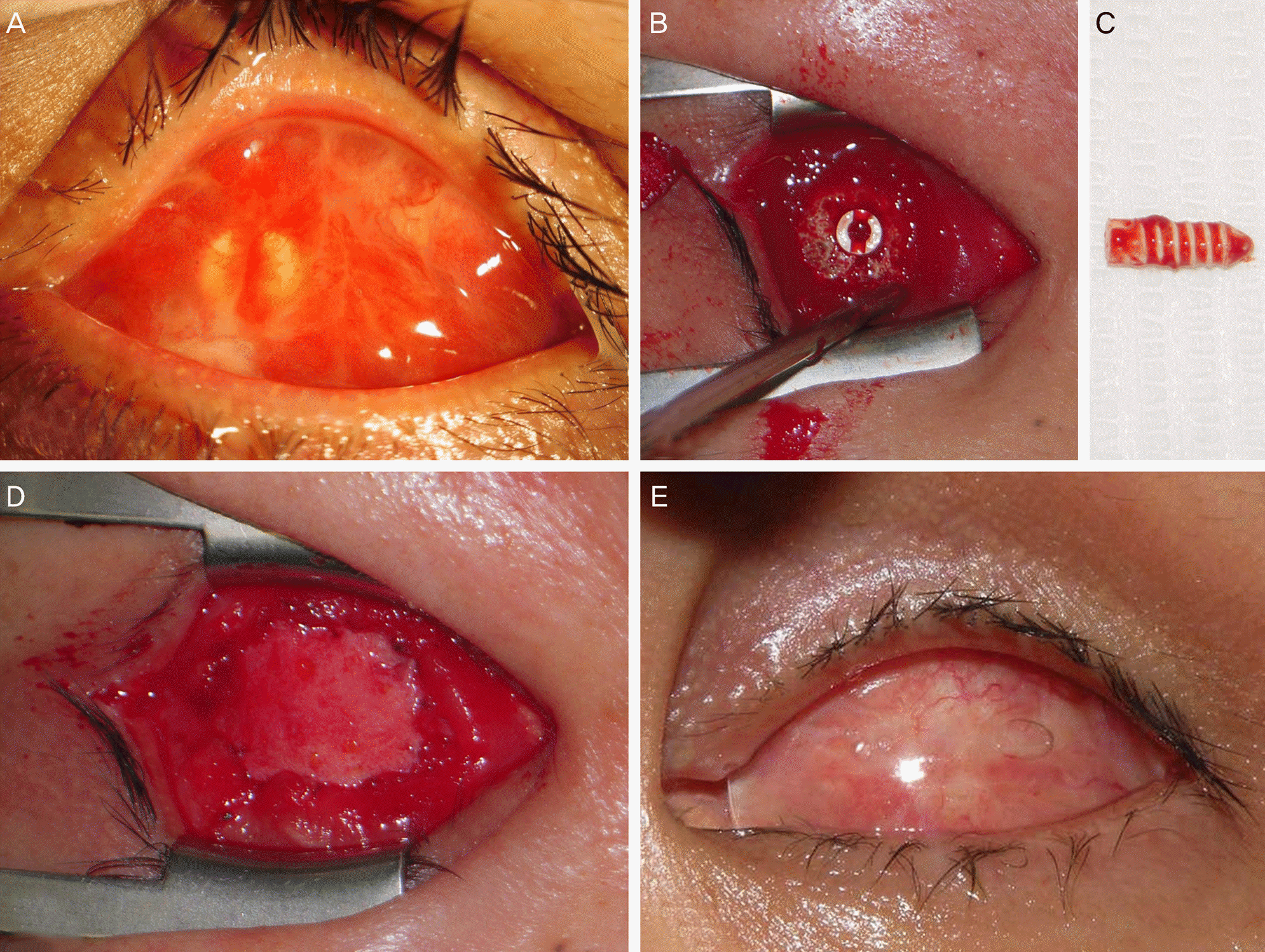 | Figure 1.Case 1. (A) A large granuloma covers the entire anterior surface of conjunctival sac at initial presentation. (B) Photograph shows a large area of hydroxyapatite implant exposure after removal of the granuloma. (C) The surface of the removed sleeve of the motility peg shows dirty gray colored materials. (D) A dermis fat graft is placed and sutured to the edge of the conjunctival defect. (E) Photograph taken 2 months after the exchange of the orbital implant and dermis fat grafting shows well healed conjunctival wounds and a conformer placed in the conjunctival sac. |
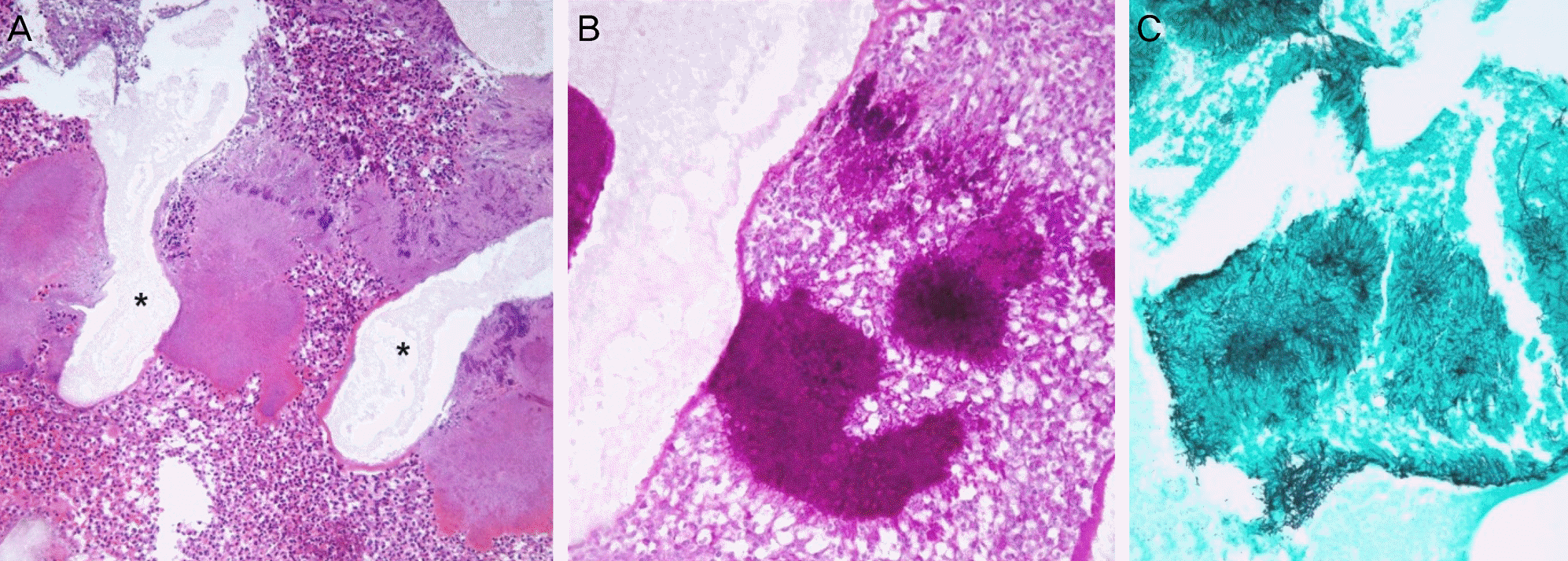 | Figure 2.Case 1. (A) Histopathological examination shows sulphur granules and surrounding inflammatory cells infiltrated between the hydroxyapatite spicules (asterisks) (hematoxylin-eosin, × 200). (B, C) Clusters of filamentous microorganisms which are characteristic findings of actinomycosis (B: periodic acid-Schiff, × 400, C: Grocott's methenamine silver, × 400). |
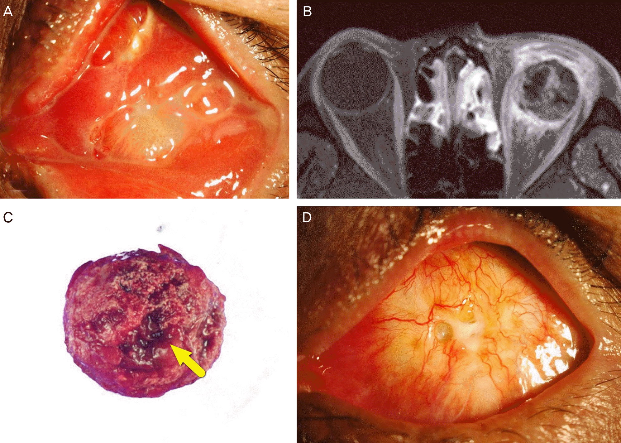 | Figure 3.Case 2. (A) Photograph shows a large conjunctival granuloma of the left eye at initial presentation. (B) A contrast-enhanced Tl-weighted axial image of orbit MRI demonstrates non-enhancing area of implant suggesting poor fibrovascular ingrowth and severe infiltration around the hydroxyapatite orbital implant of the left orbit. (C) The removed hydroxyapatite orbital implant shows darkish discoloration around the hole in which the motility peg was placed (arrow). (D) Photograph taken 6 months after the removal of the hydroxyapatite orbital implant and dermis-fat grafting shows a well-covered graft. |
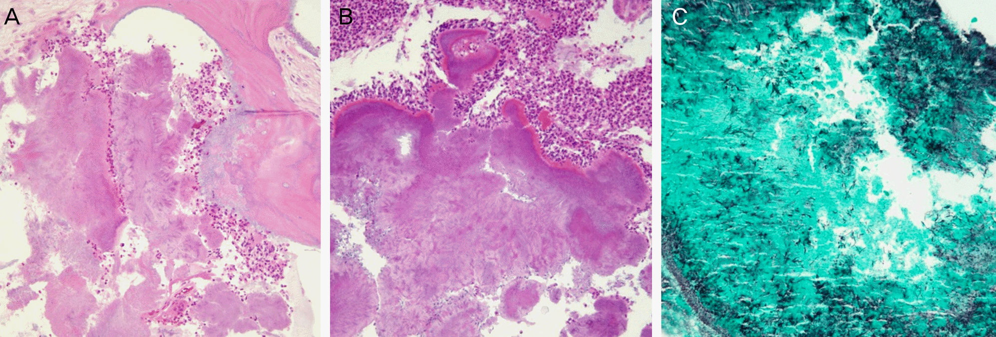 | Figure 4.Case 2. (A) Histopathological examination of the removed hydroxyapatite implant shows sulphur granules and surrounding inflammatory cells infiltrated between the hydroxyapatite spicules (hematoxylin-eosin, × 200). (B) Histopathological examination of pyogenic granuloma also shows sulphur granules and infiltrated inflammatory cells (hematoxylin-eosin, × 200). (C) Clusters of filamentous microorganisms which are characteristic findings of actinomycosis (Grocott's methenamine silver, ×400). |
 XML Download