Abstract
Purpose
To report a case of idiopathic sclerosing inflammatory tumor presenting as painful masses in the medial side of both upper eyelids.
Case summary
A 26-year-old female presented with pain in both eyes at upward gaze and progressive medial side masses on bilateral upper eyelids 3-4 months prior. Orbit MRI showed an orbital benign tumor and partial excisional biopsy was performed for the larger right upper eyelid mass. The biopsy result was chronic inflammation with fibrosis. There was no change in the masses size after an oral steroid was prescribed for 11 days. At 6 weeks after the first operation, complete excisional biopsy was performed for the bilateral upper eyelid masses and idiopathic sclerosing inflammatory tumor was diagnosed. Intravenous steroid injections were administered twice with a 1-week interval postoperatively. After 6 months of follow-up, no recurrence was evident.
Conclusions
Idiopathic sclerosing inflammatory tumor usually involves the anterior, lateral, or apex of the unilateral orbit and does not invade the inferomedial side of the orbit and typically has a chronic course. We experienced a rare case of idiopathic sclerosing inflammatory tumor that involved the medial side of both upper eyelids which was cured by complete excision.
References
1. Rootman J, McCarthy M, White V, et al. Idiopathic sclerosing in-flammation of the orbit. A distinct clinicopathologic entity. Ophthalmology. 1994; 101:570–84.
2. Liu CH, Ma L, Ku WJ, et al. Bilateral idiopathic sclerosing in-flammation of the orbit: report of three cases. Chang Gung Med J. 2004; 27:758–65.
3. Harris GJ. Idiopathic orbital inflammation: a pathogenetic con-struct and treatment strategy: The 2005 ASOPRS Foundation Lecture. Ophthal Plast Reconstr Surg. 2006; 22:79–86.
4. Kim SH, Sun DY, Kim YD. Clinical characteristics of sclerosing pseudotumor of the orbit. J Korean Ophthalmol Soc. 2000; 41:2157–67.
5. Lee IS, Kim SJ, Kim HY, et al. A case of sclerosing orbital pseudotumor. J Korean Ophthalmol Soc. 1996; 37:1321–6.
6. Schaffler GJ, Simbrunner J, Lechner H, et al. Idiopathic sclerotic inflammation of the orbit with left optic nerve compression in a patient with multifocal fibrosclerosis. AJNR Am J Neuroradiol. 2000; 21:194–7.
7. Chen YM, Hu FR, Liao SL. Idiopathic sclerosing orbital in-flammation–a case series study. Ophthalmologica. 2010; 224:55–8.
8. Lee JH, Kim YS, Yang SW, et al. Radiotherapy with or without sur-gery for patients with idiopathic sclerosing orbital inflammation refractory or intolerant to steroid therapy. Int J Radiat Oncol Biol Phys. 2012; 84:52–8.

9. On AV, Hirschbein MJ, Williams HJ, Karesh JW. CyberKnife ra-diosurgery and rituximab in the successful management of sclerosing idiopathic orbital inflammatory disease. Ophthal Plast Reconstr Surg. 2006; 22:395–7.

10. Park BC, Woo KI, Kim YD. Three cases of rituximab treatment for orbital inflammatory disease. J Korean Ophthalmol Soc. 2012; 53:721–7.

11. Plaza JA, Garrity JA, Dogan A, et al. Orbital inflammation with IgG4-positive plasma cells: manifestation of IgG4 systemic disease. Arch Ophthalmol. 2011; 129:421–8.
Figure 1.
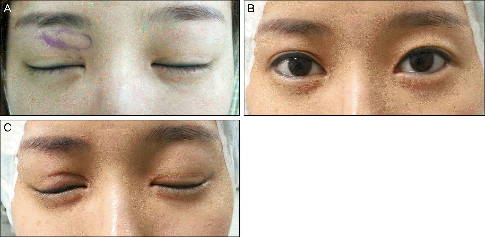
(A) The photo on the first visit does not show remarkable elevated lesions. However, there were hard palpable masses in the medial side of both upper lids. (B, C) The photos which were taken before second complete excisional biopsy show no change in the size of the masses after oral steroid treatment for 11 days.

Figure 2.
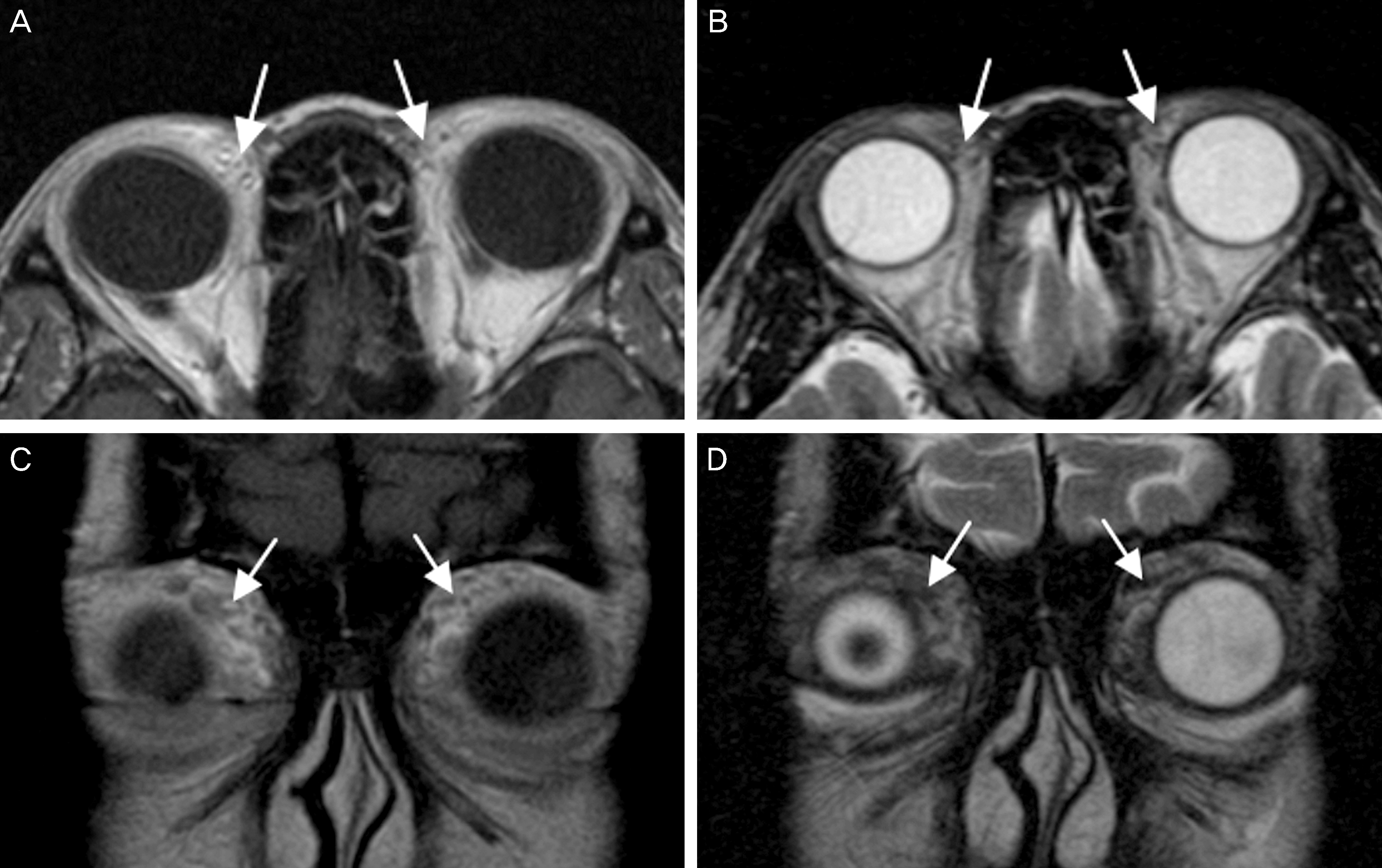
(A) T1 weighted axial image of the enhanced orbit MRI shows ill-defined moderate signal lesions (white arrows) in the medial aspect of both upper eyelids. About 1.5 cm and 2.7 mm masses are noted in the right and left upper eyelids, respectively. (B) At T2 axial view, heterogeneous low to moderate signal masses (white arrows) are shown at the same areas. (C) T1 weighted coronal images also show ill-defined lesions (white arrows) in both upper eyelids. (D) T2 coronal images show heterogeneous lesions (white arrows). Fibrotic lesions could present moderate signal in T1 and T2 images.

Figure 3.
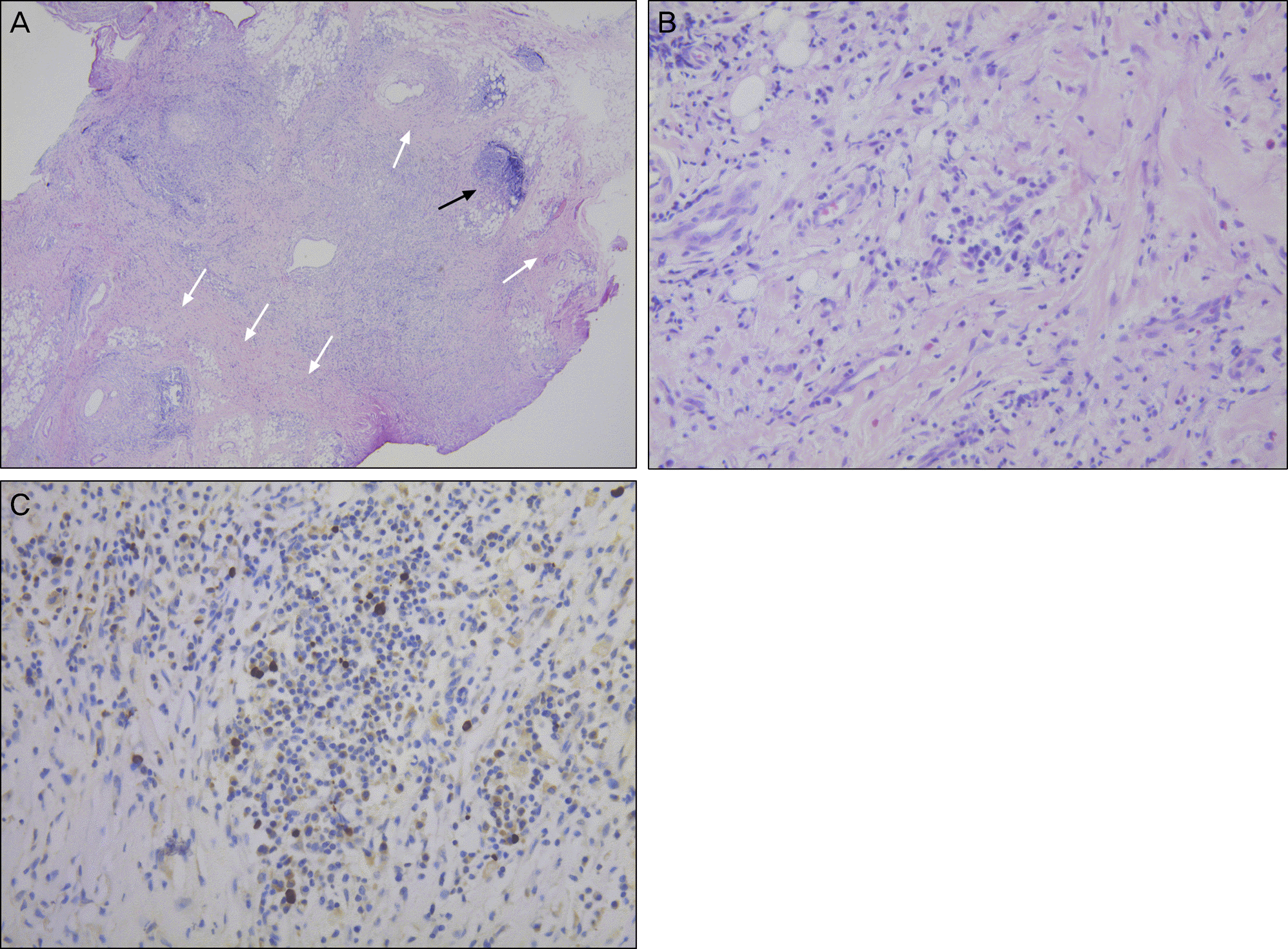
(A) Microscopic examination shows inflammatory lymphoid follicle formation (black arrow) with background fibrosis (white arrows) (Hematoxilin-eosin, ×40). (B) The biopsy specimen shows lymphoplasmacytic infiltrates composed of plasma cells, lymphocytes, neutrophils, and eosinophils with background fibrosis (Hematoxilin-eosin, ×400). (C) The IgG4 staining shows negative result (×400).


 PDF
PDF ePub
ePub Citation
 Print
Print


 XML Download