Abstract
Case summary
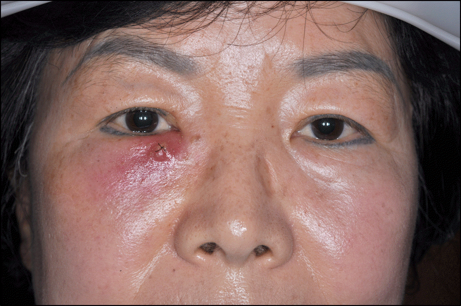
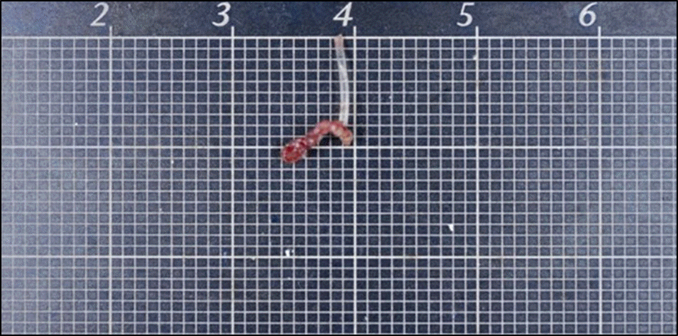
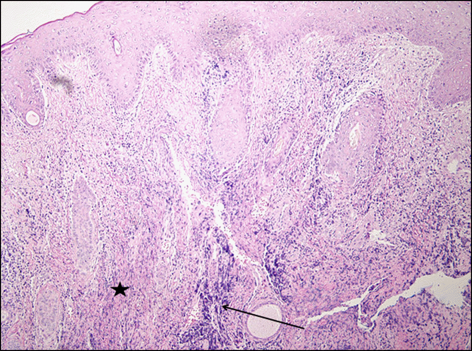
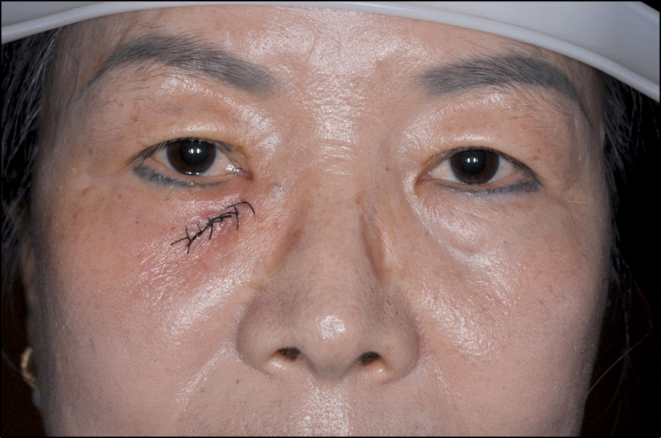
A 56-years-old female who suffered from purulent discharge in inner skin of the right lower eyelid visited our clinic. Lacrimal fistula was found in the skin at the medial side of the right lower eyelid. The patient reported that she had a silicone tube intubation operation 3 years prior due to a nasolacrimal obstruction of right eye. On syringing test, saline solution and purulent discharge were drained from the fistula skin opening and there was no nasolacrimal obstruction. After admission, antibiotic treatment and potadine soaking dressing were performed to facilitate spontaneous closing of the lacrimal fistula. However, the lacrimal fistula relapsed and lacrimal fistulectomy and bicanalicular silicone tube intubation were performed. During surgery, silicone tube remnant material not totally extubated at the lacrimal sac was found which we removed. Postoperatively, systemic antibiotic therapy was administered and the chronic inflammation improved.
Go to : 
References
1. Choi JS, Lee JH, Paik HJ. A silastic sheet found during endoscopic transnasal dacryocystorhinostomy for acute dacryocystitis. Korean J Ophthalmol. 2006; 20:65–9.

2. Lee TS, Woog JJ. Endonasal dacryocystorhinostomy in the pri-mary treatment of acute dacryocystitis with abscess formation. Ophthal Plast Reconstr Surg. 2001; 17:180–3.

3. Cahill KV, Burns JA. Management of acute dacryocystitis in adults. Ophthal Plast Reconstr Surg. 1993; 9:38–41.

4. Kim HH, Jeong BJ, Shin DS, et al. Treatment of the lacrimal fistula with punctal plug. J Korean Ophthalmol Soc. 2007; 48:589–92.
5. Wu W, Yan W, MacCallum JK, et al. Primary treatment of acute da-cryocystitis by endoscopic dacryocystorhinostomy with silicone intubation guided by a soft probe. Ophthalmology. 2009; 116:116–22.

6. Morgan S, Austin M, Whittet H. The treatment of acute dacryocys-titis using laser assisted endonasal dacryocystorhinostomy. Br J Ophthalmol. 2004; 88:139–41.

7. Song BY, Ji YS, Wu MH, et al. The clinical manifestations and treatment results of congenital lacrimal fistula. J Korean Ophthalmol Soc. 2006; 47:871–6.
Go to : 
 | Figure 1.Acute purulent dacryocystitis was treated with incision followed by drainage: there was a noticeable improvement following the incision site being sutured. However, the lacrimal fistula still persisted. |


 PDF
PDF ePub
ePub Citation
 Print
Print


 XML Download