

 PDF
PDF ePub
ePub Citation
 Print
Print


Abstract
Purpose
To compare outcomes of femtosecond laser-enabled deep anterior lamellar keratoplasty (IE-DALK) versus manual trephine using deep anterior lamellar keratoplasty (Manual DALK, M-DALK).
Methods
Seventeen eyes from 17 patients underwent manual deep anterior lamellar keratoplasty, and femtosecond la-ser-enabled deep anterior lamellar keratoplasty was performed in 13 eyes of 13 patients. Postoperative clinical outcomes such as best corrected visual acuity, refractive astigmatism, keratometric astigmatism, endothelial cell density were compared between the two groups.
Results
The mean log MAR best spectacle-corrected visual acuity (BSCVA) was 0.31 ± 0.17, 0.23 ± 0.15, 0.18 ± 0.14 in the IE-DALK group, and 0.55 ± 0.41, 0.45 ± 0.28, 0.35 ± 0.22 (p = 0.056, p = 0.025, p = 0.313) in the M-DALK group at post-operative 2, 4, and 6 months respectively. The mean keratometric cylinder was 5.35 ± 1.57, 4.24 ± 1.97, 3.65 ± 1.31 in the IE-DALK, 8.32 ± 2.75, 6.80 ± 2.50, 4.54 ± 1.25 (p = 0.031, p = 0.041, p = 0.370) in the M-DALK group at postoperative 2, 4, and 6 months respectively. Endothelial cell counts in the two groups did not differ significantly at postoperative 6 months.
References
1. Tsubota K, Kaido M, Monden Y. . A new surgical technique for deep lamellar keratoplasty with single running suture adjustment. Am J Ophthalmol. 1998; 126:1–8.

2. Stern D, Schoenlein RW, Puliafito CA. . Corneal ablation by nanosecond, picosecond, and femtosecond lasers at 532 and 625 nm. Arch Ophthalmol. 1989; 107:587–92.
3. Farid M, Kim M, Steinert RF.Results of penetrating keratoplasty performed with a femtosecond laser zigzag incision initial report. Ophthalmology. 2007; 114:2208–12.
4. Ignacio TS, Nguyen TB, Chuck RS. . Top hat wound config-uration for penetrating keratoplasty using the femtosecond laser: a laboratory model. Cornea. 2006; 25:336–40.
5. Malta JB, Soong HK, Shtein R. . Femtosecond laser-assisted keratoplasty: laboratory studies in eye bank eyes. Curr Eye Res. 2009; 34:18–25.
6. Anwar M, Teichmann KD.Deep lamellar keratoplasty: surgical techniques for anterior lamellar keratoplasty with and without baring of Descemet's membrane. Cornea. 2002; 21:374–83.
7. Archila EA.Deep lamellar keratoplasty dissection of host tissue with intrastromal air injection. Cornea. 1984-1985; 3:217–8.
8. Benson WH, Goosey CB, Prager TC, Goosey JD.Visual improve-ment as a function of time after lamellar keratoplasty for keratoconus. Am J Ophthalmol. 1993; 116:207–11.
9. Panda A, Bageshwar LM, Ray M. . Deep lamellar keratoplasty versus penetrating keratoplasty for corneal lesions. Cornea. 1999; 18:172–5.
10. Tan DT, Mehta JS.Future directions in lamellar corneal transplantation. Cornea. 2007; 26(9 Suppl 1):S21–8.
11. Watson SL, Ramsay A, Dart JKG. . Comparison of deep la-mellar keratoplasty and penetrating keratoplasty in patients with keratoconus. Ophthalmology. 2004; 111:1676–82.
12. Amayem AF, Anwar M.Fluid lamellar keratoplasty in keratoconus. Ophthalmology. 2000; 107:76–9.
13. Funnell CL, Ball J, Noble BA.Comparative cohort study of the outcomes of deep lamellar keratoplasty and penetrating kerato-plasty for keratoconus. Eye (Lond). 2006; 20:527–32.
14. Trimarchi F, Poppi E, Klersy C, Piacentini C.Deep lamellar keratoplasty. Ophthalmologica. 2001; 215:389–93.
15. Yoo SH, Kymionis GD, Koreishi A. . Femtosecond laser-as-sisted sutureless anterior lamellar keratoplasty. Ophthalmology. 2008; 115:1303–7.
16. Mosca L, Fasciani R, Tamburelli C. . Femtosecond laser-as-sisted lamellar keratoplasty: early results. Cornea. 2008; 27:668–72.
17. Noble BA, Agrawal A, Collins C. . Deep Anterior Lamellar Keratoplasty (DALK): visual outcome and complications for a het-erogeneous group of corneal pathologies. Cornea. 2007; 26:59–64.
18. Sugita J, Kondo J.Deep lamellar keratoplasty with complete re-moval of pathological stroma for vision improvement. Br J Ophthalmol. 1997; 81:184–8.
19. Buratto L, Böhm E.The use of the femtosecond laser in penetrating keratoplasty. Am J Ophthalmol. 2007; 143:737–42.
20. Bahar I, Kaiserman I, McAllum P, Rootman D.Femtosecond laser-assisted penetrating keratoplasty: stability evaluation of differ-ent wound configurations. Cornea. 2008; 27:209–11.
21. Sarayba MA, Ignacio TS, Tran DB, Binder PS.A 60 kHz IntraLase femtosecond laser creates a smoother LASIK stromal bed surface compared to a Zyoptix XP mechanical microkeratome in human donor eyes. J Refract Surg. 2007; 23:331–7.
22. Alió JL, Piñero DP.Very high-frequency digital ultrasound meas-urement of the LASIK flap thickness profile using the IntraLase femtosecond laser and M2 and Carriazo-Pendular microkeratomes. J Refract Surg. 2008; 24:12–23.
23. Bourne WM.Cellular changes in transplanted human corneas. Cornea. 2001; 20:560–9.
24. van Dooren BT, Mulder PG, Nieuwendaal CP. . Endothelial cell density after deep anterior lamellar keratoplasty (Melles tech-nique). Am J Ophthalmol. 2004; 137:397–400.
25. Arentsen JJ.Corneal transplant allograft reaction: possible predis-posing factors. Trans Am Ophthalmol Soc. 1983; 81:361–402.
Figure 1.
Settings on the FSL to create a partial-thickness mushroom configuration in the recipient and doner cornea as-suming a minimum corneal pachymetry of 550 mm. Vertical cuts overlap 0.2 mm horizontally and 20 mm vertically with the horizontal lamellar cut at a 90-degree angulation to the corneal surface to ensure completely cut intersecting wound edges. The posterior side cut depth was calculated by subtracting 100 mm of residual cornea from the minimum corneal pachymetry (solid line, FSL cuts; dotted line, manual dissection by the sur-geon).
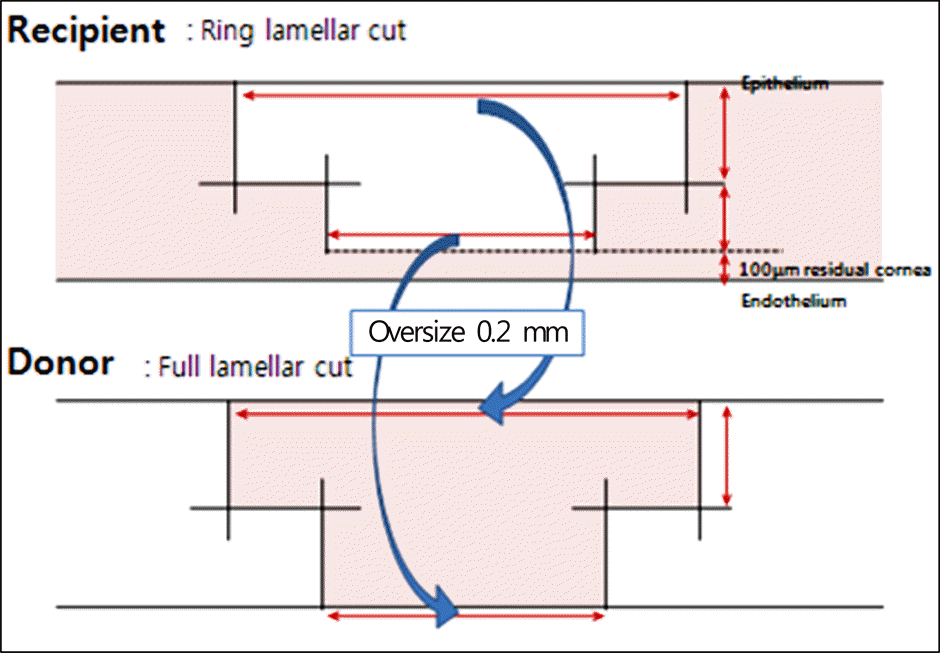
Figure 2.
Postoperative changes of log MAR mean best-spec-tacle corrected visual acuity (BCVA) after manual trephine using deep anterior lamellar keratoplasty (M-DALK) versus IntraLase-enabled deep anterior lamellar keratoplasty (IE-DALK); Postoperative BCVA improved gradually in both groups. BCVA in IE-DALK group was better than M-DALK group postoperatively, but the differences between two groups were statistically significant at 4 months (p = 0.034). The statistical analysis was performed using Mann-Whitney U test. A p-value less than 0.05 is statistically significant.
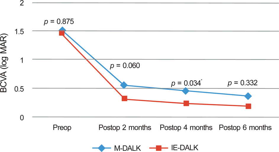
Figure 3.
The cylinder measured using autorefractor in both groups of M-DALK and IE-DALK at 2, 4 and 6 months postoperatively. The refractive cylinder showed lower value in IE-DALK during follow up periods, and the difference was statistically significant at 2 months postoperatively (p = 0.032) (A). The keratometric cylinder measured using manual keratometer showed lower value in IE-DALK than M-DALK, and the difference was statistically significant at 2 months (p = 0.031), and 4 months (p = 0.041) postoperatively (B). The statistical analysis was performed using Mann-Whitney U test. A p-value less than 0.05 is statistically significant.
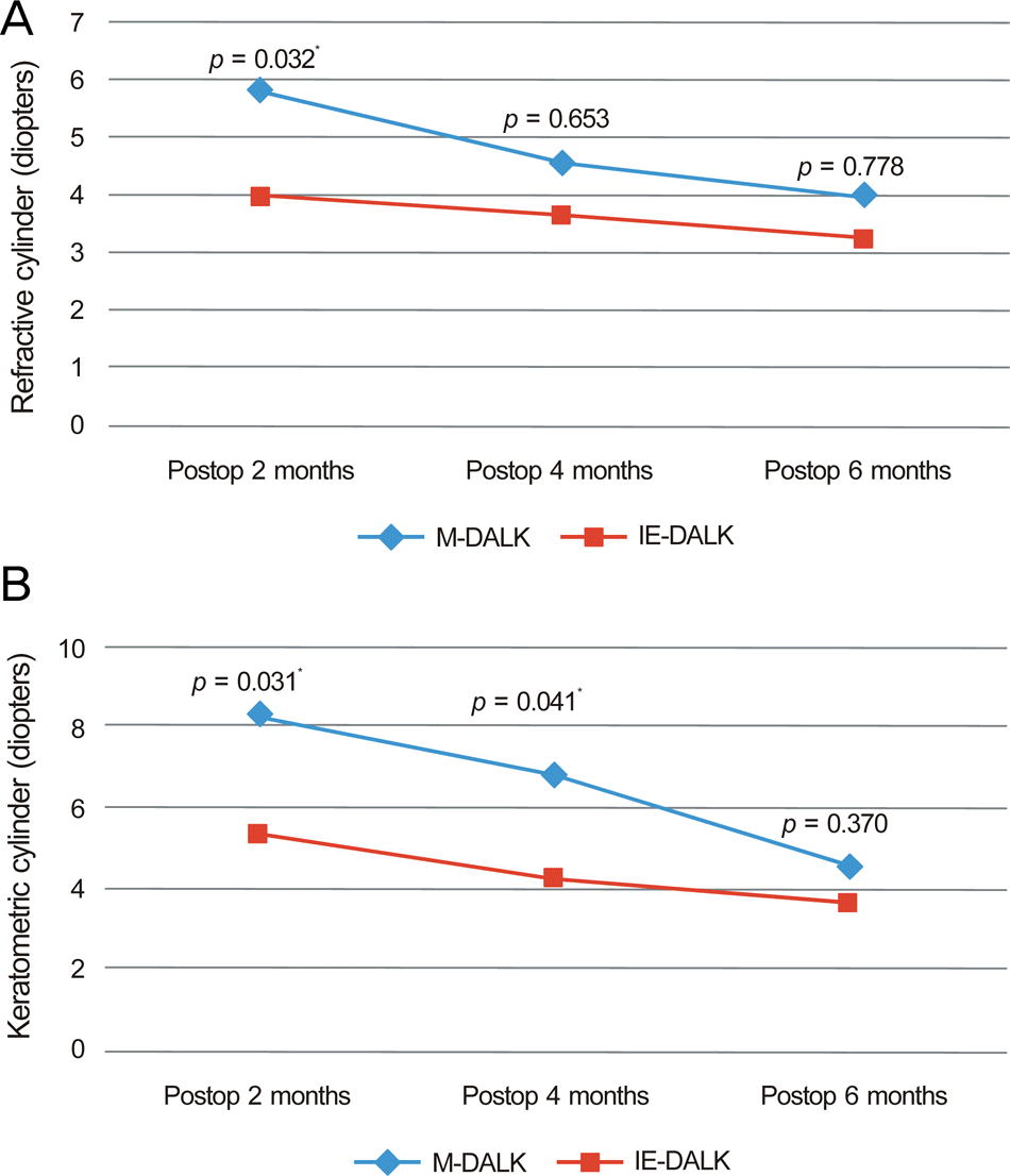
Figure 4.
Difference of postoperative central corneal thickness between M-DALK (535.75 ± 54.0) and IE-DALK (530.14 ±31.2) group (p = 0.865, Mann Whitney test). Pachymetry was performed at 6 months postoperatively (both group).
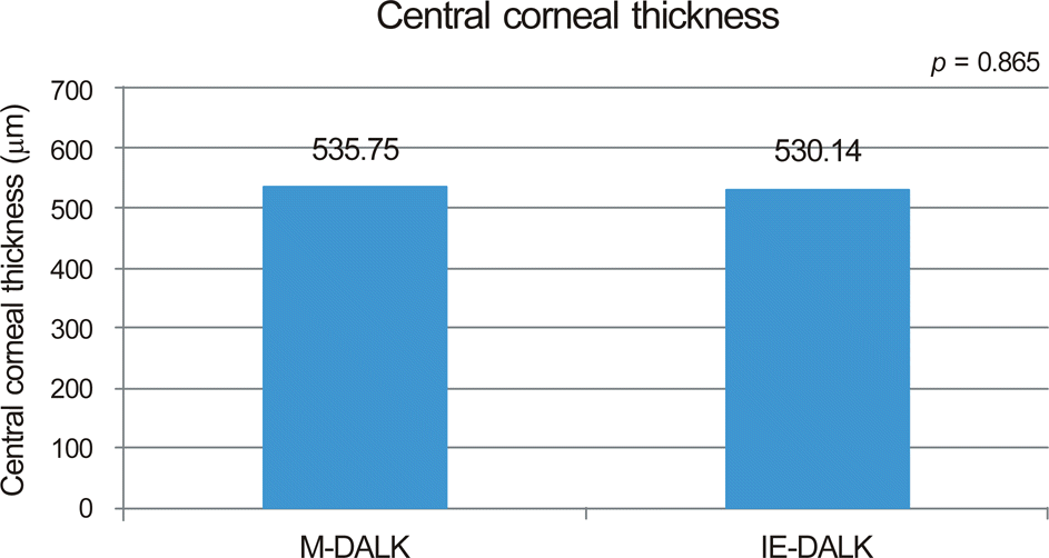
Figure 5.
Difference of postoperative endothelial cell density between M-DALK (2361.60 ± 320.5) and IE-DALK (2559.00± 531.75) group (p = 0.880, Mann Whitney test). Specular microscopy was performed at 6 months postoperatively (both group).
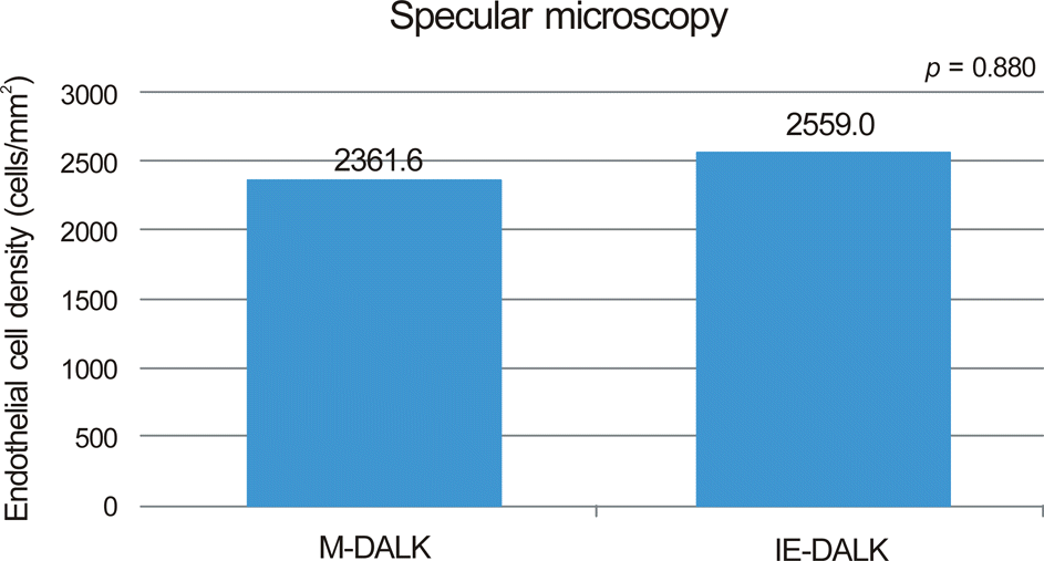
Figure 6.
Anterior segment photographs of the patients 2 months after surgery show the clear central cornea with well attached peripheral flange in IE-DALK (A), and mild edematous cornea with well attached graft in M-DALK (B).
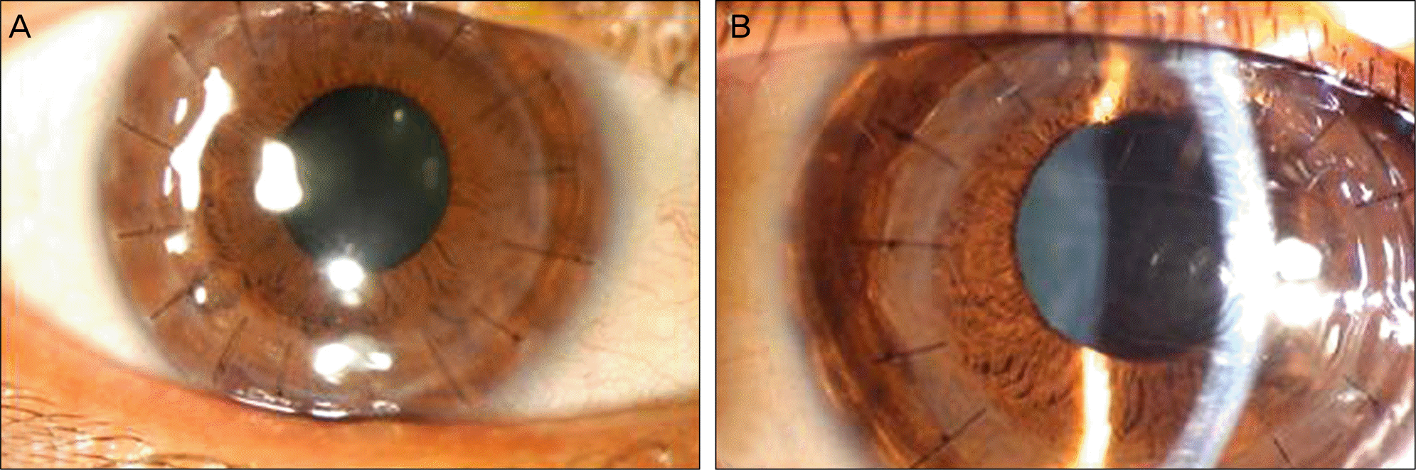
Figure 7.
Six months postoperatively, Visante optical coherence tomography of IE-DALK (mushroom shape) (A), demonstrating the perfect match of the recipient to the donor. In contrast, conventional M-DALK (B) may show the lack of precise match of cut between the two pieces of tissues and protrusions such as hills.
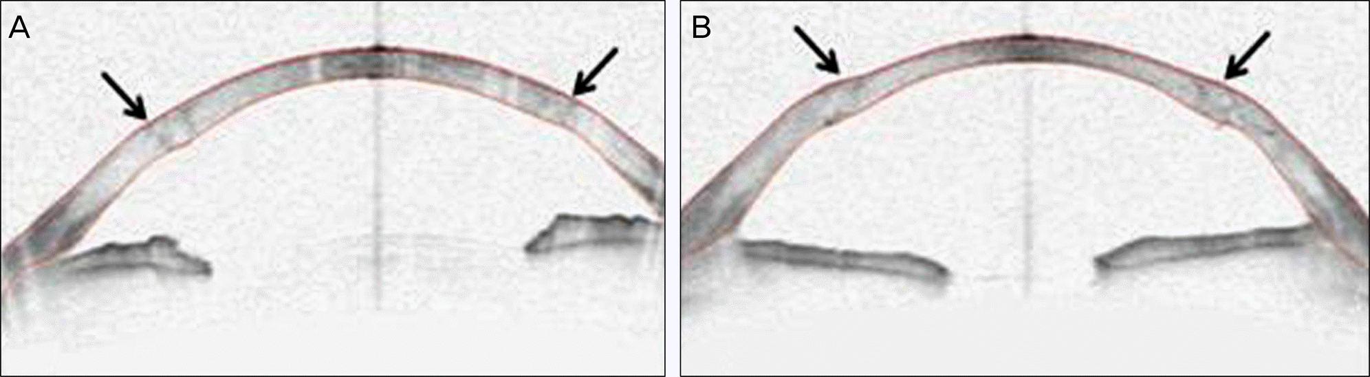
Table 1.
Demographic data of the patients who underwent M-DALK and IE-DALK
 XML Download