

 PDF
PDF ePub
ePub Citation
 Print
Print


Abstract
Purpose
To compare the results of anterior segment biometry including white-to-white (WTW) between scanning-slit topography (ORBscan llz®, Bausch & Lomb), optical low-coherence reflectometry (OLCR) biometry (Lenstar®, Haag-Streit), and Castroviejo calipers.
Methods
Measurements on 72 eyes of 36 patients that underwent refractive surgery were measured using ORBscan®, Lenstar®, and calipers and compared. Ocular biometry parameters used in this study included the WTW, central corneal thickness, anterior chamber depth (ACD), keratometry, and pupil size.
Results
The WTW measurements using ORBscan® and calipers (11.57 ± 0.35 mm and 11.58 ± 0.34 mm, respectively) were statistically similar. However, the measurement using Lenstar® (12.05 ± 0.40 mm) was significantly greater than with the other methods (p < 0.001). Central corneal thickness and keratometry measurements using ORBscan® were greater than when using Lenstar® (p =0.01 for both). ACD and pupil size measurement using Lenstar® were greater than when using ORBscan® (p < 0.001 for both).
Conclusions
Because WTW and ACD measurements using Lenstar® were greater than when using ORBscan® and calipers, unexpected high-vaulting may be observed due to the selection of a larger-sized posterior chamber phakic intraocular lens. Therefore, the differences in measurements obtained when using these methods should be considered.
Go to : 
References
1. Barraquer J. Anterior chamber plastic lenses. Results and conclusions from five year's experience. Trans Ophthalmol Soc UK. 1959; 79:393–424.
2. Chang DH, Davis EA. Phakic intraocular lenses. Curr Opin Ophthalmol. 2006; 17:99–104.

3. Lovisolo CF, Reinstein DZ. Phakic intraocular lenses. Surv Ophthalmol. 2005; 50:549–87.
4. Chun YS, Park IK, Lee HI, et al. Iris and trabecular meshwork pigment changes after posterior chamber phakic intraocular lens implantation. J Cataract Refract Surg. 2006; 32:1452–8.
5. Vetter JM, Tehrani M, Dick HB. Surgical management of acute angle-closure glaucoma after toric implantable contact lens implantation. J Cataract Refract Surg. 2006; 32:1065–7.
6. Baumeister M, Terzi E, Ekici Y, Kohnen T. Comparison of manual and automated methods to determine horizontal corneal diameter. J Cataract Refract Surg. 2004; 30:374–80.
7. Buckhurst PJ, Wolffsohn JS, Shah S, et al. Anew optical low coherence reflectometry device for ocular biometry in cataract patients. Br J Ophthalmol. 2009; 93:949–53.
8. Sanders DR, Doney K, Poco M. United States food and drug ad-ministration clinical trial of the Implantable Collamer Lens (ICL) for moderate to high myopia: Three-year follow-up. Ophthalmology. 2004; 111:1683–92.
9. Sarikkola AU, Sen HN, Uusitalo RJ, Laatikainen L. Traumatic cataract and other adverse events with implantable contact lens. J Cataract Refract Surg. 2005; 31:511–24.
10. Smallman DS, Probst L, Rafuse PE. Pupillary block glaucoma secondary to posterior chamber phakic intraocular lens implantation for high myopia. J Cataract Refract Surg. 2004; 30:905–7.
11. Kodjikian L, Gain P, Donate D, et al. Malignant glaucoma induced by a phakic posterior chamber intraocular lens for myopia. J Cataract Refract Surg. 2002; 28:2217–21.
12. Biermann J, Bredow L, Boehringer D, Reinhard T. Evaluation of ciliary sulcus diameter using ultrasound biomicroscopy in emmetropic eyes and myopic eyes. J Cataract Refract Surg. 2011; 37:1686–93.
13. Kawamorita T, Uozato H, Kamiya K, Shimizu K. Relationship between ciliary sulcus diameter and anterior chamber diameter and corneal diameter. J Cataract Refract Surg. 2010; 36:617–24.
14. Yokoyama S, Kojima T, Horai R, et al. Repeatability of the ciliary sulcus-to-sulcus diameter measurement using wide-scanning-field ultrasound biomicroscopy. J Cataract Refract Surg. 2011; 37:1251–6.
15. Pop M, Payette Y, Mansour M. Predicting sulcus size using ocular measurements. J Cataract Refract Surg. 2001; 27:1033–8.
16. Dinc UA, Oncel B, Gorgun E, et al. Assessment and comparison of anterior chamber dimensions using various imaging techniques. Ophthalmic Surgery, Lasers & Imaging. 2010; 41:115–22.
17. Jung Y, Kim KH. Comparison of white-to-white diameters measured by IOLMaster, Lenstar, Orbscan, and a manual method. J Korean Ophthalmol Soc. 2013; 54:1187–92.
18. Holzer MP, Mamusa M, Auffarth GU. Accuracy of a new partial coherence interferometry analyser for biometric measurements. Br J Ophthalmol. 2009; 93:807–10.
19. Chen YA, Hirnschall N, Findl O. Evaluation of 2 new optical biometry devices and comparison with the current gold standard biometer. J Cataract Refract Surg. 2011; 37:513–7.
20. Bjelos RM, Busic M, Cima I, et al. Intraobserver and interobserver repeatability of ocular components measurement in cataract eyes using a new optical low coherence reflectometer. Graefes Arch Clin Exp Ophthalmol. 2011; 249:83–7.
21. Shin JW, Seong M, Kang MH, et al. Comparison of ocular biometry and postoperative refraction in cataract patients between Lenstar® and IOL Master®. J Korean Ophthalmol Soc. 2012; 53:833–8.
Go to :
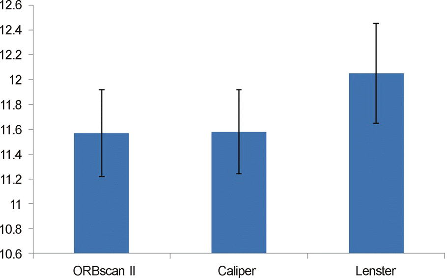 | Figure 1.Comparison of the white to white measurements between the ORBscan®, Lenstar®, and caliper. Measurements by ORBscan® and caliper were 11.57 ± 0.35 mm, 11.58 ± 0.34 mm respectively, and Lenstar®, which was 12.05 ± 0.40 mm measured significantly larger (p < 0.001). |
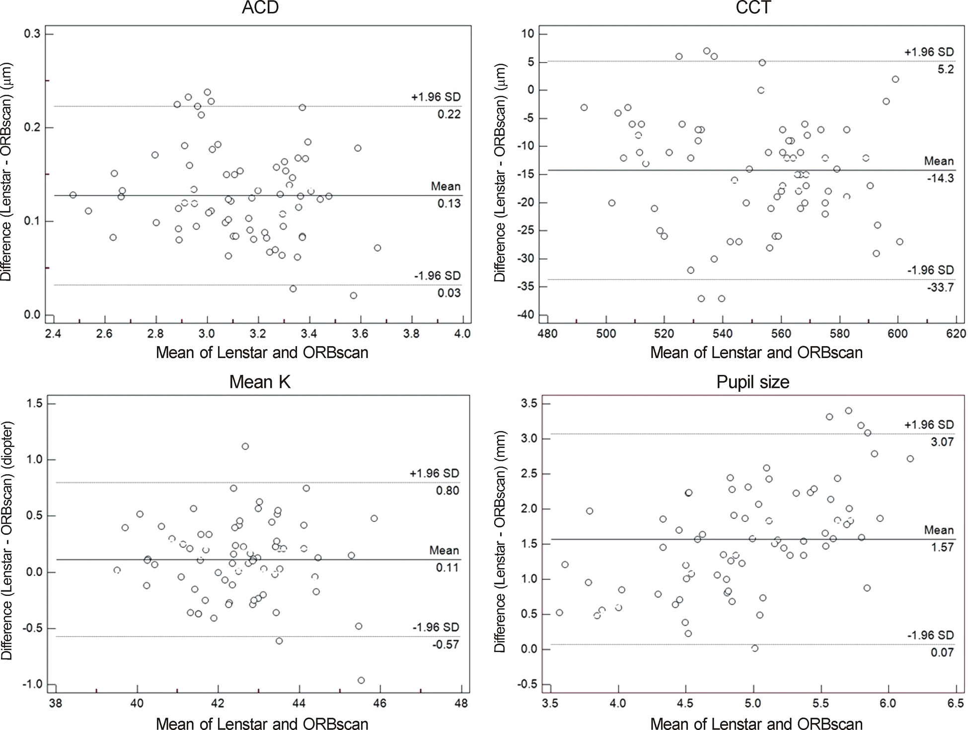 | Figure 2.Bland-Altman plots of agreement between Lenstar® against ORBscan® biometry measurements. The solid line indicates the mean difference (bias). The upper and lower lines represent the 95% LoA. Results show that the observed differences in each measurement were unrelated to the mean difference. ACD = anterior chamber depth; CCT = central corneal thickness; K = keratometry; LoA = limits of agreement. |
Table 1.
Mean ACD, CCT, pupil size, and K readings
 XML Download