

 PDF
PDF ePub
ePub Citation
 Print
Print


Abstract
Case summary
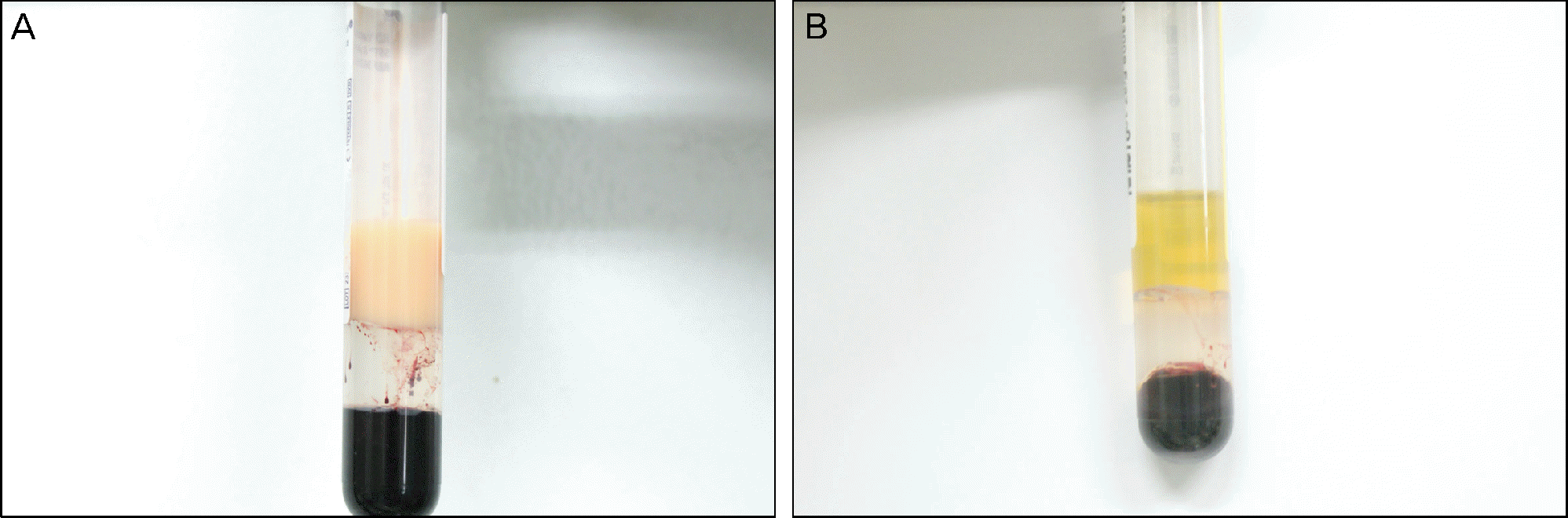
A 27-year-old female with type 2 diabetes visited our clinic with visual disturbance in her left eye while being followed up from a pars plana vitrectomy in her right eye for proliferative diabetic retinopathy. On fundus examination of both eyes, the retinal vessels were creamy white and the retinal veins were undistinguishable from the retinal arteries. The serum triglyceride level was 2,676 mg/dL. The patient was asymptomatic except for visual impairment due to vitreous hemorrhage in her left eye. The patient was diagnosed with lipemia retinalis and chylomicronemia syndrome. After controlling the triglyceride level, funduscopic findings in the both eyes were improved. However, the visual acuity in her right eye remained unchanged.
References
1. Brunzell JD, Bierman EL. Chylomicronemia syndrome. Interaction of genetic and acquired hypertriglyceridemia. Med Clin North Am. 1982; 66:455–68.

2. Citkowitz E. Hypertriglyceridemia. Emedicine Online. Updated November 2013.http://emedicine.medscape.com/article/126568-overview.
3. Mostaza JM, Vega GL, Snell P, Grundy SM. Abnormal metabolism of free fatty acids in hypertriglyceridaemic men: apparent insulin resistance of adipose tissue. J Intern Med. 1998; 243:265–74.
4. Yuan G, Al-Shali KZ, Hegele RA. Hypertriglyceridemia: its etiology, effects and treatment. CMAJ. 2007; 176:1113–20.
5. Fredrickson DS, Levy RI, Lees RS. Fat transport in lipoproteinsdan integrated approach to mechanisms and disorders. N Engl J Med. 1967; 276:34–44.
6. Vinger PF, Sachs BA. Ocular manifestations of hyperlipoproteinemia. Am J Ophthalmol. 1970; 70:563–73.
7. Gopal L, Sunder KS, Rao SK, et al. Hyperlipidemia in a poorlycontrolled diabetic presenting with lipemic aqueous and lipemia retinalis. Retina. 2004; 24:312–5.
8. Lu CK, Chen SJ, Niu DM, et al. Electrophysiological Changes in lipaemia retinalis. Am J Ophthalmol. 2005; 139:1142–5.
9. Leaf DA. Chylomicronemia and the chylomicronemia syndrome: a practical approach to management. Am J Med. 2008; 121:10–2.
Figure 1.
(A) Fundus photograph shows preretinal hemorrhage involving the macula. (B) Fundus photograph after pars plana vitrectomy.
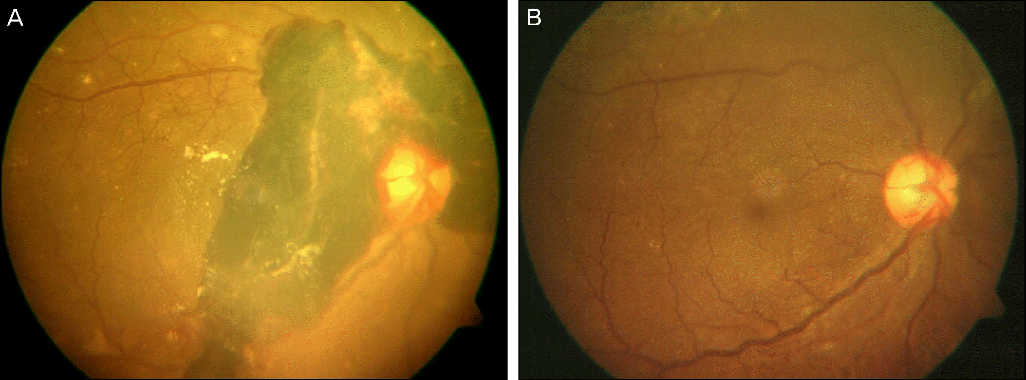
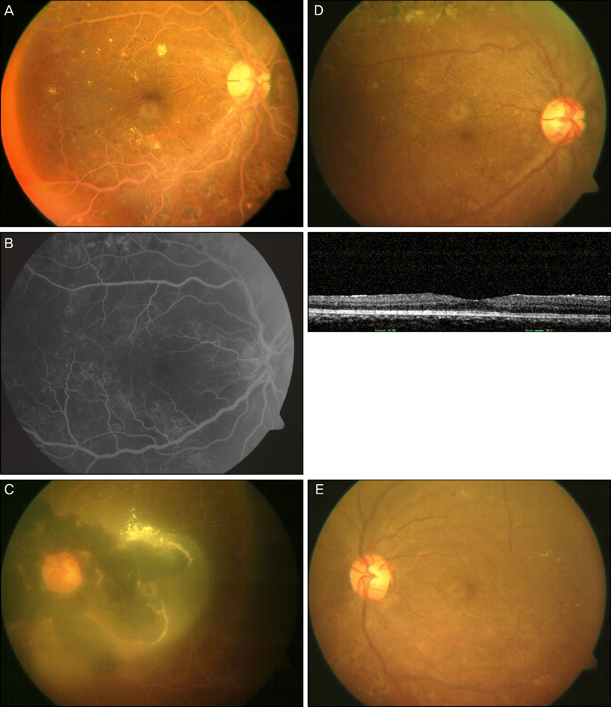
Figure 2.
Fundus photography, fluorescein angiogram and optical coherence tomography before (A, B, C) and after (D, E) resolution of elevated triglyceride. (A) Retinal vessels are orange-colored and arterioles and venules are indistinguishable. (B) Fluorescein angiogram (left) shows tortuous and dilated large retinal vessels without leakage. Optical coherence tomography (right) reveals normal retinal thickness with foveal depression. (C) Vitreous hemorrhage and preretinal hemorrhage with suspected lipid materials were seen. (D, E) In both eyes retinal vessels have recovered their own colors.
 XML Download