

 PDF
PDF ePub
ePub Citation
 Print
Print


Abstract
Purpose
To investigate the measurement repeatability of peripapillary atrophy (PPA) parameters obtained by optic disc stereophotography (ODP) and evaluate the usefulness of PPA parameters to differentiate open-angle glaucoma (OAG) from normal eyes in patients with PPA.
Methods
Sixty-five eyes of 65 patients with PPA were examined. Disc area, cup area, rim area, vertical cup to disc (CD) ratio, CD area ratio, PPA area, zone beta (β) area and zone alpha (α) area were obtained by ODP using intrinsic algorithms. The area under the receivers operating characteristic (AUROC) curves was used to compare the PPA parameters with that of the disc parameters to differentiate OAG from normal eyes. Two examiners analyzed PPA parameters to confirm reproducibility and repeatability of ODP.
Results
Vertical CD ratio (VCD), area CD ratio, zone β area, zone β area per PPA area, zone β area per disc area and axial length were significantly larger in OAG patients (p < 0.05). Among PPA parameters, zone β area per PPA area was strongly correlated with MD (r = -0.431) and PSD (r = 0.411). In addition, PPA (area) to disc area ratio showed the best diagnostic ability (AUROC curve value of 0.786) when comparing the OAG group to the normal group. PPA area and zone β area obtained by ODP showed good reproducibility and repeatability (ICC > 0.997).
Go to : 
References
1. Park KH, Tomita G, Liou SY, Kitazawa Y. Correlation between peripapillary atrophy and optic nerve damage in normal-tension glaucoma. Ophthalmology. 1996; 103:1899–906.

2. Wilensky JT, Kolker AE. Peripapillary changes in glaucoma. Am J Ophthalmol. 1976; 81:341–5.
3. Jonas JB, Naumann GO. Parapapillary chorioretinal atrophy in normal and glaucoma eyes. II. Correlations. Invest Ophthalmol Vis Sci. 1989; 30:919–26.
4. Jonas JB, Nguyen XN, Gusek GC, Naumann GO. Parapapillary chorioretinal atrophy in normal and glaucoma eyes. I. Morphometric data. Invest Ophthalmol Vis Sci. 1989; 30:908–18.
5. Xu L, Li Y, Wang S, et al. Characteristics of highly myopic eyes: the Beijing Eye Study. Ophthalmology. 2007; 114:121–6.
6. Grødum K, Heijl A, Bengtsson B. Refractive error and glaucoma. Acta Ophthalmol Scand. 2001; 79:560–6.
7. Mitchell P, Hourihan F, Sandbach J, Wang JJ. The relationship between glaucoma and myopia: the Blue Mountains Eye Study. Ophthalmology. 1999; 106:2010–5.
8. Chihara E, Liu X, Dong J, et al. Severe myopia as a risk factor for progressive visual field loss in primary open-angle glaucoma. Ophthalmologica. 1997; 211:66–71.
9. Tuulonen A, Jonas JB, Välimäki S, et al. Interobserver variation in the measurements of peripapillary atrophy in glaucoma. Ophthalmology. 1996; 103:535–41.
10. Park KH, Park SJ, Lee YJ, et al. Ability of peripapillary atrophy parameters to differentiate normal-tension glaucoma from glaucoma-like disk. J Glaucoma. 2001; 10:95–101.
11. Uchida H, Ugurlu S, Caprioli J. Increasing peripapillary atrophy is associated with progressive glaucoma. Ophthalmology. 1998; 105:1541–5.
12. Kono Y, Jonas JB, Zangwill L, et al. Agreement of measurement of parapapillary atrophy with confocal scanning laser ophthalmo-scopy and planimetry of photographs. J Glaucoma. 1999; 8:105–10.
13. Anderson D, Patella V. Automated static perimetry. St. Louis: Mosby;1999. p. 117.
14. Lee M, Yoo H, Ahn J. Comparison of disc analysis algorithms provided by cirrus OCT and stereo optic-disc photography in normal and open angle glaucoma patients. Curr Eye Res. 2013; 38:605–13.
15. Buus DR, Anderson DR. Peripapillary crescents and halos in normal-tension glaucoma and ocular hypertension. Ophthalmology. 1989; 96:16–9.
16. Jonas JB, Fernández MC, Naumann GO. Glaucomatous parapapillary atrophy. Occurrence and correlations. Arch Ophthalmol. 1992; 110:214–22.
17. Tezel G, Kass MA, Kolker AE, Wax MB. Comparative optic disc analysis in normal pressure glaucoma, primary open-angle glaucoma, and ocular hypertension. Ophthalmology. 1996; 103:2105–13.
18. Araie M, Sekine M, Suzuki Y, Koseki N. Factors contributing to the progression of visual field damage in eyes with normal-tension glaucoma. Ophthalmology. 1994; 101:1440–4.
19. Ahn JK, Kang JH, Park KH. Correlation between a disc hemorrhage and peripapillary atrophy in glaucoma patients with a unilateral disc hemorrhage. J Glaucoma. 2004; 13:9–14.
20. Tomais G, Georgopoulos G, Koutsandrea C, Moschos M. Correlation of central corneal thickness and axial length to the optic disc and peripapillary atrophy among healthy individuals, glaucoma and ocular hypertension patients. Clin Ophthalmol. 2008; 2:981–8.
21. Ehrlich JR, Radcliffe NM. The role of clinical parapapillary atrophy evaluation in the diagnosis of open angle glaucoma. Clin Ophthalmol. 2010; 4:971–6.
22. Wang YX, Hu LN, Yang H, et al. Frequency and associated factors of structural progression of open-angle glaucoma in the Beijing Eye Study. Br J Ophthalmol. 2012; 96:811–5.
23. Park SC, Lee DH, Lee HJ, Kee C. Risk factors for normal-tension glaucoma among subgroups of patients. Arch Ophthalmol. 2009; 127:1275–83.
Go to :
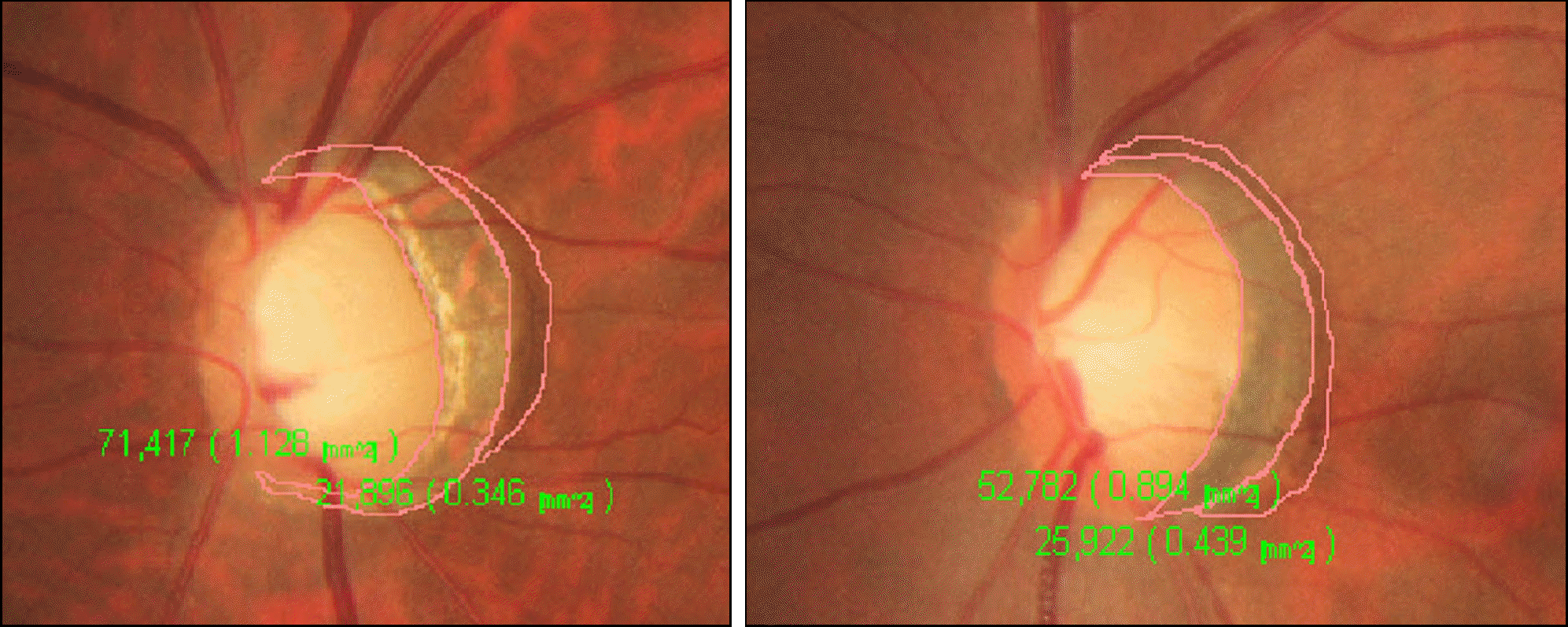 | Figure 1.Peripapillary atrophy measurement by AFC-210 stereophotographs (ODP). After physician made free drawing aground the suspected margin of Zone β and Zone α, the area was calculated by its own intrinsic algorithms. |
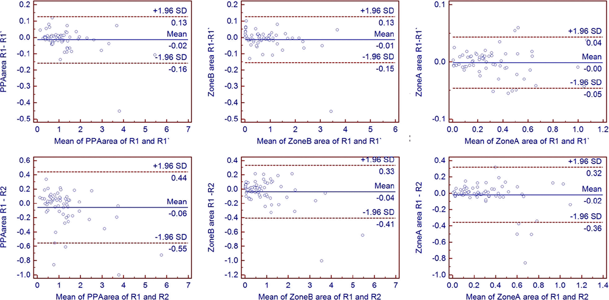 | Figure 2.Upper column: Bland-Altman plot showing the intra-rater repeatability of measuring of the peripapillary atrophy area, Zone β area and Zone α (From left to right). Lower column: Bland-Altman plot showing the inter-rater repeatability of measuring of the Peripapillary atrophy area, Zone β area and Zone α (From left to right). |
Table 1.
Limits of agreement and intraclass coefficient of correlation between two separate examinations
Table 2.
Comparison of characteristics of disc parameters and PPA parameters between OAG and normal group
Table 3.
Correlation of PPA parameters with severity of visual field
Table 4.
Comparison of PPA parameters with disc parameters in differentiating OAG from normal
 XML Download