

 PDF
PDF ePub
ePub Citation
 Print
Print


Abstract
Purpose
To measure the diameter of the retinal arterial and venous caliber of normal Korean subjects and evaluate the factors affecting these diameters.
Methods
Fundus photography was performed on 152 normal Korean subjects. Central retinal artery equivalent (CRAE) and central retinal vein equivalent (CRVE) were measured with a computer-based program (IVAN), and were used to investigate the relationship between the diameter and factors including age, gender, hypertension, diabetes, and smoking.
Results
Among the study subjects, the CRVE was 209.33 ± 12.40 μm and the CRAE was 149.70 ± 9.01 μm. The CRVE and CRAE decreased with increasing age in all study subjects (both p < 0.001). There were significant gender differences in the CRVE and CRAE (p = 0.002, p = 0.042, respectively). After adjusting for age and gender, the hypertension group showed smaller CRVE and CRAE than the non-hypertension group (p = 0.038, p = 0.032, respectively). Smoking and diabetes were not significant factors affecting the CRAE and CRVE (both p > 0.05).
Go to : 
References
1. Wong TY, Kamineni A, Klein R, et al. Quantitative retinal venular caliber and risk of cardiovascular disease in older persons: the cardiovascular health study. Arch Intern Med. 2006; 166:2388–94.
2. Ikram MK, de Jong FJ, Vingerling JR, et al. Are retinal arteriolar or venular diameters associated with markers for cardiovascular dis-orders? The rotterdam study. Invest Ophthalmol Vis Sci. 2004; 45:2129–34.

3. Wong TY, Islam FM, Klein R, et al. Retinal vascular caliber, cardiovascular risk factors, and inflammation: the multiethnic study of atherosclerosis (MESA). Invest Ophthalmol Vis Sci. 2006; 47:2341–50.
4. McGeechan K, Liew G, Macaskill P, et al. Meta-analysis: retinal vessel caliber and risk for coronary heart disease. Ann Intern Med. 2009; 151:404–13.
5. Ikram MK, de Jong FJ, Bos MJ, et al. Retinal vessel diameters and risk of stroke: the rotterdam study. Neurology. 2006; 66:1339–43.
6. McGeechan K, Liew G, Macaskill P, et al. Prediction of incident stroke events based on retinal vessel caliber: a systematic review and individual-participant meta-analysis. Am J Epidemiol. 2009; 170:1323–32.
7. Jeganathan VS, Sabanayagam C, Tai ES, et al. Retinal vascular caliber and diabetes in a multiethnic asian population. Microcirculation. 2009; 16:534–43.
8. Islam FM, Nguyen TT, Wang JJ, et al. Quantitative retinal vascular calibre changes in diabetes and retinopathy: the Singapore Malay eye study. Eye (Lond). 2009; 23:1719–24.
9. Ikram MK, Janssen JA, Roos AM, et al. Retinal vessel diameters and risk of impaired fasting glucose or diabetes: the rotterdam study. Diabetes. 2006; 55:506–10.
10. Wong TY, Duncan BB, Golden SH, et al. Associations between the metabolic syndrome and retinal microvascular signs: the atherosclerosis risk in communities study. Invest Ophthalmol Vis Sci. 2004; 45:2949–54.
11. Liew G, Sharrett AR, Wang JJ, et al. Relative importance of systemic determinants of retinal arteriolar and venular caliber: The atherosclerosis risk in communities study. Arch Ophthalmol. 2008; 126:1404–10.
12. Klein R, Klein BE, Knudtson MD, et al. Are inflammatory factors related to retinal vessel caliber? The beaver dam eye study. Arch Ophthalmol. 2006; 124:87–94.
13. Nguyen TT, Wang JJ, Sharrett AR, et al. Relationship of retinal vascular caliber with diabetes and retinopathy: the multiethnic study of atherosclerosis (MESA). Diabetes Care. 2008; 31:544–9.
14. Myers CE, Klein R, Knudtson MD, et al. Determinants of retinal venular diameter: the beaver dam eye study. Ophthalmology. 2012; 119:2563–71.
15. Zheng Y, Cheung N, Aung T, et al. Relationship of retinal vascular caliber with retinal nerve fiber layer thickness: the Singapore Malay eye study. Invest Ophthalmol Vis Sci. 2009; 50:4091–6.
16. Schoenborn CA, Adams PE. Health behaviors of adults: United states, 2005-2007. Vital Health Stat 10. 2010; 245:1–132.
17. Scott WK, Schmidt S, Hauser MA, et al. Independent effects of complement factor H Y402H polymorphism and cigarette smoking on risk of age-related macular degeneration. Ophthalmology. 2007; 114:1151–6.
18. Grading diabetic retinopathy from stereoscopic color fundus pho-tographs--an extension of the modified airlie house classification. ETDRS report number 10. Early treatment diabetic retinopathy study research group. Ophthalmology. 1991; 98:786–806.
19. Leung H, Wang JJ, Rochtchina E, et al. Computer-assisted retinal vessel measurement in an older population: correlation between right and left eyes. Clin Experiment Ophthalmol. 2003; 31:326–30.
20. Hubbard LD, Brothers RJ, King WN, et al. Methods for evaluation of retinal microvascular abnormalities associated with hyper-tension/sclerosis in the atherosclerosis risk in communities study. Ophthalmology. 1999; 106:2269–80.
21. Knudtson MD, Lee KE, Hubbard LD, et al. Revised formulas for summarizing retinal vessel diameters. Curr Eye Res. 2003; 27:143–9.
22. Bewick V, Cheek L, Ball J. Statistics review 10: further non-parametric methods. Critical Care. 2004; 8:196–9.
23. McBane RD 2nd, Hardison RM, Sobel BE. Comparison of plasminogen activator inhibitor-1, tissue type plasminogen activator anti-gen, fibrinogen, and D-dimer levels in various age decades in patients with type 2 diabetes mellitus and stable coronary artery disease (from the BARI 2D trial). Am J Cardiol. 2010; 105:17–24.
24. Wong TY, Klein R, Klein BE, et al. Retinal vessel diameters and their associations with age and blood pressure. Invest Ophthalmol Vis Sci. 2003; 44:4644–50.
25. Leung H, Wang JJ, Rochtchina E, et al. Relationships between age, blood pressure, and retinal vessel diameters in an older population. Invest Ophthalmol Vis Sci. 2003; 44:2900–4.
26. Wong TY, Knudtson MD, Klein BE, et al. Estrogen replacement therapy and retinal vascular caliber. Ophthalmology. 2005; 112:553–8.
27. Wong TY, Klein R, Sharrett AR, et al. Retinal arteriolar narrowing and risk of coronary heart disease in men and women. The atherosclerosis risk in communities study. JAMA. 2002; 287:1153–9.
28. Wong TY, Hubbard LD, Klein R, et al. Retinal microvascular abnormalities and blood pressure in older people: The cardiovascular health study. Br J Ophthalmol. 2002; 86:1007–13.
29. Iida M, Iida H, Dohi S, et al. Mechanisms underlying cere-brovascular effects of cigarette smoking in rats in vivo. Stroke. 1998; 29:1656–65.
30. Sharrett AR, Ding J, Criqui MH, et al. Smoking, diabetes, and blood cholesterol differ in their associations with subclinical atherosclerosis: the multiethnic study of atherosclerosis (MESA). Atherosclerosis. 2006; 186:441–7.
Go to :
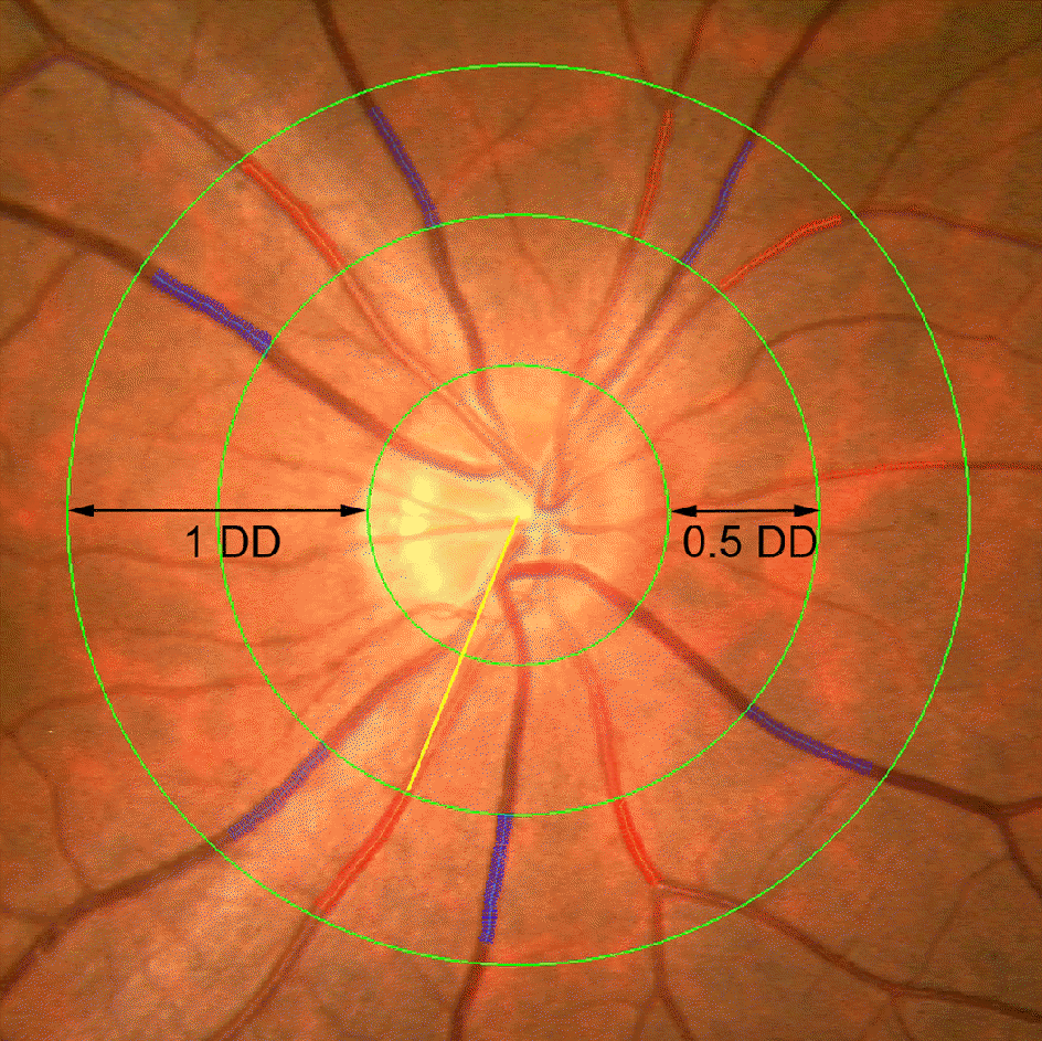 | Figure 1.Fundus photograph centered at the optic disc of right eye, grader analysis measures arteries and veins, the largest six arteries (red) and veins (blue) are used to calculate the central retinal artery equivalent (CRAE) and central retinal vein equivalent (CRVE) by the “Big 6” method. Yellow line is the vessel indicator of the IVAN. DD = disc diameter. |
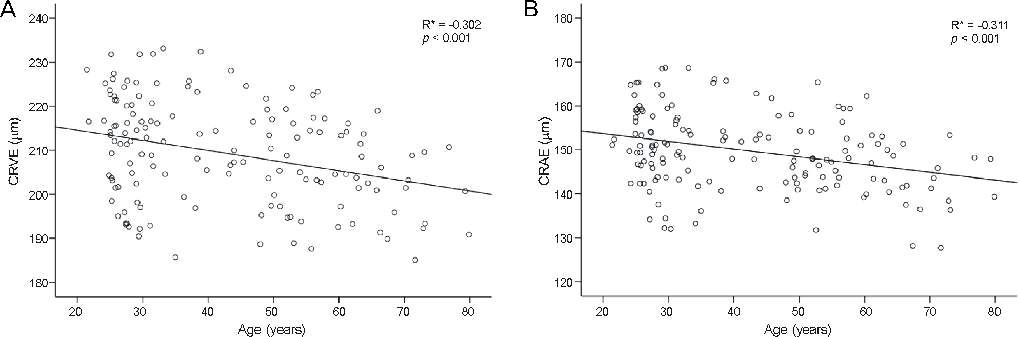 | Figure 2.(A) Central retinal vein caliber (CRVE) for all participants at a given age, (B) Central artery caliber (CRAE) for all participants at a given age. Scattering plot shows negative correlation with age and retinal vascular caliber. |
Table 1.
Demographic characteristics of study participants stratified by age group
| Age group (years) | 20-29 | 30-39 | 40-49 | 50-59 | 60-69 | 70-79 | All | p-value |
|---|---|---|---|---|---|---|---|---|
| Number of patients | 54 | 26 | 18 | 26 | 18 | 10 | 152 | |
| Sex (male:female) | 35:19 | 14:12 | 8:10 | 13:13 | 8:10 | 5:5 | 83:69 | 0.537* |
| HTN (n (%)) | 0 | 0 | 2 (11.1) | 8 (30.8) | 11 (61.1) | 5 (50.0) | 26 (17.1) | <0.001† |
| DM (n (%)) | 0 | 0 | 2 (11.1) | 2 (7.7) | 4 (22.2) | 4 (40.0) | 12 (7.9) | <0.001† |
| Smoker (n (%)) | 4 (7.4) | 4 (15.4) | 2 (11.1) | 4 (15.4) | 5 (27.8) | 1 (10.0) | 20 (13.2) | 0.363† |
| Axial length (mm) | 24.01 ± 0.99 | 24.24 ± 0.60 | 23.94 ± 0.52 | 23.89 ± 0.57 | 23.78 ± 0.32 | 23.91 ± 0.47 | 23.97 ± 0.67 | 0.425‡ |
Table 2.
Comparison of retinal vascular calibers with age group
| Age group (years) | CRVE (μm) | CRAE (μm) | AVR | |
|---|---|---|---|---|
| All | 20-29 | 212.36 ± 12.34 | 152.18 ± 9.11 | 0.72 ± 0.05 |
| 30-39 | 211.14 ± 13.95 | 151.04 ± 9.93 | 0.72 ± 0.04 | |
| 40-49 | 210.34 ± 10.32 | 149.50 ± 7.36 | 0.71 ± 0.03 | |
| 50-59 | 207.53 ± 12.01 | 148.61 ± 7.56 | 0.72 ± 0.04 | |
| 60-69 | 204.60 ± 8.86 | 146.28 ± 9.01 | 0.72 ± 0.04 | |
| 70-79 | 199.59 ± 8.86 | 142.18 ± 7.28 | 0.71 ± 0.02 | |
| Total | 209.33 ± 12.19 | 149.70 ± 9.01 | 0.72 ± 0.04 | |
| p-value* | <0.001 | <0.001 | 0.657 | |
| Male | 20-29 | 214.25 ± 10.12 | 152.98 ± 8.41 | 0.72 ± 0.04 |
| 30-39 | 213.65 ± 11.95 | 151.61 ± 10.28 | 0.71 ± 0.05 | |
| 40-49 | 213.31 ± 8.06 | 150.92 ± 7.10 | 0.71 ± 0.02 | |
| 50-59 | 210.98 ± 9.08 | 150.09 ± 7.01 | 0.71 ± 0.03 | |
| 60-69 | 207.04 ± 9.75 | 147.66 ± 8.33 | 0.71 ± 0.05 | |
| 70-79 | 203.27 ± 6.54 | 144.04 ± 6.28 | 0.71 ± 0.01 | |
| Total | 212.19 ± 10.14 | 151.05 ± 8.44 | 0.71 ± 0.04 | |
| p-value* | 0.016 | 0.012 | 0.827 | |
| Female | 20-29 | 208.88 ± 15.34 | 150.69 ± 10.35 | 0.72 ± 0.05 |
| 30-39 | 208.21 ± 16.00 | 150.37 ± 9.91 | 0.72 ± 0.03 | |
| 40-49 | 207.96 ± 11.68 | 148.37 ± 7.75 | 0.71 ± 0.03 | |
| 50-59 | 204.09 ± 13.86 | 147.13 ± 8.07 | 0.72 ± 0.04 | |
| 60-69 | 202.65 ± 8.06 | 145.17 ± 9.81 | 0.72 ± 0.03 | |
| 70-79 | 195.91 ± 9.99 | 140.32 ± 8.44 | 0.72 ± 0.02 | |
| Total | 205.88 ± 13.55 | 148.07 ± 9.45 | 0.72 ± 0.04 | |
| p-value* | 0.048 | 0.023 | 0.26 |
Table 3.
Relationship of retinal vascular calibers with association factors
| CRVE (μm) | CRAE (μm) | AVR | |||||||
|---|---|---|---|---|---|---|---|---|---|
| HTN (yes/no) | 203.93 ± 2.60 | 209.65 ±2.31 | 146.44 ± 1.93 | 1 | 150.82 ± 1.72 | 0.71 ± 0.0 | 05 0 | .72 ± 0.04 | |
| p-value* | 0.038 | 0.032 | 0.988 | ||||||
| DM (yes/no) | 205.20 ± 3.52 | 208.38 ± 1.69 | 148.30 ± 2.61 | 148.96 ± 1.26 | 0.72 ± 0.0 | 04 0 | .72 ± 0.05 | ||
| p-value* | 0.393 | 0.811 | 0.985 | ||||||
| p-value Smoker/non smoker | 208.08 ± 3.04 | 0.393 2 | 205.50 ± 1.97 | 149.68 ± 2.26 | 0.811 | 147.59 ± 1.46 | 0.72 ± 0.0 | 0.98504 0 | .72 ± 0.04 |
| p-value* | 0.403 | 0.361 | 0.994 | ||||||
 XML Download