

 PDF
PDF ePub
ePub Citation
 Print
Print


Abstract
Purpose
To evaluate the cause of dark arcuate striae observed in infrared photographs in idiopathic epiretinal membrane (ERM) and macular hole patients after internal limiting membrane (ILM) peeling.
Methods
Forty patients (41 eyes) of idiopathic ERM and macular hole who underwent trans pars plana vitrectomy with ILM peeling and gas tamponade were included in the present study. The best corrected visual acuity (BCVA) was recorded at pre-operative and postoperative 6 months. Infrared (IR) photography with spectral domain optical coherence tomography (SD-OCT) were obtained at preoperative and postoperative 1 week, 1 month, 3 months, and 6 months. When abnormal findings were visible on IR photographs, additional SD-OCT was performed at the corresponding sites.
Results
Of 40 patients, 4 patients demonstrated dark striae extending from the optic nerve to near macular area in IR photographs at 1 week postoperatively. SD-OCT images of the dark striae region revealed the swelling of retinal nerve fiber layer (RNFL). At postoperative 6 months, however, RNFL swelling previously observed subsided in all 4 cases, while temporal retinal thinning and dimples were observed in 3 cases. The preoperative and postoperative BCVA did not show any statistical difference between the patients with the RNFL swelling and the patients without the swelling.
Go to : 
References
1. Fine BS. Limiting membranes of the sensory retina and pigment epithelium. An electron microscopic study. Arch Ophthalmol. 1961; 66:847–60.
2. Poliner LS, Olk RJ, Grand MG, et al. Surgical management of pre-macular fibroplasia. Arch Ophthalmol. 1988; 106:761–4.

3. de Bustros S, Thompson JT, Michels RG, et al. Vitrectomy for idiopathic epiretinal membranes causing macular pucker. Br J Ophthalmol. 1988; 72:692–5.
4. Gass JD. Idiopathic senile macular hole. Its early stages and pathogenesis. Arch Ophthalmol. 1988; 106:629–39.
5. Johnson RN, Gass JD. Idiopathic macular holes. Observations, stages of formation, and implications for surgical intervention. Ophthalmology. 1988; 95:917–24.
6. Tognetto D, Grandin R, Sanguinetti G, et al. Internal limiting membrane removal during macular hole surgery: results of a multicenter retrospective study. Ophthalmology. 2006; 113:1401–10.
7. Kwok AKh, Lai TY, Yuen KS. Epiretinal membrane surgery with or without internal limiting membrane peeling. Clin Experiment Ophthalmol. 2005; 33:379–85.
8. Park DW, Dugel PU, Garda J, et al. Macular pucker removal with and without internal limiting membrane peeling: pilot study. Ophthalmology. 2003; 110:62–4.
9. Kwok AK, Lai TY, Li WW, et al. Indocyanine green-assisted internal limiting membrane removal in epiretinal membrane surgery: a clinical and histologic study. Am J Ophthalmol. 2004; 138:194–9.
10. Bainbridge J, Herbert E, Gregor Z. Macular holes: vitreoretinal relationships and surgical approaches. Eye (Lond). 2008; 22:1301–9.
11. Gandorfer A, Haritoglou C, Gass CA, et al. Indocyanine green-as-sisted peeling of the internal limiting membrane may cause retinal damage. Am J Ophthalmol. 2001; 132:431–3.
12. Haritoglou C, Gass CA, Schaumberger M, et al. Macular changes after peeling of the internal limiting membrane in macular hole surgery. Am J Ophthalmol. 2001; 132:363–8.
13. Wolf S, Schnurbusch U, Wiedemann P, et al. Peeling of the basal membrane in the human retina: ultrastructural effects. Ophthalmology. 2004; 111:238–43.
14. Tadayoni R, Paques M, Massin P, et al. Dissociated optic nerve fiber layer appearance of the fundus after idiopathic epiretinal membrane removal. Ophthalmology. 2001; 108:2279–83.
15. Mitamura Y, Ohtsuka K. Relationship of dissociated optic nerve fiber layer appearance to internal limiting membrane peeling. Ophthalmology. 2005; 112:1766–70.
16. Miura M, Elsner AE, Osako M, et al. Dissociated optic nerve fiber layer appearance after internal limiting membrane peeling for idiopathic macular hole. Retina. 2003; 23:561–3.
17. Mitamura Y, Suzuki T, Kinoshita T, et al. Optical coherence tomo-graphic findings of dissociated optic nerve fiber layer appearance. Am J Ophthalmol. 2004; 137:1155–6.
18. Ito Y, Terasaki H, Takahashi A, et al. Dissociated optic nerve fiber layer appearance after internal limiting membrane peeling for idiopathic macular holes. Ophthalmology. 2005; 112:1415–20.
19. Imai H, Ohta K. Microperimetric determination of retinal sensitivity in areas of dissociated optic nerve fiber layer following internal limiting membrane peeling. Jpn J Ophthalmol. 2010; 54:435–40.
20. Clark A, Balducci N, Pichi F, et al. Swelling of the arcuate nerve fiber layer after internal limiting membrane peeling. Retina. 2012; 32:1608–13.
21. Pendergast SD, McCuen BW 2nd. Visual field loss after macular hole surgery. Ophthalmology. 1996; 103:1069–77.
22. Paques M, Massin P, Santiago PY, et al. Visual field loss after vitrectomy for full-thickness macular holes. Am J Ophthalmol. 1997; 124:88–94.
23. Hutton WL, Fuller DG, Snyder WB, et al. Visual field defects after macular hole surgery. A new finding. Ophthalmology. 1996; 103:2152–8.
24. Terasaki H, Miyake Y, Nomura R, et al. Focal macular ERGs in eyes after removal of macular ILM during macular hole surgery. Invest Ophthalmol Vis Sci. 2001; 42:229–34.
25. Hollander H, Makarov F, Dreher Z, et al. Structure of the macroglia of the retina: sharing and division of labour between astrocytes and Muller cells. J Comp Neurol. 1991; 313:587–603.
26. Haritoglou C, Gandorfer A, Kampik A. NFL appearance after peeling. Ophthalmology. 2006; 113:1690.
Go to :
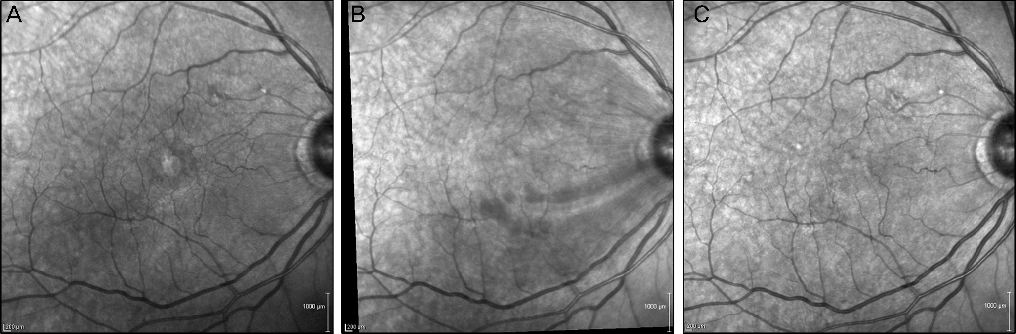 | Figure 1.Pre- and post-operative infrared (IR) images of the left eye of a 78-year-old man who underwent vitrectomy and ILM peeling for epiretinal membrane. (A) Preoperative IR image with signs of macular pucker due to epiretinal membrane. (B) IR image of postoperative 7 days demonst rating dark arcuate striae radiating from the optic nerve head toward the macula. Several round dark spots are visible at temporal ends of the striae, which are considered as signs of trauma from the ILM forceps. (C) IR image of post-operative 6 months revealing subsided swelling of the retinal nerve fiber layer. |
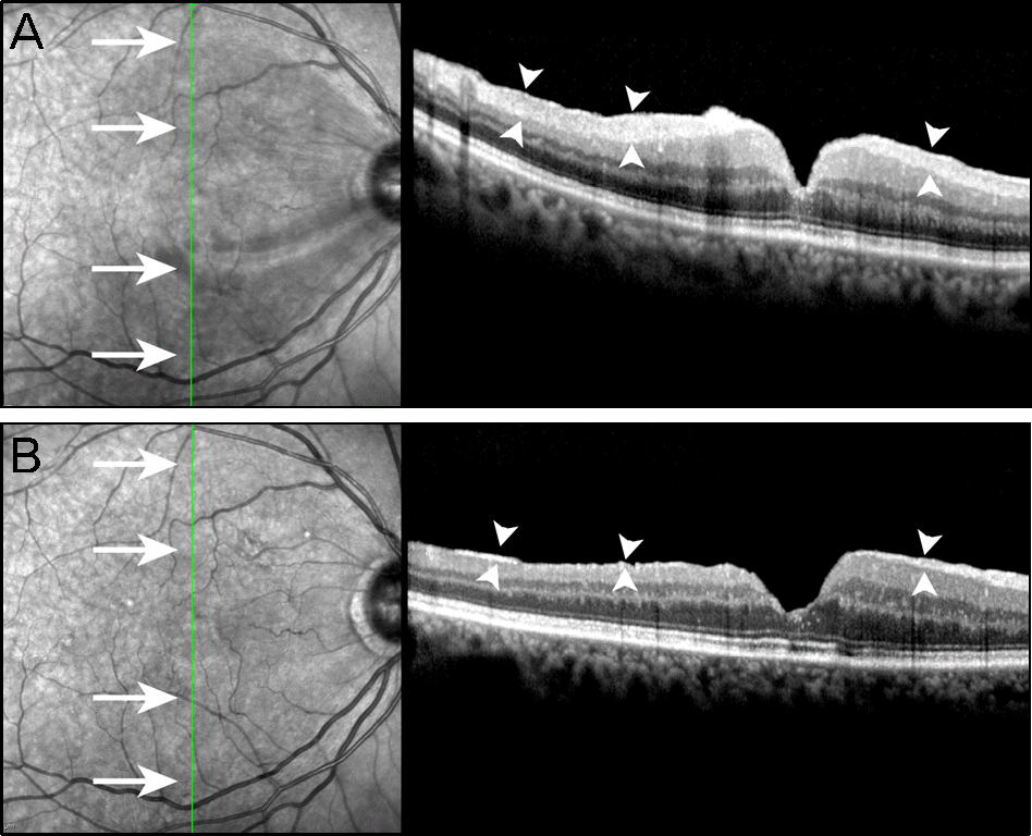 | Figure 2.Postoperative infrared (IR) photographs and spectral domain optical coherent tomography (SD-OCT) images of the patient who demonstrated swelling of retinal nerve fiber layer (RNFL). (A) SD-OCT image at 1-week postoperatively demonstrates swelling of the RNFL (arrowheads), which corre-sponds to the dark striae on the IR image (arrow). (B) Postoperative 6 months follow-up IR and SD-OCT image at the same plane reveals resolution of the dark striae (arrow) and the corresponding swelling of RNFL (arrowheads). |
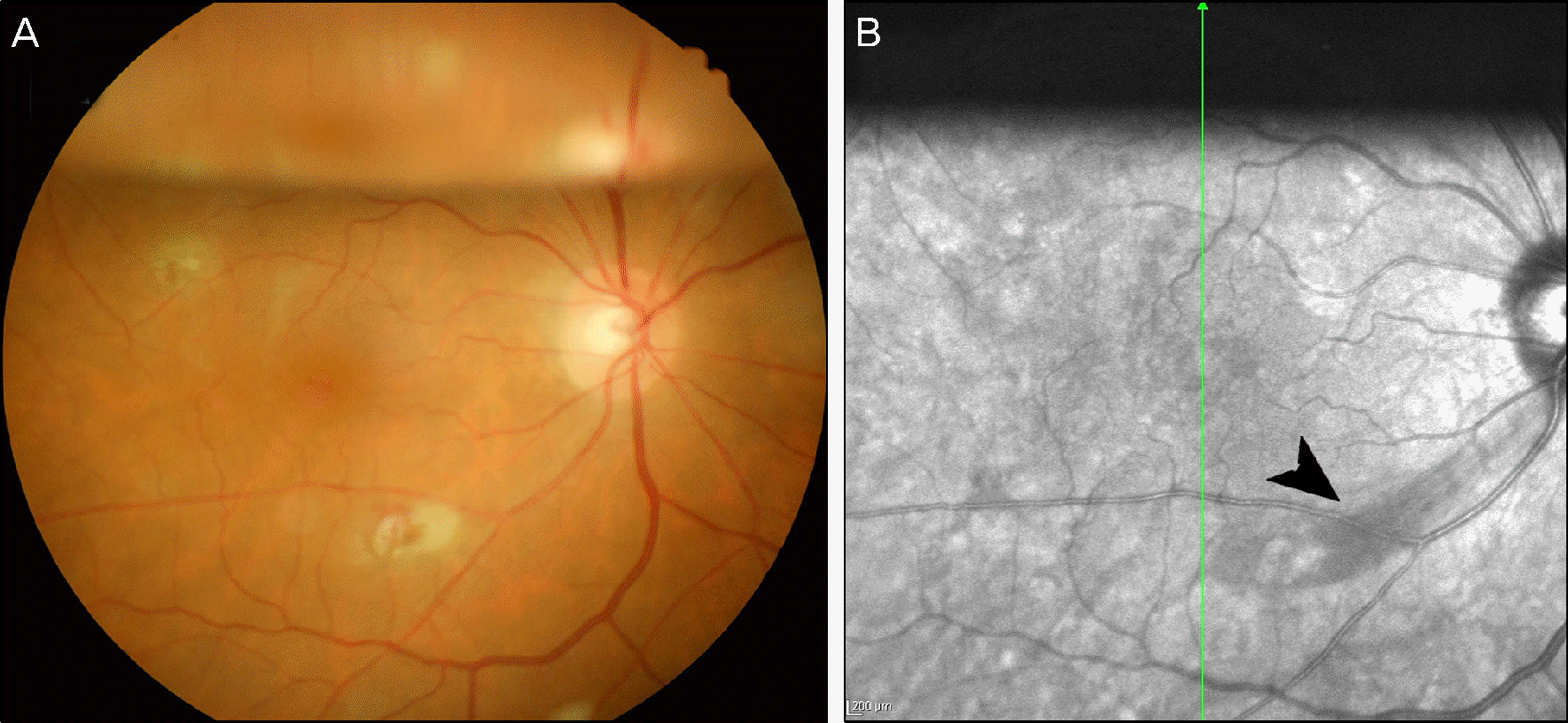 | Figure 3.Color and infrared (IR) images of postoperative day 7 in a 69-year-old woman who underwent vitrectomy and ILM peeling for a epiretinal membrane. (A) The color photograph demonstrates the gas bubble above and endodiathermy site inferior to the macula, but no visible striae. (B) The IR image demonstrates one thick dark striae, radiating from optic nerve head to below the papillomacular bundle (arrowhead), which was not visible in the color fundus photograph. A gas bubble is seen above, and endodiathermy site is visible at the end of the dark striae. |
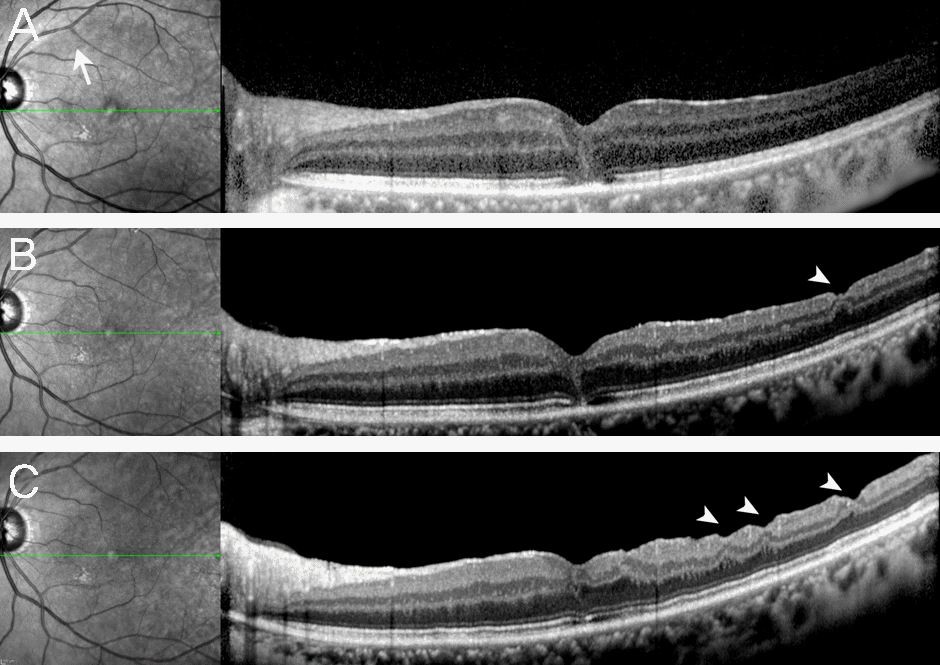 | Figure 4.Postoperative images of infrared (IR) photographs and spectral domain optical coherent tomography (SD-OCT) at 1 week, 3 months, and 6 months postvitrectomy for macular hole. (A) One week postoperative IR image illustrates swelling of the retinal nerve fiber layer (RNFL) radiating above the macular area (arrow), and the SD-OCT image demonstrates a relatively normal temporal macula after surgery. (B) Three-month postoperative IR image illustrates resolution of RNFL swelling, and SD-OCT image reveals slight thinning of the temporal retina and the development of “dimples” similar to that described in dissociated optic nerve fiber layer (arrowhead). (C) Six-month postoperative images demonstrate further thinning and deep-ening of “dimples” in the temporal macula (arrowhead). |
Table 1.
Demographic characteristics of subjects in this study
| SRNFL | No SRNFL | Total | |
|---|---|---|---|
| Sex (male/female) | 3/1 | 12/24 | 15/25 |
| Retinal pathology (ERM/MH) | 3/1 | 23/14 | 26/15 |
| Age* (years) | 68.75 ± 11.15 | 61.81 ± 9.42 | 62.49 ± 9.67 |
| Range (years) | 53-78 | 43-83 | 43-83 |
Table 2.
Pre- and postoperative visual acuities in patients with and without swelling of retinal nerve fiber layer
|
ERM |
MH |
Total |
|||||||
|---|---|---|---|---|---|---|---|---|---|
| SRNFL | No SRNFL | p* | SRNFL | No SRNFL | p* | SRNFL | No SRNFL | p* | |
| Preoperative BCVA (log MAR) | 0.30 ± 0.42 | 0.49 ± 0.37 | 0.64 | 0.53 ± 0.67 | 0.64 ± 0.29 | 0.79 | 0.41 ± 0.48 | 0.55 ± 0.36 | 0.62 |
| 6 month postoperative BCVA (log MAR) | 0.15 ± 0.21 | 0.32 ± 0.33 | 0.51 | 0.30 ± 0.28 | 0.43 ± 0.36 | 0.92 | 0.23 ± 0.22 | 0.36 ± 0.34 | 0.59 |
 XML Download