

 PDF
PDF ePub
ePub Citation
 Print
Print


Abstract
Purpose
To report the therapeutic efficacy of plasma exchange therapy on steroid-unresponsive neuromyelitisoptica (NMO) patients.
Case summary
Three patients who had not achieved improvement of visual acuity and visual field after high steroid pulse therapy after optic neuritis visited our clinic with decreasing visual acuity combined with eye pain in the other eye. All patients were diagnosed as neuromyelitisoptica (NMO) based on the presence of NMO-IgG antibody and optic nerve en-hancing in contrast-enhanced orbital magnetic resonance imaging (MRI). Recurrent optic neuritis was observed. Steroid pulse retreatment was started but visual acuity was not improved in all patients. The patients received plasma exchange therapy, followed by immune suppression therapy. All patients showed improved visual acuity and restored visual field promptly without recurrence of neuromyelitisoptica.
Go to : 
References
1. Sellnera J, Boggild M, Clanet M, et al. EFNS guidelines on diagnosis and management of neuromyelitis. Eur Neurol. 2010; 17:1019–32.
2. Watanabe S, Nakashima I, Misu T, et al. Therapeutic efficacy of plasma exchange in NMO-IgG-positive patients with neuromyelitis optica. Mult Scler. 2007; 13:128–32.
3. Keegan M, Pineda AA, McCelland RL, et al. Plasma exchange for severe attacks of CNS demyeliation: predictors of response. Neurology. 2002; 58:143–6.
4. Llufriu S, Castillo J, Blanco Y, et al. Plasma exchange for acute attacks of CNS demyelination: predictors of improvement at 6 months. Neurology. 2009; 73:949–53.

5. Khatri BO, Kramer J, Dukic M, et al. Maintenance plasma exchange therapy for Steroid-refractory neuromyelitis optica. J Clin Apher. 2012; 27:183–92.
6. Wingerchuk DM, Hogancamp WF, O’Brien PC, Weinshenker BG. The clinical course of neuromyelitis optica (Devic's syndrome). Neurology. 1999; 22:1107–14.
7. Lennon VA, Wingerchuk DM, Kryzer TJ, et al. A serum autoanti-body marker of neuromyelitis optica: distinction from multiple sclerosis. Lancet. 2004; 364:2106–12.
8. Wingerchuk DM, Pittock SJ, Lucchinetti CF, et al. A secondary progressive clinical course is uncommon in neuromyelitis optica. Neurology. 2007; 68:603–5.
9. Venero JL, Vizuete ML, Ilunda'in AA, et al. Detailed localization of aquaporin-4 messenger RNA in the CNS: preferential expression inperiventricular organs. Neuroscience. 1999; 94:239–50.
10. Vitellaro-Zuccarello L, Mazzetti S, Bosisio P, et al. Distribution of Aquaporin 4 in rodent spinal cord: relationship with astrocyte markers and chondroitin sulfate proteoglycans. Glia. 2005; 51:148–59.
11. Nakashima I, Fujihara K, Miyazawa I, et al. Clinical and MRI features of Japanese patients with NMO-IgG. J Neurol Neurosurg Psychiatry. 2006; 77:1073–5.
12. Lucchinetti CF, Mandler RN, McGavern D, et al. A role for humor-al mechanisms in the pathogenesis of Devic's neuromyelitis optica. Brain. 2002; 125:1450–61.
13. Narikawa K, Misu T, Fujihara K, et al. CSF chemokine levels in relapsing neuromyelitis optica and multiple sclerosis. J Neuroimmunol. 2004; 49:182–6.
14. Yaguchl H, Sakushima K, Takahashi I, et al. Efficacy of intra-venous cyclophosphamide therapy for neuromyelitis optica spectrum disorder. Intern Med. 2013; 52:969–72.
15. Mandler RN, Ahmed W, Dencoff JE. Devic's neuromyelitis optica: a prospective study of seven patients treated with prednisone and azathioprine. Neurology. 1998; 51:1219–20.
16. Roesner S, Appel R, Gbadamosi J, et al. Treatment of steroid-un-responsive optic neuritis with plasma exchange. Acta Neurol Scand. 2012; 126:103–8.
17. Munemoto M, Otaki Y, Kasma S, et al. Therapeutic efficacy of double filtration plasmapheresis in patients with anti-aquaporin-4 antibody-positive multiple sclerosis. J Clin Neurosci. 2011; 18:478–80.
18. Shin CW, Kim SH, Cho SY, et al. Steroid unresponsive neuromyelitis optica improved with plasmapheresis. J Korean Neurol Assoc. 2009; 27:417–20.
19. Seo JD, Kim SH, Park KP. Complete recovery of visual acuity by plasmapheresis in neuromyelitis optica. Korean J Clin Neurophysiol. 2011; 13:114–6.
20. Shin CW, Kim SH, Cho SY, et al. Steroid unresponsive neuromyelitis optica improved with plasmapheresis. J Korean Neurol Assoc. 2009; 27:417–20.
21. Cortese I, Chaudhry V, So YT, et al. Evidence-based guideline update: plasmapheresis in neurologic disorders: report of the Therapeutics and Technology Assessment Subcommittee of the Academy of Neurology. Neurology. 2011; 76:294–300.
22. Gartzena K, Limmrothb V, Putzkia N. Relapsing neuromyelitis optica responsive to glatiramer acetate treatment. Eur J Neurol. 2007; 14:e12–3.
23. Cree BA, Lamb S, Morgan K, et al. An open label study of the effects of rituximab in neuromyelitis optica. Neurology. 2005; 64:1270–2.
Go to :
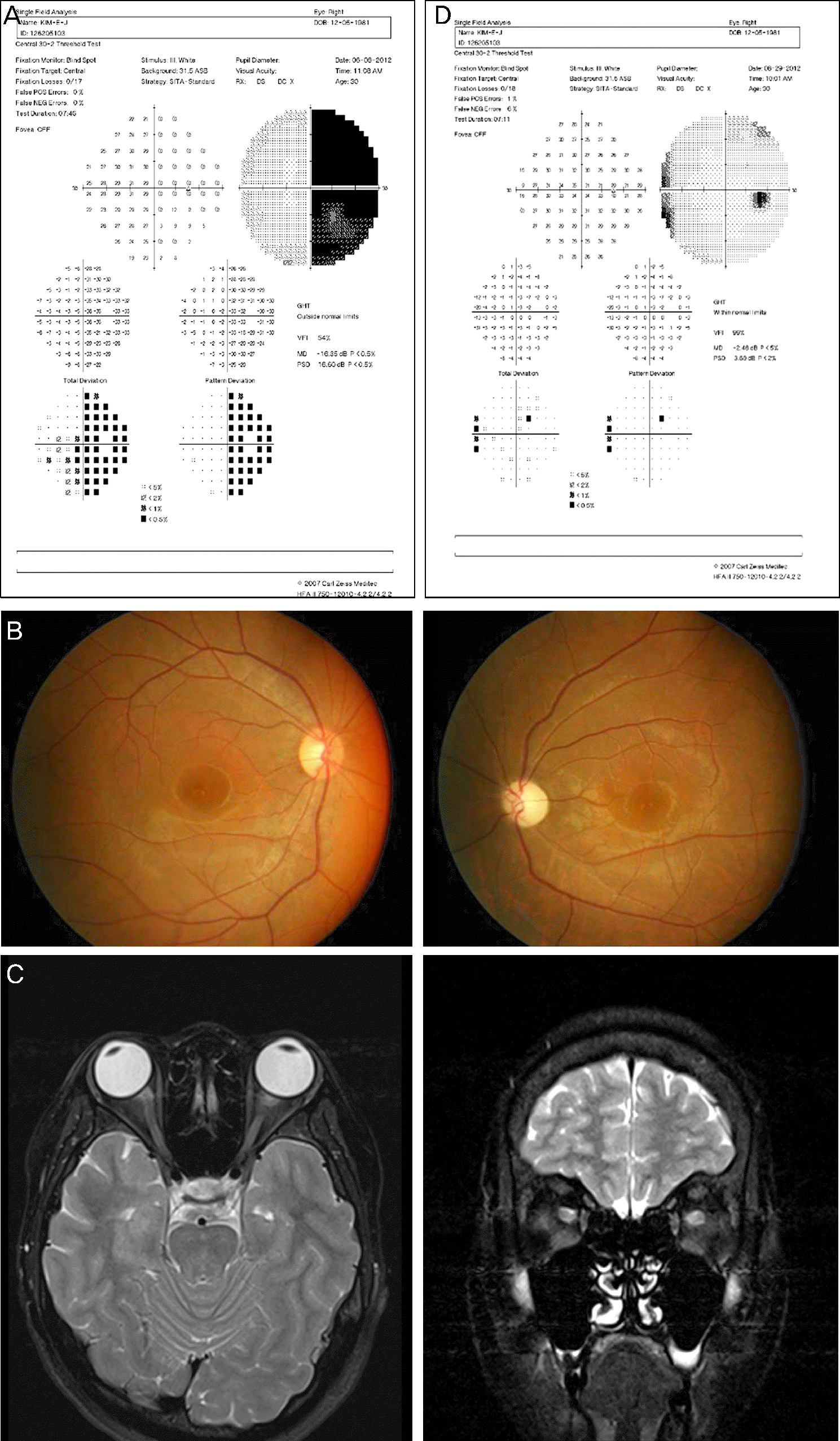 | Figure 1.(A) Visual field test shows right hemianopsia in the right eye. (B) Fundus photo shows optic atrophy in the left eye. (C) In orbit MRI enhanced by omniscan, both optic nerves are enhanced. (D) Visual field test shows improvement of visual field defects in the right eye after plasma exchange at last visit visit (1.5 years after onset time). |
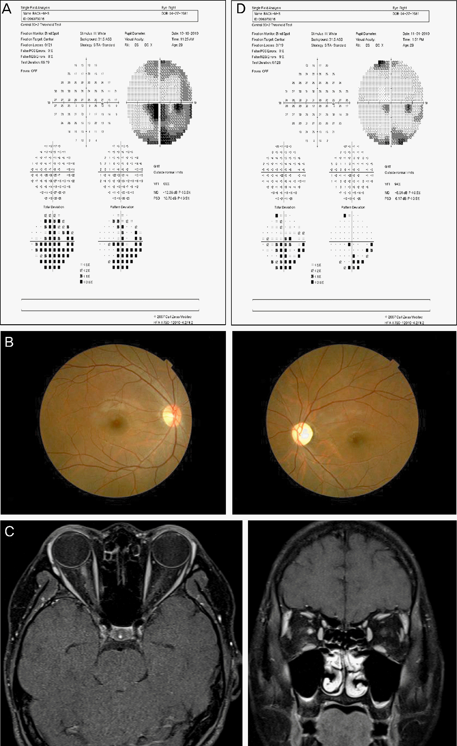 | Figure 2.(A) Visual field test shows diffuse visual field defects in the right eye. (B) Fundus photo shows optic atrophy in the left eye. (C) In orbit MRI enhanced by megaray, optic nerve of the right eye is enhanced. (D) Visual field test shows improvement of visual field defects in the right eye after plasma exchange at last visit (3 years after onset time). |
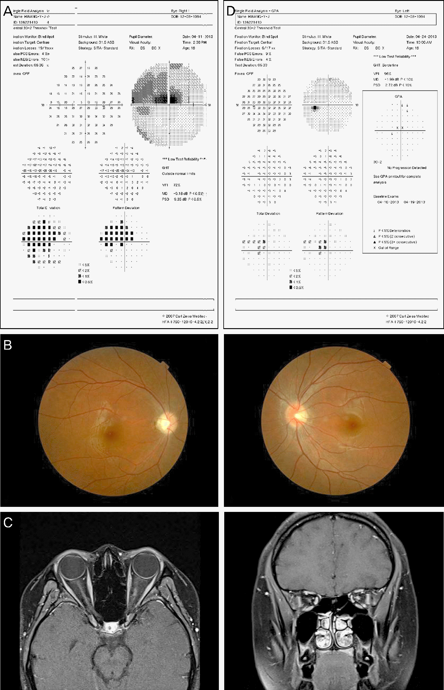 | Figure 3.(A) Visual field test shows partial visual field defect in the left eye. (B) Fundus photo shows optic atrophy in the right eye. (C) In orbit MRI enhanced by megaray, optic nerve of the left eye is enhanced. (D) Visual field test shows improvement of visual field defect in the left eye after plasma exchange at last visit (6 months after onset time). |
 XML Download