Abstract
Case summary
A 52-year-old man visited our hospital with a recurrent mass on his right upper eyelid, which had developed 4 years prior. Initially, he received laser therapy at a dermatologic clinic to remove the mass. Two years later, the mass recurred and was excised at another clinic. At the time the patient visited our institution, the lesion had developed into multiple erythematous nodules at the margin of the right upper eyelid. The results of excisional biopsy performed under local anesthesia revealed hidradenoma papilliferum. One month after excision, recurred multiple elevated nodules were found at the margin of the excision, and thus total excision of the mass and reconstruction of the upper eyelid was performed. Biopsy confirmed that the mass was apocrine adenocarcinoma. Five months have passed since the excision and no evidence of recurrence has been observed.
Go to : 
References
1. Paties C, Taccagni GL, Papotti M, et al. Apocrine carcinoma of the skin: a clinicopathologic, immunocytochemical and ultrastructural study. Cancer. 1993; 71:375–81.

2. Cooper PH. Carcinomas of sweat glands. Pathol Annu. 1987; 22:83–124.
3. Shintaku M, Tsuta K, Yoshida H, et al. Apocrine adenocarcinoma of the eyelid with aggressive biological behavior: report of a case. Pathol Int. 2002; 52:169–73.

4. Thomson SJ, Tanner NS. Carcinoma of the apocrine glands at the base of eyelashes; a case report and discussion of histological diagnostic criteria. Br J Plast Surg. 1989; 42:598–602.

5. Valenzuela AA, Cupp DG, Heathcote JG. Primary apocrine adenocarcinoma of the eyelid. Orbit. 2012; 31:316–8.

6. Kage M, Nakamura Y, Ozumi K. A case report of equivocal neo-plasm originating from an apocrine gland on the eyelid. Acta Pathol Jpn. 1990; 40:431–4.
7. Mazoujian G, Pinkus GS, Davis S, Haagensen DE Jr. Immunohistochemistry of a gross cystic disease fluid protein (GCDFP-15) of the breast. A marker of apocrine epithelium and breast carcinomas with apocrine features. Am J Pathol. 1983; 110:105–12.
10. Kipkie GF, Haust MD. Carcinoma of apocrine gland; report of case. AMA Arch Derm. 1958; 78:440–5.
11. Mazoujian G, Margolis R. Immunohistochemistry of gross cystic disease fluid protein (GCDFP-15) in 65 benign sweat gland tumors of the skin. Am J Dermatopathol. 1988; 10:28–35.

12. Tsubura A, Senzaki H, Sasaki M, et al. Immunohistochemical dem-onstration of breast-derived and/or carcinoma-associated glyco-proteins in normal skin appendages and their tumors. J Cutan Pathol. 1992; 19:73–9.

13. Qureshi HS, Ormsby AH, Lee MW, et al. The diagnostic utility of p63, CK5/6, CK7, and CK20 in distinguishing primary cutaneous adnexal neoplasms from metastatic carcinomas. J Cutan Pathol. 2004; 31:145–52.
Go to : 
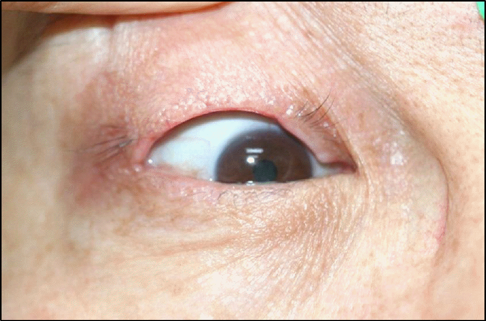
 | Figure 1.Clinical gross photograph of right eye showing the multiple erythematous nodules at the margin of right upper eyelid (A) and recurred nodules at 1 month after excisional biopsy (B). |
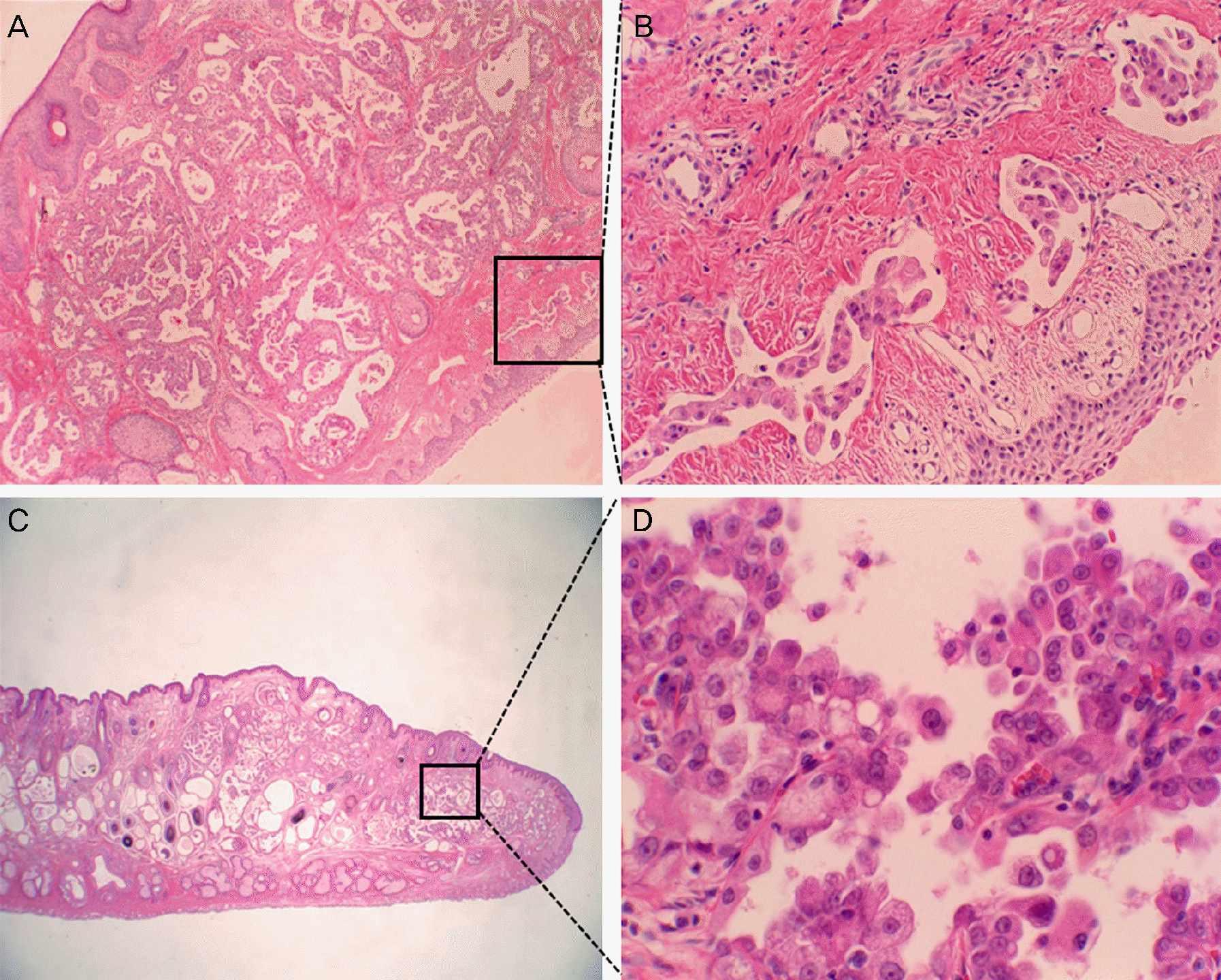
 | Figure 2.(A) Intraluminal papillary growth of apocrine gland epithelium with stratification and lymphatic spreading in the first biopsy. (B) High power view of lymphatic invasion of the tumor cell nests. (C) The same tumor is noted in the second biopsy and (D) High power views of the stratified carcinoma cells. (A) H&E, ×40, (B) ×200, (C) ×10, (D) ×400. |


 PDF
PDF ePub
ePub Citation
 Print
Print


 XML Download