

 PDF
PDF ePub
ePub Citation
 Print
Print


Abstract
Methods
We retrospectively reviewed stereo fundus photographs of 590 glaucoma patients and 273 non-glaucoma patients. An experienced investigator evaluated the presence or absence of the gray crescent (a crescent-shaped, slate-gray pigmentation on the periphery of the neuroretinal rim) which is entirely inside the scleral crescent. Correlations with age, gender, refractive error, disc diameters, and the presence of glaucoma or peripapillary atrophy were also analyzed.
Results
Out of 863 patients, the gray crescent was observed in 166 patients and was found in 19.0% of glaucoma patients and 19.8% of non-glaucoma patients. The gray crescent was most often located temporally (30.1%) and most frequently occurred within only 1 quadrant (63.9%). The prevalence of the gray crescent was not correlated with refractive error (p = 0.61) or the occurrence of glaucomatous optic neuropathy (p = 0.25), but was significantly related to peripapillary atrophy (p < 0.001) and the horizontal diameter of the optic disc (p = 0.001).
Conclusions
The gray optic disc crescent is a common finding within a glaucomatous or non-glaucomatous eye and factors significantly related to occurrence of the gray crescent include peripapillary atrophy and the horizontal diameter of the optic disc. Patients with gray crescent require special attention when the optic disc is examined.
Go to : 
References
1. Shields MB. Gray crescent in the optic nerve head. Am J Ophthalmol. 1980; 89:238–44.

2. Higginbotham L, Shafranov G, Shields MB. Gray optic disc crescent influence of ethnicith in a glaucoma population. J Glaucoma. 2007; 16:572–6.
3. Jonsson O, Damji KF, Jonasson F, et al. Epidemiology of the optic nerve gray crescent in the Reykjavik Eye Study. Br J Ophthalmol. 2005; 89:36–9.
4. Fournier AV, Damji KF, Epstein DL, Pollock SC. Disc excavation in dominant optic atrophy: differentiation from normal tension glaucoma. Ophthalmology. 2001; 108:1595–602.
5. Araie M, Sekine M, Suzuki Y, Koseki N. Factors contributing to the progression of visual field damage in eyes with normal-tension glaucoma. Ophthalmology. 1994; 101:1440–4.
6. Jonas JB, Nguyen XN, Gusek GC, Naumann GO. Parapapillary chorioretinal atrophy in normal and glaucoma eyes. I. Morphometric data. Invest Ophthalmol Vis Sci. 1989; 30:908–18.
7. Jonas JB, Naumann GO. Parapapillary chorioretinal atrophy in normal and glaucoma eyes. II. Correlations. Invest Ophthalmol Vis Sci. 1989; 30:1604–11.
8. Park KH, Tomita G, Liou SY, Kitazawa Y. Correlation between peripapillary atrophy and optic nerve damage in normal-tension glaucoma. Ophthalmology. 1996; 103:1899–906.
9. Jonas JB. Clinical implications of peripapillary atrophy in glaucoma. Curr Opin Ophthalmol. 2005; 16:84–8.
10. Teng CC, De Moraes CG, Prata TS, et al. The region of largest β-zone parapapillary atrophy area predicts the location of most rapid visual field progression. Ophthalmology. 2011; 118:2409–13.
Go to :
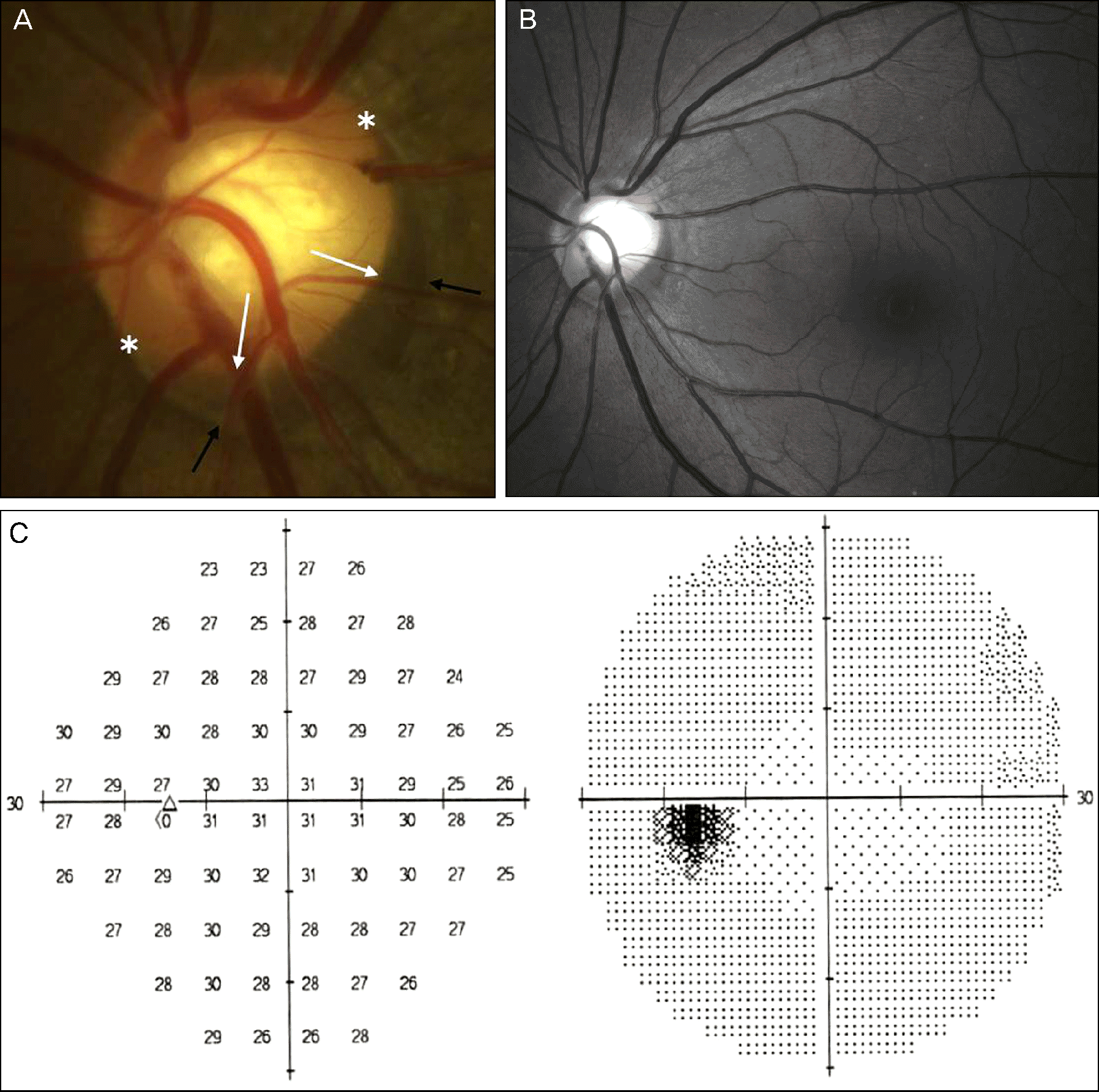 | Figure 1.A non-glaucoma patient with a wide gray optic disc crescent (white arrows) in the inferior quadrants of optic nerve head. The gray crescent is inside the narrow, white scleral crescent (black arrows). The extent of the gray crescent is between asterisks (A). The vertical cup-to-disc ratio of this patient is 0.5 and inferior-temporal neuroretinal rim is intact. However if the gray crescent is mistaken for PPA, vertical cup-to-disc ratio is 0.6 and the inferior-temporal neuroretinal rim looks thinner than the superior or nasal rim. The retinal nerve fiber layer photography (B) and visual field test (C) are normal in this patient. |
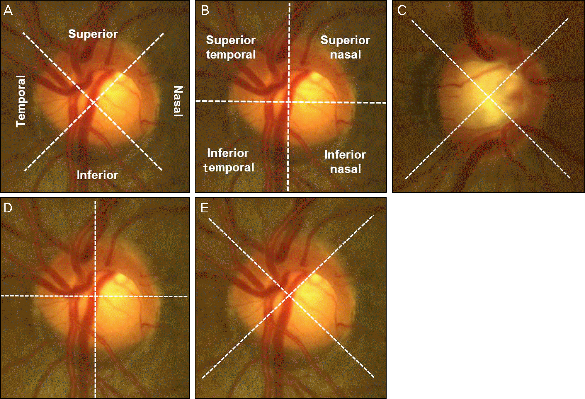 | Figure 2.The definition of the location of the gray crescent (A, B). Example of temporal located (C) and inferior located (bottom) gray crescent. The center of gray crescent in second example (D, E) was included in the inferior nasal and inferior quadrant. In this case the center of the gray crescent is closer to the midline of the inferior quadrant, we defined this location as the inferior quadrant. |
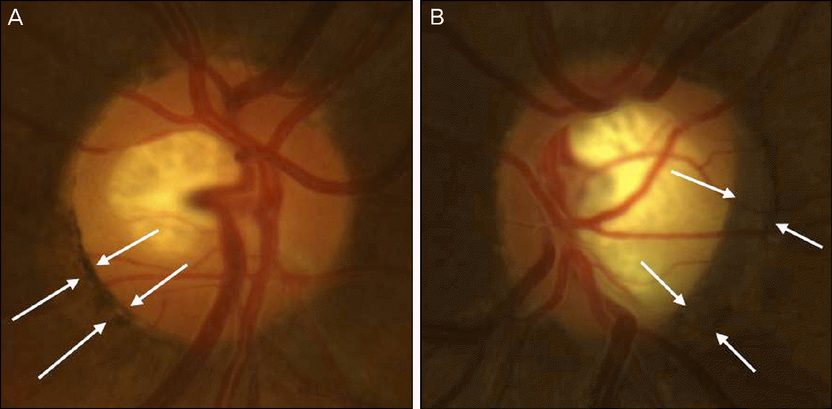 | Figure 3.The optic disc with conus pigmentosus in the inferior temporal quadrant (A) and the optic disc with large gray crescent in the temporal quadrant (B). Note that the conus pigmentosus has more darker and irregular borders and the gray crescent appears to be located more deeply on the disc. |
Table 1.
The location of the gray crescent
Table 2.
The circumferential extent of the gray crescent
| Extent | Number of eyes (right) | Percentage of eyes (right) | Number of eyes (left) | Percentage of eyes (left) |
|---|---|---|---|---|
| <90° | 64 | 63.4 | 64 | 61.0 |
| 90-180° | 31 | 30.7 | 34 | 32.4 |
| 180-270° | 6 | 5.9 | 7 | 6.7 |
| >270° | 0 | 0.0 | 0 | 0.0 |
Table 3.
Associations between gray crescent and other parameters
 XML Download