

 PDF
PDF ePub
ePub Citation
 Print
Print


Abstract
Purpose
To evaluate the characteristics of choroidal nevus using the enhanced depth imaging spectral domain optical coherence tomography (EDI SD-OCT), with a comparison to the B scan ultrasound (BUS) findings.
Methods
Medical records of 124 eyes of 124 choroidal nevus patients were reviewed retrospectively. All patients underwent fundus photography (FP), EDI SD-OCT, and BUS.
Results
Of 124 eyes with choroidal nevus examined by EDI SD-OCT, 43 eyes (35%) displayed good images to study. The most common EDI-OCT imaging features included choroidal shadowing, choriocapillary thinning, retinal pigment epithelial changes, and overlying subretinal fluid. The mean nevus thickness was 817 μm (120-1850 μm) by EDI-OCT compared 1295 μm (780-2400 μm) by BUS. The mean difference in the tumor thickness between two techniques was 475 μm (27-1319 μm) (p < 0.05).
Conclusions
These results have suggested that imaging of choroidal nevus with EDI-OCT shows superior measurement of its characteristics compared with ultrasonography. The clinical utility of this modality is emerging. EDI-OCT is useful in distinguishing suspicious nevi from other chorioretinal lesions, detecting tumor re-growth along the treatment margin, and demonstrating retinal or choroid tumor location.
Go to : 
References
1. Ng CH, Wang JJ, Mitchell P, et al. Prevalence and characteristics of choroidal nevi in an Asian vs white population. Arch Ophthalmol. 2009; 127:314–9.

2. Singh AD, Kalyani P, Topham A. Estimating the risk of malignant transformation of a choroidal nevus. Ophthalmology. 2005; 112:1784–9.
3. Shields CL, Cater J, Shields JA, et al. Combination of clinical factors predictive of growth of small choroidal melanocytic tumors. Arch Ophthalmol. 2000; 118:360–4.
4. Sakata LM, Deleon-Ortega J, Sakata V, Girkin CA. Optical coherence tomography of the retina and optic nerve - a review. Clin Experiment Ophthalmol. 2009; 37:90–9.
5. Spaide RF, Koizumi H, Pozzoni MC. Enhanced depth imaging spectral-domain optical coherence tomography. Am J Ophthalmol. 2008; 146:496–500.
6. Kiernan DF, Mieler WF, Hariprasad SM. Spectral-domain optical coherence tomography: a comparison of modern high-resolution retinal imaging systems. Am J Ophthalmol. 2010; 149:18–31.
7. Shields CL, Materin MA, Shields JA. Review of optical coherence tomography for intraocular tumors. Curr Opin Ophthalmol. 2005; 16:141–54.
8. Shields CL, Mashayekhi A, Materin MA, et al. Optical coherence tomography of choroidal nevus in 120 patients. Retina. 2005; 25:243–52.
9. Muscat S, Parks S, Kemp E, Keating D. Secondary retinal changes associated with choroidal naevi and melanomas documented by optical coherence tomography. Br J Ophthalmol. 2004; 88:120–4.
10. Schaudig U, Hassenstein A, Bernd A, et al. Limitations of imaging choroidal tumors in vivo by optical coherence tomography. Graefes Arch Clin Exp Ophthalmol. 1998; 236:588–92.
11. Shields CL, Furuta M, Berman EL, et al. Choroidal nevus transformation into melanoma: analysis of 2514 consecutive cases. Arch Ophthalmol. 2009; 127:981–7.
12. Espinoza G, Rosenblatt B, Harbour JW. Optical coherence tomography in the evaluation of retinal changes associated with suspicious choroidal melanocytic tumors. Am J Ophthalmol. 2004; 137:90–5.
13. Materin MA, Raducu R, Bianciotto C, Shields CL. Fundus auto-fluorescence and optical coherence tomography findings in choroidal melanocytic lesions. Middle East Afr J Ophthalmol. 2010; 17:201–6.
14. Shah SU, Kaliki S, Shields CL, et al. Enhanced depth imaging optical coherence tomography of choroidal nevus in 104 cases. Ophthalmology. 2012; 119:1066–72.
15. Collaborative Ocular Melanoma Study Group. Comparison of clinical, echographic, and histopathological measurements from eyes with medium-sized choroidal melanoma in the collaborative ocular melanoma study: COMS report no. 21. Arch Ophthalmol. 2003; 121:1163–71.
Go to :
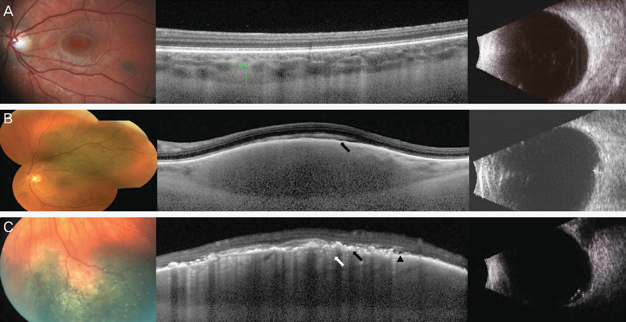 | Figure 1.Color fundus photograph (FP), EDI-OCT and BUS of choroidal nevi. (A) FP showing pigmented choroidal nevus. EDI-OCT showing no drusen, RPE thinning, and thinning of choriocapillaris (CC). (B) FP showing pigmented choroidal nevus. EDI-OCT showing RPE thinning (black arrow), and normal CC. (C) FP showing pigmented choroidal nevus with overlying drusen. EDI-OCT showing drusen, trace subretinal fluid (arrowhead), RPE thinning (black arrow), and marked thinning of CC (white arrow). |
 XML Download