

 PDF
PDF ePub
ePub Citation
 Print
Print


Abstract
Purpose
In this study we analyzed and objectified the characteristics of the Avellino corneal dystrophy patients consider-ing disease severity using in vivo confocal microscopy (IVCM).
Methods
Each corneal layer of 36 eyes in 18 patients with Avellino corneal dystrophy was examined using IVCM (ConfoScan 4.0, NIDEK, Co. Ltd., Albignasego, Italy). Patients were classified into 3 groups based on disease severity (mild, moderate, or severe).
Results
In the mild group, hyper-reflective granular deposits without dark shadows were observed in the anterior stroma. As the disease progressed, corneal deposits were also found at the posterior stroma and epithelium, and clusters of hyper-reflective corneal deposits resembling stromal opacity were noted. The range of corneal deposits measured using Z-scan optical pachymeter was 111.14 ± 30.95 um in the mild group, 157.47 ± 25.00 um in the moderate group, and 193.42 ± 52.23 um in the severe group (p < 0.05).
Go to : 
References
1. Klintworth GK. Advances in the molecular genetics of corneal dystrophies. Am J Ophthalmol. 1999; 128:747–54.

2. Kim TI, Pak JH, Chae JB, et al. Mitomycin C inhibits recurrent Avellino dystrophy after phototherapeutic keratectomy. Cornea. 2006; 25:220–3.
3. Ferry AP, Benson WH, Weinberg RS. Combined granular-lattice (‘Avellino’) corneal dystrophy. Trans Am Ophthalmol Soc. 1997; 95:61–77.
4. Konishi M, Mashima Y, Nakamura Y, et al. Granular-lattice (Avellino) corneal dystrophy in Japanese patients. Cornea. 1997; 16:635–8.
5. Akiya S, Takahashi H, Nakano N, et al. Granular-lattice (Avellino) corneal dystrophy. Ophthalmologica. 1999; 213:58–62.
6. Yi JH, Ha BJ, Kim SW, et al. The number of cases, cause and treatment of avellino corneal dystrophy exacerbated after LASIK. J Korean Ophthalmol Soc. 2008; 49:1415–24.
7. Chung SH, Kim CY, Kim EK. The classification and clinical characteristics in Korean patients with avellino corneal dystrophy. J Korean Ophthalmol Soc. 2005; 46:938–44.
8. Akiya S, Takahashi H, Nakano N, et al. Granular-lattice (Avellino) corneal dystrophy. Ophthalmologica. 1999; 213:58–62.
9. Korvatska E, Munier FL, Chaubert P, et al. On the role of kera-to-epithelin in the pathogenesis of 5q31-linked corneal dystrophies. Invest Ophthalmol Vis Sci. 1999; 40:2213–9.
10. Kanai A, Yamaguchi T, Nakajima A. [The histochemical and analytical electron microscopy studies of the corneal granular dystrophy (author's transl)]. Nihon Ganka Gakkai Zasshi. 1977; 81:145–54.
11. Munier FL, Korvatska E, Djemaï A, et al. Kerato-epithelin mutations in four 5q31-linked corneal dystrophies. Nat Genet. 1997; 15:247–51.
Go to :
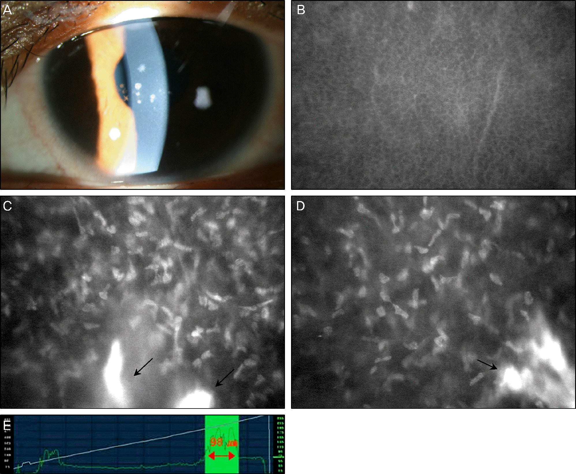 | Figure 1.(Case 1) Corneal images obtained from left eye of a “mild” Avellino corneal dystrophy patient by confocal microscopy. (A) Slit lamp photograph reveals mild granular corneal deposits. (B) Normal wing cell layer of the corneal epithelium. (C) Deposition of hyper-reflective materials in the anterior stroma (black arrows). (D) Some hyper-reflective deposits in the mid-stroma (black arrow). (E) The range of corneal deposits measured by Z-scan optical pachymeter is 98 um. |
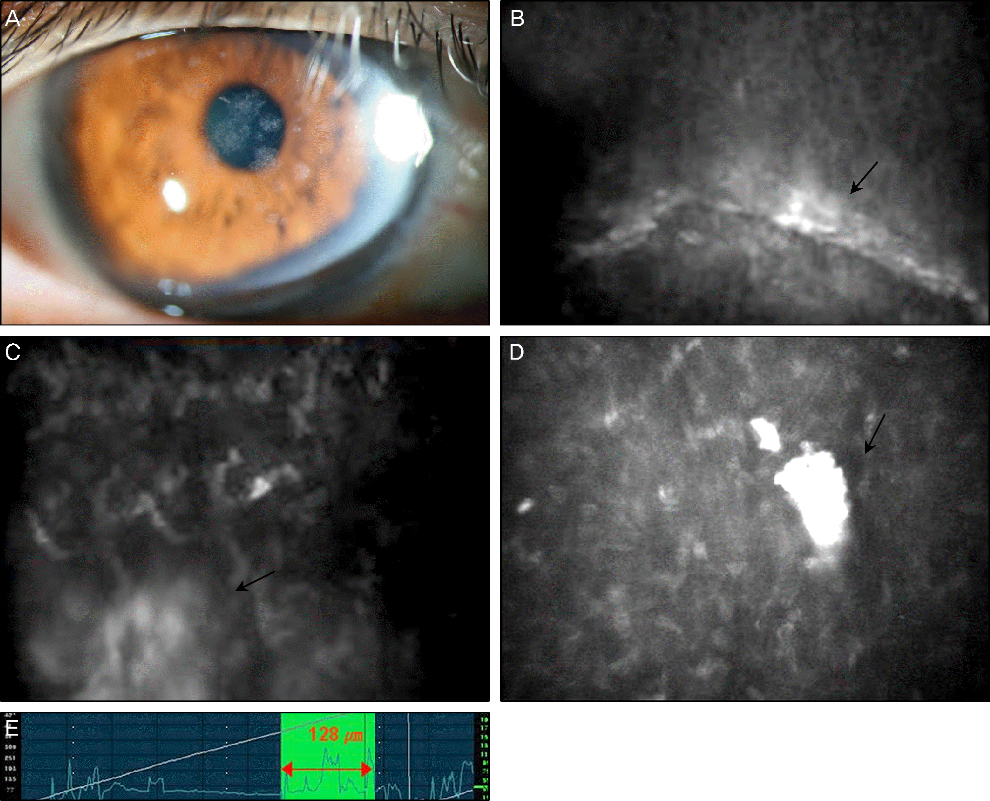 | Figure 2.(Case 2) Corneal images obtained from left eye of a “moderate” Avellino corneal dystrophy patient by confocal microscopy. (A) Slit lamp photograph reveals round white corneal deposits and scattered stellate opacities. (B) Hyper-reflective deposits are found in the area of epithelium (black arrow). (C) Hyper-reflective materials deposit in the anterior stroma (white arrow). (D) Cluster of hyper-reflective deposits in the posterior stroma (black arrow). (E) The range of corneal deposits measured by Z-scan optical pachymeter is 128 um. |
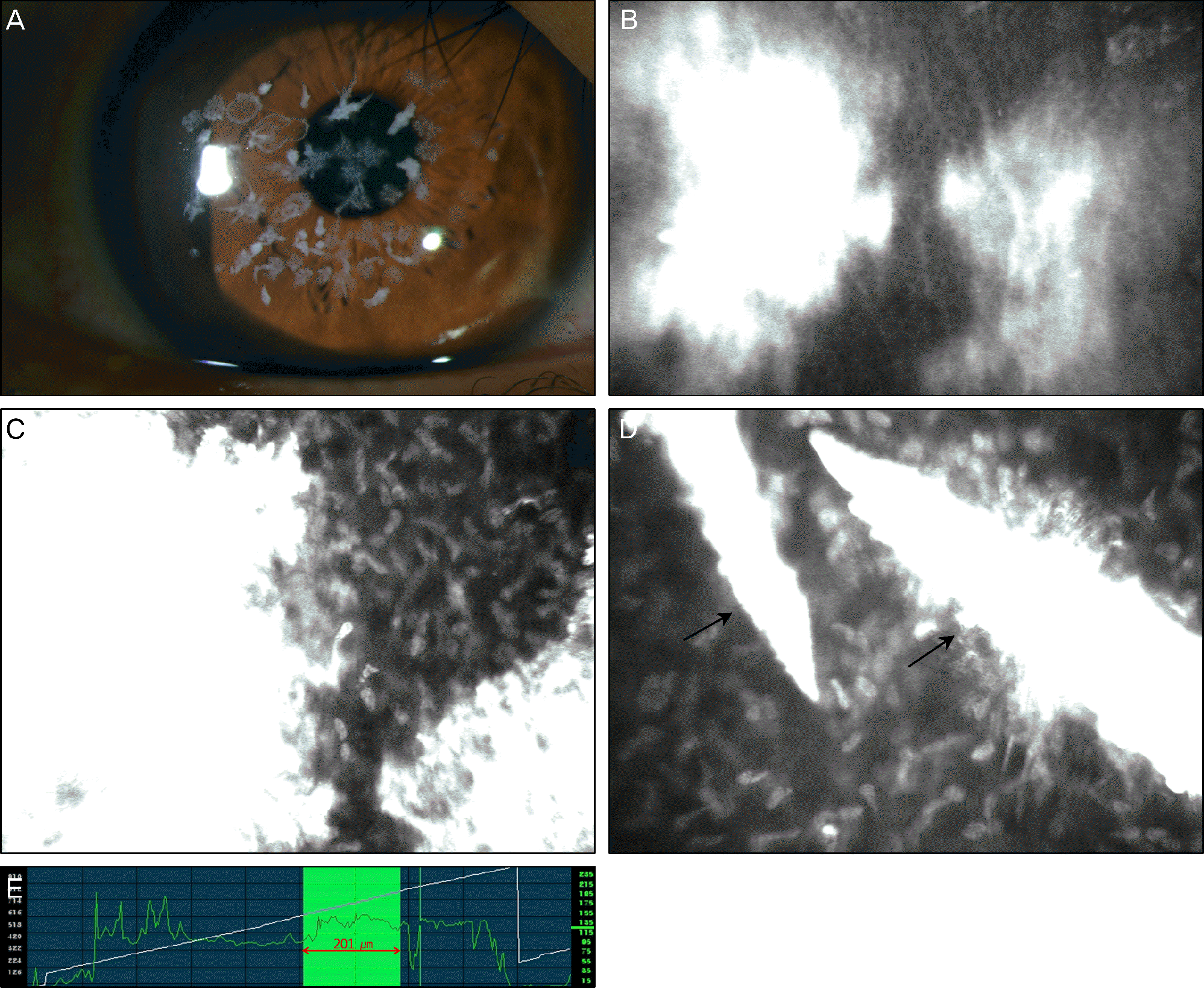 | Figure 3.(Case 3) Corneal images obtained from the left eye of a “severe” Avellino corneal dystrophy patient by confocal microscopy. (A) Slit lamp photograph reveals multiple round white-gray corneal deposits. (B) Hyper-reflective corneal deposits in the wing cell layer of corneal epithelium. (C) Cluster of hyper-reflective deposits in the anterior stroma, resembling stromal scar. (D) Linear shaped hyper-reflective deposits are observed in the posterior stroma (black arrows). (E) The range of corneal deposits measured by Z-scan optical pachymeter is 201 um. |
Table 1.
Baseline information of the Avellino corneal dystrophy patients
 XML Download