

 PDF
PDF ePub
ePub Citation
 Print
Print


Abstract
Purpose
We report a case of a full-thickness macular hole which occurred many years after a blunt eye trauma leading to choroidal rupture.
Case summary
A 50-year-old male visited our clinic with a complaint of decreased vision in his left eye 2 years in duration. He experienced a blunt trauma to his left eye with a baseball when he was 6 years old, although he did not complain of any visual disturbance in the left eye at that time. Fundus examination revealed a full-thickness macular hole with vertical fibrotic scar at the temporal side of the macula, which was thought to be a choroidal rupture induced by the previous blunt eye trauma. We performed vitrectomy and intravitreal tamponade injection. Two months later, the full-thickness macular hole completely closed and visual acuity of the left eye improved.
References
1. Arana B, Fonollosa A, Artaraz J. . Macular hole secondary to toxoplasmic retinochoroiditis. Int Ophthalmol. 2013. Mar 12; [Epub ahead of print].

2. Gaudric A, Haouchine B, Massin P. . Macular hole formation: new data provided by optical coherence tomography. Arch Ophthalmol. 1999; 117:744–51.
3. Colucciello M, Nachbar JG.Macular hole following ruptured reti-nal arterial macroaneurysm. Retina. 2000; 20:94–6.
4. Beatty S, Harrison RJ, Roche P.Bilateral macular holes resulting from septic embolization. Am J Ophthalmol. 1997; 123:557–9.
5. Gass JD.Lamellar macular hole: a complication of cystoid macular edema after cataract extraction. Arch Ophthalmol. 1976; 94:793–800.
6. Morgan CM, Schatz H.Involutional macular thinning. A pre-macular hole condition. Ophthalmology. 1986; 93:153–61.
7. Huang J, Liu X, Wu Z, Sadda S.Comparison of full-thickness trau-matic macular holes and idiopathic macular holes by optical coher-ence tomography. Graefes Arch Clin Exp Ophthalmol. 2010; 248:1071–5.
8. Yamada H, Sakai A, Yamada E. . Spontaneous closure of trau-matic macular hole. Am J Ophthalmol. 2002; 134:340–7.
9. Gass JD.Idiopathic senile macular hole. Its early stages and pathogenesis. Arch Ophthalmol. 1988; 106:629–39.
10. Bainbridge J, Herbert E, Gregor Z.Macular holes: vitreoretinal re-lationships and surgical approaches. Eye (Lond). 2008; 22:1301–9.
11. Azzolini C, Patelli F, Brancato R.Correlation between optical co-herence tomography data and biomicroscopic interpretation of idi-opathic macular hole. Am J Ophthalmol. 2001; 132:348–55.
12. Tanner V, Chauhan DS, Jackson TL, Williamson TH.Optical co-herence tomography of the vitreoretinal interface in macular hole formation. Br J Ophthalmol. 2001; 85:1092–7.
13. Kobayashi H, Kobayashi K, Okinami S.Macular hole and myopic refraction. Br J Ophthalmol. 2002; 86:1269–73.
14. Zografos L, Chamero J.[Long-term course of indirect traumatic ruptures of the choroid]. J Fr Ophtalmol. 1990; 13:269–75.
15. Hirata A, Tanihara H.Ruptured internal limiting membrane associated with blunt trauma revealed by indocyanine green staining. Graefes Arch Clin Exp Ophthalmol. 2004; 242:527–30.
16. Korobelnik JF, Bernard JA, Chauvaud D, Pouliquen Y.Choroid rupture, macular hole and subretinal neovessels following contusions. J Fr Ophtalmol. 1993; 16:549–51.
17. Chauhan DS, Antcliff RJ, Rai PA. . Papillofoveal traction in macular hole formation: the role of optical coherence tomography. Arch Ophthalmol. 2000; 118:32–8.
18. Ito Y, Terasaki H, Suzuki T. . Mapping posterior vitreous de-tachment by optical coherence tomography in eyes with idiopathic macular hole. Am J Ophthalmol. 2003; 135:351–5.
19. Sebag J.Anomalous posterior vitreous detachment: a unifying concept in vitreo-retinal disease. Graefes Arch Clin Exp Ophthalmol. 2004; 242:690–8.
Figure 1.
At the initial visit, fundus examination shows normal in the right eye and multiple linear parallel fibrotic scar temporal to the fovea with macular hole and an 1 disc diameter sized round RPE change at superotemporal proximal major vascular arcade were observed. Best corrected visual acuity was 20/50 in the left eye.
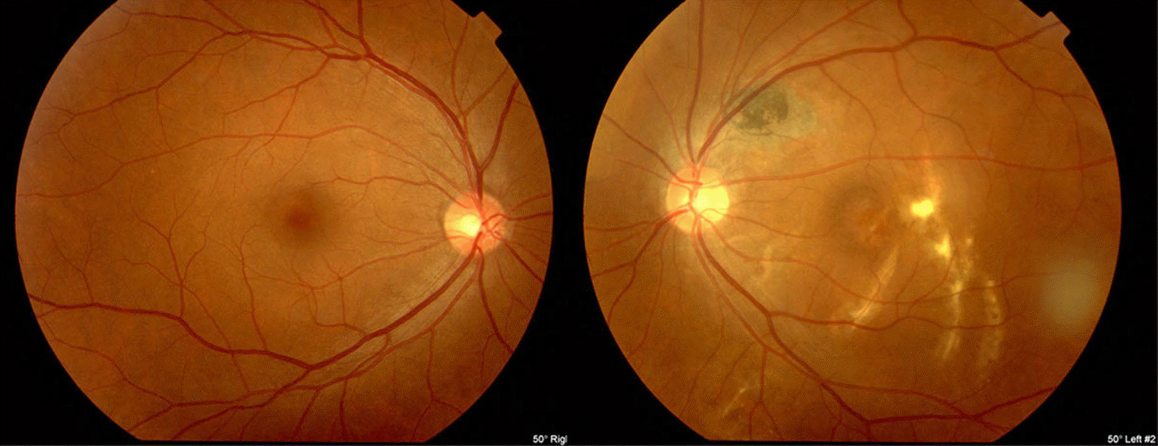
Figure 2.
(A) Foveal vertical section, surface, and outer retinal images spectral-domain optical coherence tomography (SD-OCT) of the left eye. Stage 2 full thickness macular hole (MH) with retinal schisis at the margin of MH are observed. (B) Oblique section by spectral-domain optical coherence tomography (SD-OCT) of fibrotic scar of left eye. Several hyperreflective protrusion are observed at the subretinal pigment epithelial level.
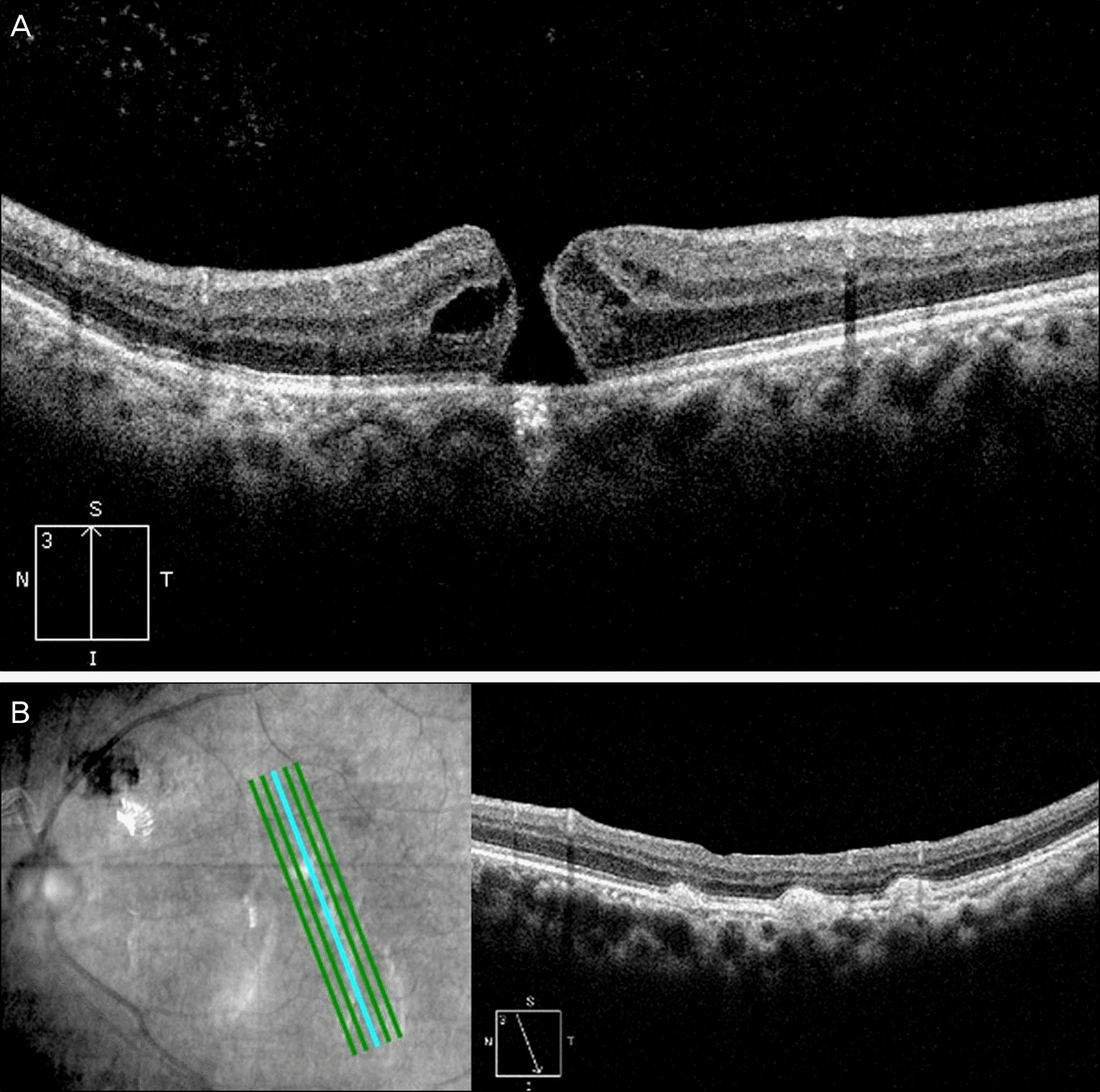
Figure 3.
Fluorescein angiography showed hyperfluorescence temporal to the fovea and the lesion of superotemporal proximal major vascular arcade due to atrophy of the retinal pigment epithelium in the early phase (A), stain of the dye in the late phase (B).
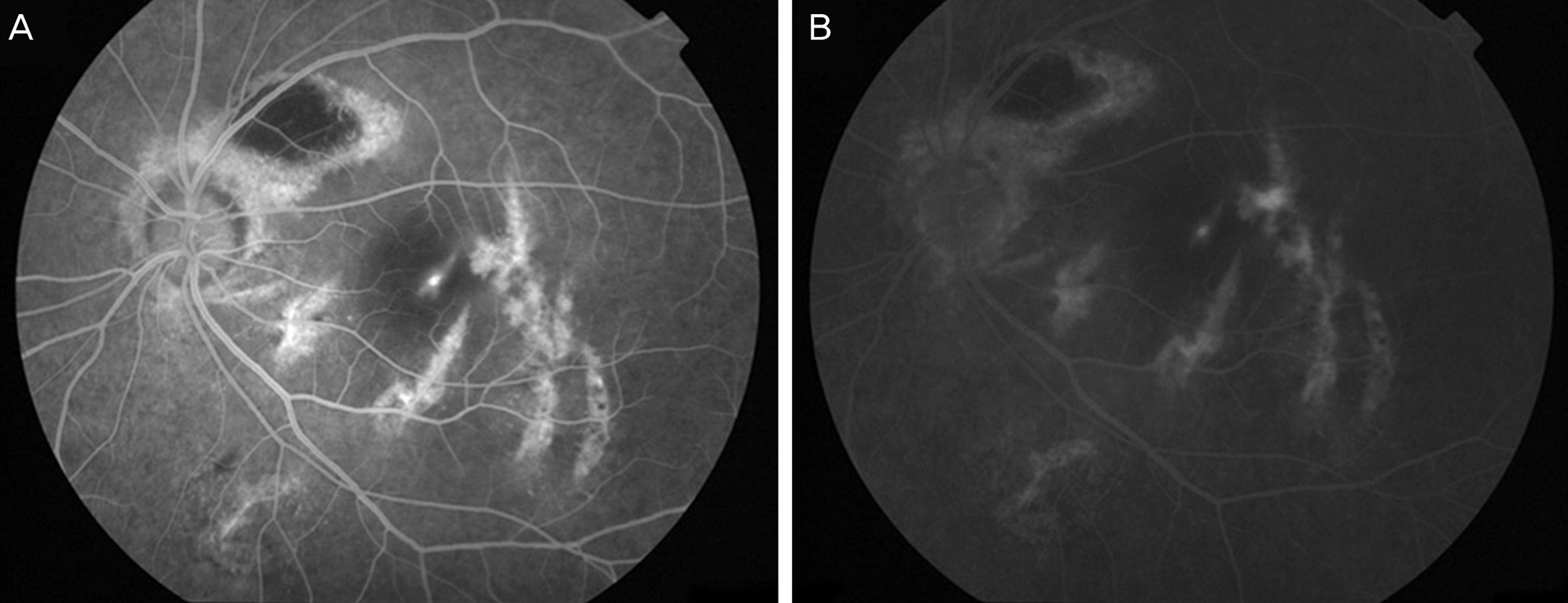
Figure 4.
The comparison of 3 directional macular images and ILM-RPE thickness (μm) between the case of present study (A) and idiopathic macular hole (B). Macular thickness is increased with asymmetric traction line in the superonasal direction in the present case (A). But Symmetrically increased macular thickness is observed with anteroposterior traction of the vitreous in idiopathic macular hole (B).
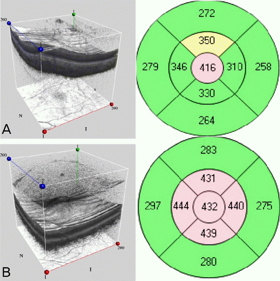
Figure 5.
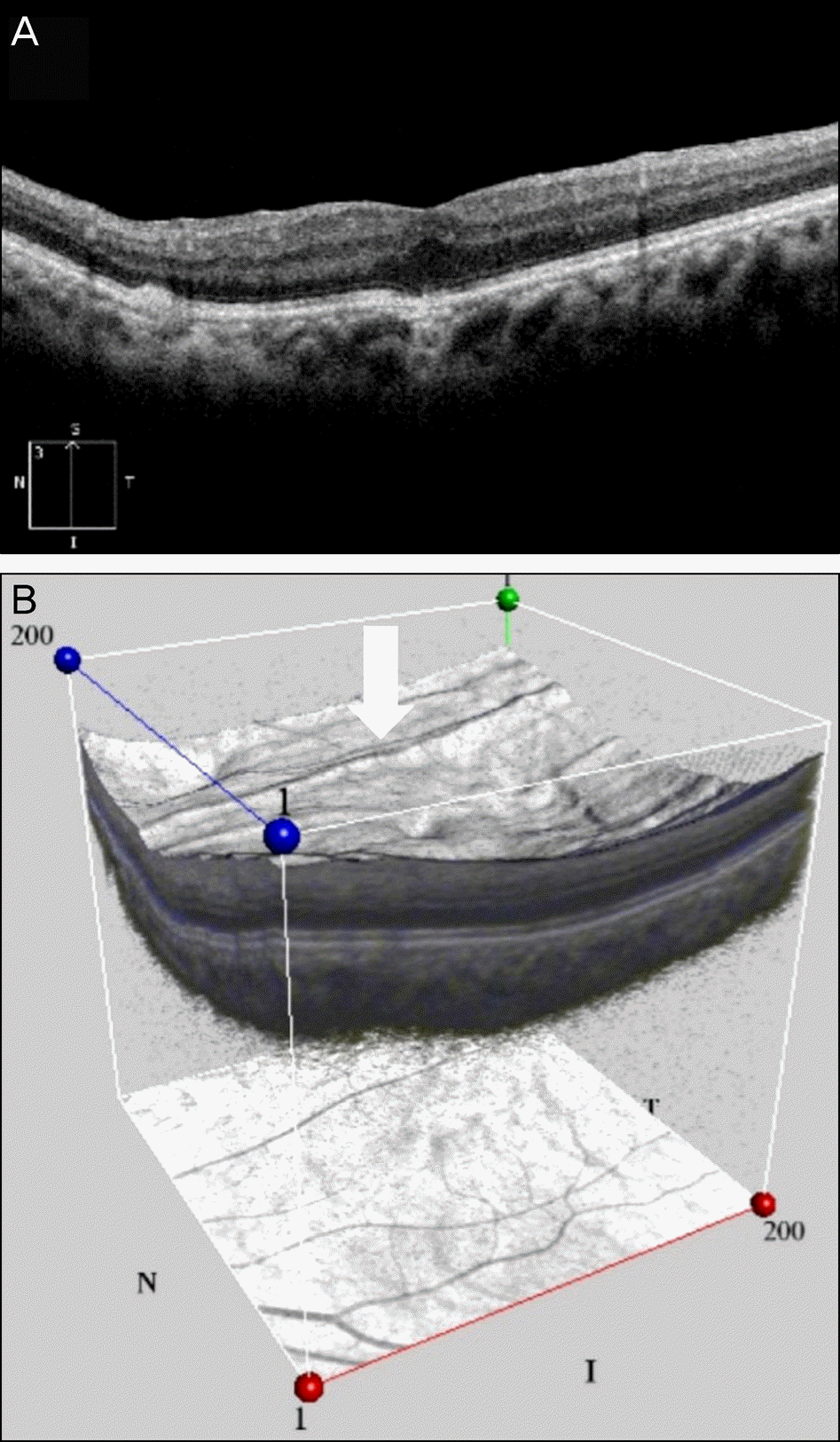
Three months after the surgery, a closed MH and remnant foveal photoreceptor disruption are observed (A). Best corrected visual acuity improved to 20/32 in the left eye. Even after macular hole had been closed successfully, horizontal traction line (white arrow) was seen superior to the macular (B).
 XML Download