Abstract
Purpose
To report a case of streptoococcus gordonii endophthalmitis after pneumaic retinopexy in a patient with rhegmatogenous retinal detachment.
Case summary
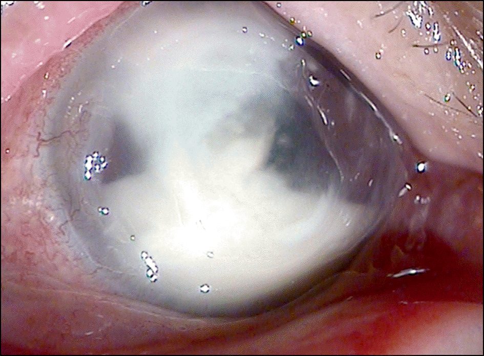
A 40-year-old man presented with a right eye macula-on retinal detachment extending from 9 to 1 o’clock with one-clock-hour hole at 11 o’clock. After sterilizing with a Betadine solution, 0.6 cc 100% SF6 gas was injected into the vitreous through the pars plana at 11 o’clock. Two days after the injection, eyeball pain, cell and flare, and pupillary membrane developed. Under the diagnosis of endophthalmitis, vitreous tap and intravitreous vancomycin (1.0 mg/0.1 cc) and ceftazime (2.0 mg/0.1 cc) were administered. However the symptoms and signs worsened, so vitrectomy was performed, and intravitrous injections of silicone, vancomycin and ceftazime were administered. Streptococcus gordonii was identified from the excised vitreous. Visual acuity was light perception due to severe retinal necrosis.
References
1. Hilton GF, Grizzard WS.Pneumatic retinopexy. A two-step out-patient operation without conjunctival incision. Ophthalmology. 1986; 93:626–41.
2. Kim KS, Kim JO, Lee SY.Comparison of pneumatic retinopexy with scleral buckling in the management of rhegmatogenous reti-nal detachments. J Korean Ophthalmol Soc. 1995; 36:1954–63.
4. Hilton GF, Tornambe PE.Pneumatic retinopexy. An analysis of in-traoperative and postoperative complications. The retinal detach-ment study group. Retina. 1991; 11:285–94.
5. Eckardt C.Staphylococcus epidermidis endophthalmitis after pneu-matic retinopexy. Am J Ophthalmol. 1987; 103:720–1.

6. Baek CM, Kim JW, Park WT, Kim KS.The factors influencing to the results to pneumatic retinopexy. J Korean Opthalmol Soc. 2003; 44:2242–9.
7. KimTY Kwon OW, Kim YR. Treatment of retinal detachments by pneumatic retinopexy. J Korean Ophthalmol Soc. 1988; 29:689–95.
8. Seo MS, Kim CR.Clinical evaluation of pneumatic retinopexy. J Korean Ophthalmol Soc. 2000; 41:831–7.
9. Lee SH, Kim KS, Oh JS.Clinical report of pneumatic retinopexy. J Korean Ophthalmol Soc. 1992; 33:827–33.
10. Cho WH, Lee DC, Chang MH.Three cases of pneumoretinopexy for rhegmatogenous retinal detachment by multiple retinal tears over 1 hour in distance. J Korean Ophthalmol Soc. 2005; 46:2110–4.
11. Lee SE, Chang MH.The success rate and factors influencing the results of pneumatic retinopexy. J Korean Ophthalmol Soc. 2013; 54:1241–7.

12. Tornambe PE, Hilton GF.Pneumatic retinopexy. A multicenter randomized controlled clinical trial comparing pneumatic reti-nopexy with scleral buckling. The retinal detachment study group. Ophthalmol. 1989; 96:772–83.
Table 1.
Culture and antibiotic sensitivity test of aspirated vitreous


 PDF
PDF ePub
ePub Citation
 Print
Print


 XML Download