

 PDF
PDF ePub
ePub Citation
 Print
Print


Abstract
Purpose
We present a case with conjunctival inclusion cyst at inferior fornix treated by marsupialization.
Case summary
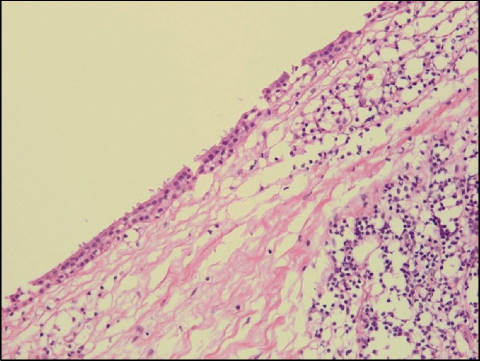
A 23-year-old woman visited our clinic complaining of left lower eyelid swelling. Ophthalmologic examination and CT scan showed a cystic mass from inferior conjunctival fornix to anterior orbit with shallow fornix and focal symblepharon. The cyst was effectively removed with marsupializaion. Postoperatively, there was no recurrence of cyst and the fornix was deepened.
Go to : 
References
1. Sameshima SS, Beyer-Machule CK.Acquired ptosis associated with a conjunctival cyst. Ophthal Plast Reconstr Surg. 1988; 4:159–62.

2. Memarzadeh F, Chuck RS, McCulley TJ.Fornix reconstruction with conjunctival inclusion cyst marsupialization in Stevens-Johnson syndrome. Ophthal Plast Reconstr Surg. 2006; 22:475–6.
3. Goodglick TA, Mertz P, Wolfley D. . Ciliated respiratory-like epithelium forming cystic conjunctival lesions in a patient with Stevens-Johnson syndrome. Ophthalmic Surg. 1992; 23:557–9.
4. Lee J, Kwak AY, Chung WS, Ha BJ.A new simple technique for re-moval of subconjunctival cyst under the slit lamp microscope. J Korean Ophthalmol Soc. 2011; 52:1531–6.
5. Kim B, Kang NY.Successful removal of apocrinehydrocytoma us-ing indocyanine green and sodium hyaluronate. J Korean Ophthalmol Soc. 2011; 52:994–8.
6. Desai VN, Shields CL, Shields JA.Orbital cyst in a patient with Stevens-Johnson syndrome. Cornea. 1992; 11:592–4.
7. Harris GJ.Marsupialization of a lacrimal gland cyst. Ophthalmic Surg. 1983; 14:75–8.
8. McCulley TJ, Kersten RC, Yip CC, Kulwin DR.Dacryocystoceles in the aftermath of Stevens-Johnson syndrome. Ophthal Plast Reconstr Surg. 2005; 21:159–61.
Go to :
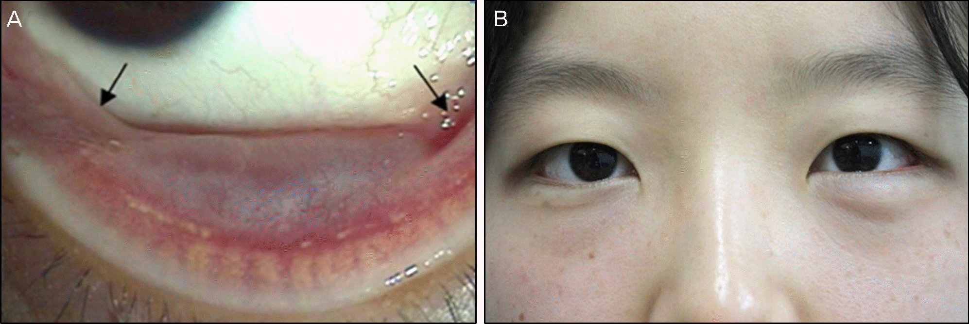 | Figure 1.Anterior segment photograph of conjunctival inclusion cyst at initial presentation. (A) About 1.5 × 1 cm sized subcon-junctival cyst is noted at inferior fornix in the left eye. There is symblepharon at medial and lateral end of cyst (black arrows). (B) External photographs showed contour of the mass, which is transcutaneously visible at left lower eyelid. |
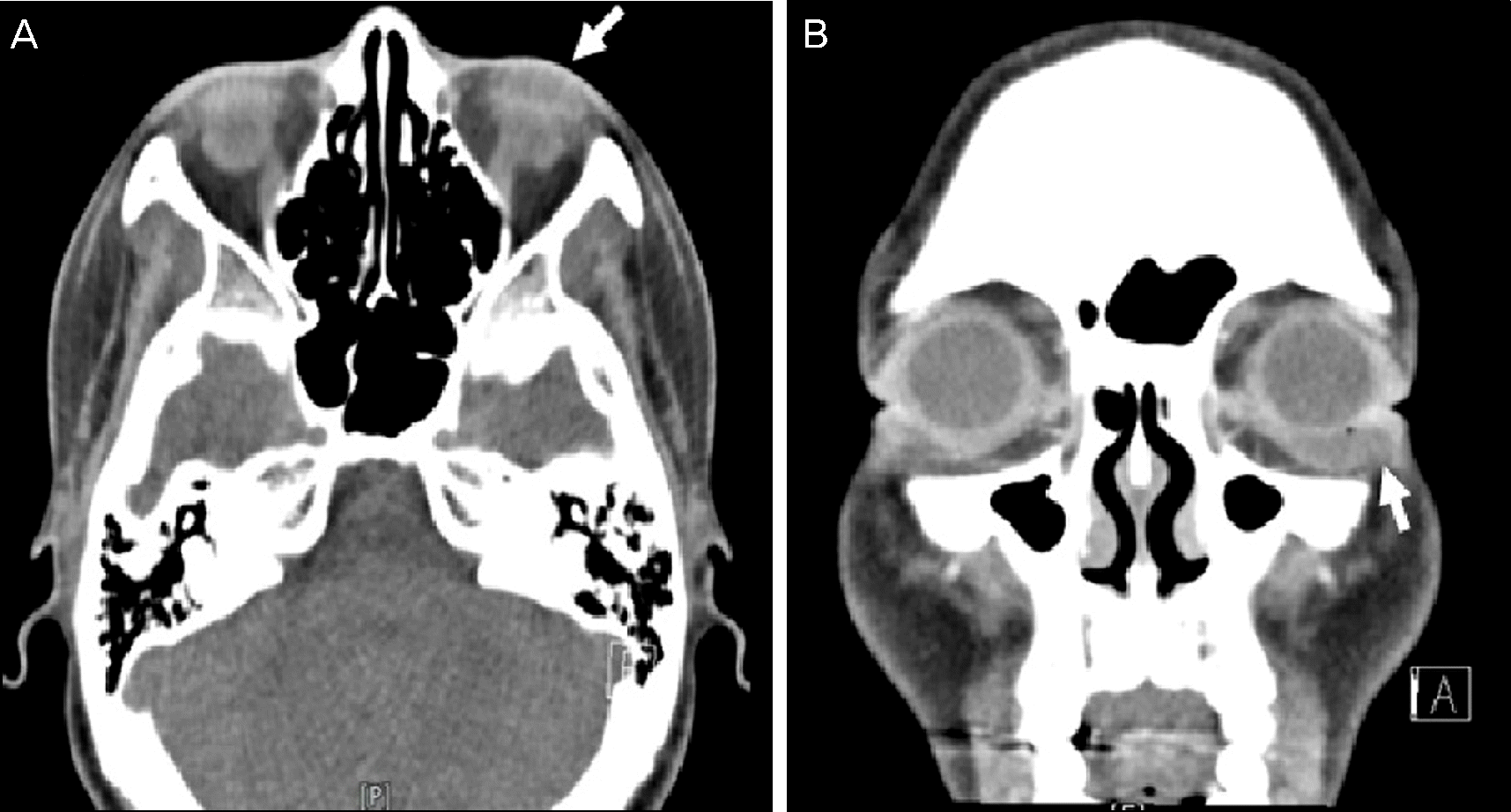 | Figure 2.Axial (A) and coronal (B) view enhanced computed tomography images demonstrate 1.5 × 0.8 cm sized ovoid shaped cystic mass (white arrow) in anterior– inferior part of the left orbit. |
 XML Download