

 PDF
PDF ePub
ePub Citation
 Print
Print


Abstract
Purpose
To evaluate the outcomes and prognostic factors of trabeculectomy with mitomycin C in Korean patients with juvenile open-angle glaucoma (JOAG).
Methods
A retrospective review was performed on 29 eyes with JOAG who had undergone trabeculectomy between January 2004 and January 2013. Intraocular pressure (IOP) and postoperative complications were monitored at 1 day pre-operatively, at 1 day, 1 week, 1 month, 3 months, 6 months, and 12 months postoperatively, and at final follow-up after postoperative 18 months. Surgical success was defined as a final IOP of <21 mm Hg or <80% of preoperative IOP, regard-less of the use of anti-glaucoma medication. Prognostic factors for surgical success or failure were analyzed by the Cox proportional hazards model.
Results
Mean (± standard deviation) age at trabeculectomy was 24.6 (± 8.9) years, and mean follow-up period was 24.3 (± 20.8) months. The overall success rate was 82.8% at final follow-up. The Kaplan-Meier cumulative probabilities of surgical success were 100% at 1 year, 87.4% at 2 years, and 78.7% at 3 years postoperatively. The Cox proportional hazards model failed to determine any significant factors that were associated with surgical failure. The three most frequent post-operative complications were immediately postoperative hypotony (n = 19, 65.5%), hypotony maculopathy (n = 8, 27.6%), and encapsulated bleb (n = 8, 27.6%), most of which were resolved after a minor procedure or observation. Two of 8 eyes with encapsulated bleb required a second surgery. There were no vision-threatening severe complications.
References
1. Turalba AV, Chen TC.Clinical and genetic characteristics of pri-mary juvenile-onset open-angle glaucoma (JOAG). Semin Ophthalmol. 2008; 23:19–25.

2. Wiggs JL, Del Bono EA, Schuman JS. . Clinical features of five pedigrees genetically linked to the juvenile glaucoma locus on chromosome 1q21-q31. Ophthalmology. 1995; 102:1782–9.
3. Costa VP, Katz LJ, Spaeth GL. . Primary trabeculectomy in young adults. Ophthalmology. 1993; 100:1071–6.
4. Stewart RH, Kimbrough RL, Bachh H, Allbright M.Trabeculectomy and modifications of trabeculectomy. Ophthalmic Surg. 1979; 10:76–80.
5. Levene RZ.Glaucoma filtering surgery: Factors that determine pressure control. Ophthalmic Surg. 1984; 15:475–83.
6. D'Ermo F, Bonomi L, Doro D.A critical analysis of the long-term results of trabeculectomy. Am J Ophthalmol. 1979; 88:829–35.
7. Inaba Z.Long-term results of trabeculectomy in the Japanese: an analysis by life-table method. Jpn J Ophthalmol. 1982; 26:361–73.
8. Borisuth NS, Phillips B, Krupin T.The risk profile of glaucoma filtration surgery. Curr Opin Ophthalmol. 1999; 10:112–6.
9. Sidoti PA, Belmonte SJ, Liebmann JM, Ritch R. Trabeculectomy with mitomycin-C in the treatment of pediatric glaucomas. Ophthalmology. 2000; 107:422–9.
10. Cairns JE. Trabeculectomy Preliminary report of a new method. Am J Ophthalmol. 1968; 66:673–9.
11. Mermoud A, Salmon JF, Murray AD.Trabeculectomy with mito-mycin C for refractory glaucoma in blacks. Am J Ophthalmol. 1993; 116:72–8.
12. Tsai JC, Chang HW, Kao CN. . Trabeculectomy with mitomy-cin C versus trabeculectomy alone for juvenile primary open-angle glaucoma. Ophthalmologica. 2003; 217:24–30.
13. Yalvac IS, Nurözler A, Kahraman C. . The results of trabeculectomy with and without mitomycin C in young patients. Ophthalmologica. 1998; 212:399–403.
14. Koraszewska-Matuszewska B, Samochowiec-Donocik E, Filipek E.[Prognosis in juvenile glaucoma after trabeculectomy]. Klin Oczna. 2002; 104:115–8.
15. Jacobi PC, Dietlein TS, Krieglstein GK.Primary trabeculectomy in young adults: long-term clinical results and factors influencing the outcome. Ophthalmic Surg Lasers. 1999; 30:637–46.
16. Gressel MG, Heuer DK, Parrish RK 2nd. Trabeculectomy in young patients. Ophthalmology. 1984; 91:1242–6.
17. Pathania D, Senthil S, Rao HL. . Outcomes of trabeculectomy in juvenile open angle glaucoma. Indian J Ophthalmol. 2013; Epub ahead of print.
18. Jung HR, Kim YY.Initial mitomycin C trabeculectomy in young patients. J Korean Ophthalmol Soc. 1997; 38:424–9.
19. Kim HK, Lee SH.Long-term surgical outcome of juvenile glucoma. J Korean Ophthalmol Soc. 2003; 44:870–5.
20. Perkins ES.Glaucoma in the younger age groups. Arch Ophthalmol. 1960; 64:882–91.
21. Lotufo D, Ritch R, Szmyd L Jr, Burris JE.Juvenile glaucoma, race, and refraction. JAMA. 1989; 261:249–52.
22. Gupta V, Ov M, Rao A. . Long-term structural and functional outcomes of therapy in juvenile-onset primary open-angle glauco-ma: a five-year follow-up. Ophthalmologica. 2012; 228:19–25.
23. Stürmer J, Broadway DC, Hitchings RA.Young patient trabe- culectomy. Assessment of risk factors for failure. Ophthalmology. 1993; 100:928–39.
24. Jacobi PC, Dietlein TS, Krieglstein GK.Adjunctive mitomycin C in primary trabeculectomy in young adults: a long-term study of case-matched young patients. Graefes Arch Clin Exp Ophthalmol. 1998; 236:652–7.
25. Heuer DK, Barton K, Grehn F. . Consensus on definitions of success. Shaarawy TM, Sherwood MB, Grehn F, editors. Guidelines on design and reporting of glaucoma surgical trials. Amsterdam: Kugler publications;2009. chap. 2.
26. Eslami Y, Mohammadi M, Khodaparast M. . Sutureless tunnel trabeculectomy without peripheral iridectomy: a new modification of the conventional trabeculectomy. Int Ophthalmol. 2012; 32:449–54.
27. Jea SY, Francis BA, Vakili G. . Ab interno trabeculectomy ver-sus trabeculectomy for open-angle glaucoma. Ophthalmology. 2012; 119:36–42.
28. Rotchford AP, King AJ.Needling revision of trabeculectomies bleb morphology and long-term survival. Ophthalmology. 2008; 115:1148–53.e4.
29. Palanca-Capistrano AM, Hall J, Cantor LB. . Long-term out-comes of intraoperative 5-fluorouracil versus intraoperative mito-mycin C in primary trabeculectomy surgery. Ophthalmology. 2009; 116:185–90.
30. Quigley HA.Reappraisal of the mechanisms of glaucomatous op-tic nerve damage. Eye (Lond). 1987; 1(Pt 2):318–22.
31. Cahane M, Bartov E.Axial length and scleral thickness effect on susceptibility to glaucomatous damage: a theoretical model im-plementing Laplace's law. Ophthalmic Res. 1992; 24:280–4.
32. Broadway DC, Grierson I, O'Brien C, Hitchings RA.Adverse ef-fects of topical antiglaucoma medication. II. The outcome of filtration surgery. Arch Ophthalmol. 1994; 112:1446–54.
33. Mietz H, Jacobi PC, Welsandt G, Krieglstein GK.Trabeculectomies in fellow eyes have an increased risk of tenon's capsule cysts. Ophthalmology. 2002; 109:992–7.
34. Richter CU, Shingleton BJ, Bellows AR. . The development of encapsulated filtering blebs. Ophthalmology. 1988; 95:1163–8.
Figure 1.
Kaplan-Meier survival curve of surgical outcome of trabeculectomy in patients with juvenile open angle glaucoma. The probabilities of surgical success were 100% at 1 year, 87.4% at 2 years, and 78.7% at 3 years postoperatively.
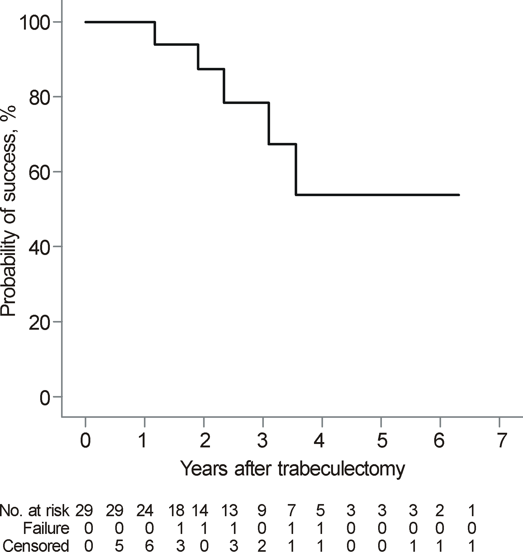
Figure 2.
Mean intraocular pressure (IOP) change profile after trabeculectomy in juvenile open angle glaucoma patients. The postoperative IOP decreased significantly compared to the preoperative level during the whole follow-up period (mean final follow-up, 24.3 ± 20.8 months; all p < 0.001). Error bars indicate ± standard deviation.
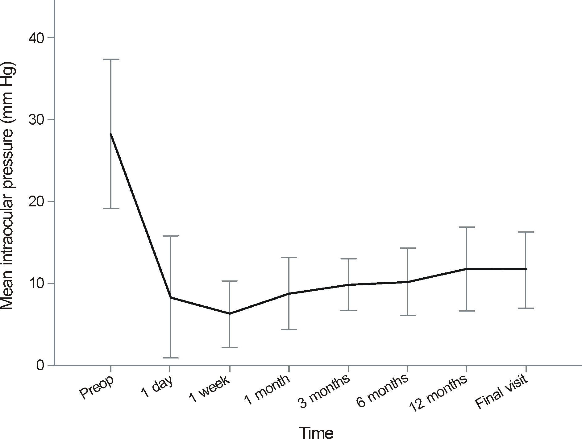
Figure 3.
Inter-eye comparison of Kaplan-Meier survival curves on the outcome of trabeculectomy in bilateral juvenile open angle glaucoma patients who underwent trabeculectomy in both eyes. The respective values of probabilities for surgical success were 53.9% and 88.9% in first- and second-operated eye at the end of the follow-up. Log-rank test revealed no statistical difference in the surgical outcome between two groups (p = 0.425).
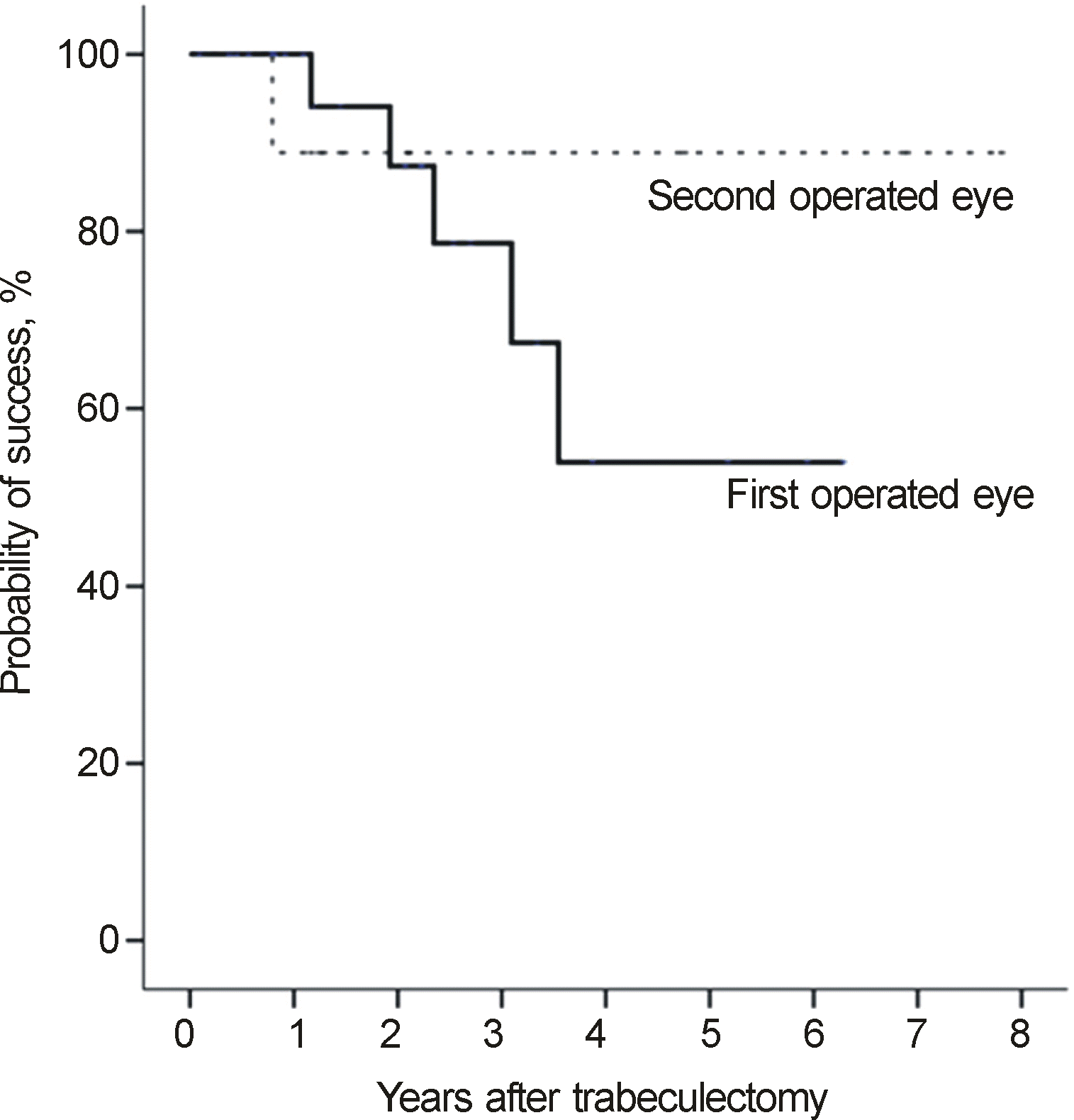
Table 1.
Patients clinical characteristics (n = 29)
Table 2.
Comparison of pre- and postoperative visual acuity, intraocular pressure, number of topical medication and visual field mean deviation
| Preoperative | Postoperative | p-value | |
|---|---|---|---|
| Visual acuity (log MAR scale) | 0.56 ± 0.71 | 0.64 ± 0.78 | 0.276* |
| IOP (mm Hg) | 28.2 ± 9.1 | 12.0 ± 5.0 | <0.001† |
| Number of anti-glaucoma medication (n) | 4.0 ± 0.8 | 1.2 ± 6 | <0.001* |
| Visual field MD (dB) | -17.9 ± 8.9 | -18.2 ± 8.6 | 0.929* |
Table 3.
Comparison between first operated eye and second operated eye
| First operated eye (n = 13) | Second operated eye (n = 13) | p-value | |
|---|---|---|---|
| Preoperative IOP (mm Hg) | 29.5 ± 8.3 | 26.2 ± 6.9 | 0.028* |
| Highest office IOP (mm Hg) | 37.9 ± 8.3 | 33.8 ± 6.9 | 0.081* |
| Final IOP (mm Hg) | 12.9 ± 5.5 | 12.0 ± 4.3 | 0.344* |
| Percent IOP reduction (%) | 53.5 ± 22.8 | 52.4 ± 17.9 | 0.742* |
| Number of topical medication before surgery (n) | 3.5 ± 0.2 | 3.7 ± 0.2 | 0.480† |
| Visual field MD (dB) | -20.9 ± 9.4 | -8.3 ± 10.8 | 0.025† |
| Global RNFL thickness (μm) | 52.9 ± 17.8 | 83.2 ± 26.7 | 0.019† |
| Spherical equivalent (diopters) | -6.4 ± 2.5 | -5.4 ± 2.6 | 0.013* |
| Axial length (mm) | 26.3 ± 1.0 | 26.1 ± 1.4 | 0.506* |
| Central corneal thickness (μm) | 555.0 ± 26.5 | 555.7 ± 30.6 | 0.799† |
Table 4.
Cox proportional hazards model determining likelihood of surgical failure for patients with juvenile open angle glaucoma who underwent trabeculectomy with mitomycin C
Table 5.
Comparison between eyes that achieved surgical success and eyes that did not achieved surgical success
| Success group (n = 24) | Failure group (n = 5) | p-value | |
|---|---|---|---|
| Age (years) | 23.6 ± 8.6 | 29.4 ± 9.5 | 0.222* |
| Female Sex (n, %) | 5 (20.8) | 1 (20) | 0.967† |
| Familial history of glaucoma (n, %) | 3 (12.5) | 1 (20) | 0.658† |
| History of steroid usage (n, %) | 10 (41.7) | 0 (0) | 0.075† |
| Preoperative IOP (mm Hg) | 28.1 ± 8.6 | 28.8 ± 12.5 | 0.674* |
| Highest office IOP (mm Hg) | 36.4 ± 9.1 | 32.4 ± 10.5 | 0.448* |
| Final IOP (mm Hg) | 10.5 ± 3.8 | 18.8 ± 4.1 | 0.001* |
| Percent IOP reduction (%) | 58.9 ± 19.6 | 28.9 ± 21.5 | 0.016* |
| Number of topical medication before surgery (n) | 4.2 ± 0.6 | 3.2 ± 0.8 | 0.032* |
| Visual field MD (dB) | -16.3 ± 9.5 | -22.9 ± 4.5 | 0.168* |
| Global RNFL thickness (μm) | 57.5 ± 19.1 | 59.8 ± 6.2 | 0.409* |
| Spherical equivalent (diopters) | -5.6 ± 1.9 | -7.2 ± 2.4 | 0.313* |
| Axial length (mm) | 26.2 ± 1.5 | 26.7 ± 0.0 | 0.610* |
| Central corneal thickness (μm) | 536.8 ± 49.7 | 576.0 ± 24.1 | 0.076* |
Table 6.
Postoperative complications in the first and second operated eye
| First operated eye (n = 29) | Second operated eye (n = 13) | p-value* | |
|---|---|---|---|
| Postoperative hypotony | 19 (65.5%) | 7 (53.8%) | 0.510 |
| Bleb leakage | 8 (27.6%) | 2 (15.4%) | 0.466 |
| Bleb hyperfiltration | 1 (3.4%) | 1 (7.7%) | 0.528 |
| Hypotony maculopathy | 8 (27.6%) | 2 (15.4%) | 0.466 |
| Bleb encapsulation | 8 (27.6%) | 2 (15.4%) | 0.466 |
| Decompression retinopathy | 1 (3.4%) | 0 (0%) | 1.000 |
| Hyphema | 0 (0%) | 1 (7.7%) | 0.310 |
| Uveitis | 1 (3.4%) | 0 (0%) | 1.000 |
| Iris incarceration | 0 (0%) | 1 (7.7%) | 0.310 |
| Retinal detachment | 0 (0%) | 0 (0%) | |
| Endophthalmitis | 0 (0%) | 0 (0%) |
 XML Download