

 PDF
PDF ePub
ePub Citation
 Print
Print


Abstract
Purpose
To investigate diurnal change in blood pressure (BP), intraocular pressure (IOP), and ocular perfusion pressure (OPP) in patients with unilateral branch retinal vein occlusion (BRVO) and compare the results with healthy controls.
Methods
We conducted a prospective case‐ control study which included 50 patients with unilateral BRVO and 50 age‐ matched volunteers as controls. Each participant underwent a comprehensive ophthalmic examination including optical coherence tomography (OCT). BP and IOP were evaluated 4 times daily at 8 AM, 10 AM, 2 PM and 6 PM. The mean and fluctuation of BP, IOP, mean arterial pressure (MAP), mean OPP (MOPP), and diastolic OPP (DOPP) were compared between fellow eyes of BRVO patients and normal control eyes.
Results
The average retinal nerve fiber layer (RNFL) thickness was significantly reduced in the fellow eyes of BRVO patients compared to control eyes (p < 0.001). Mean IOP and IOP fluctuation did not differ, but BP fluctuation (systolic BP fluctuation, p = 0.045; diastolic BP fluctuation, p = 0.037) and MAP fluctuation (p = 0.011) were greater in the fellow eyes of BRVO patients compared to normal eyes. The mean MOPP and DOPP did not differ between groups, however, the fluctuation of MOPP (p < 0.001) and DOPP (p < 0.001) were significantly increased in the fellow eyes of BRVO patients. The mean and fluctuation of BP, MAP, MOPP and DOPP were associated with reduced average RNFL thickness in the fellow eyes of BRVO patients.
References
1. David R, Zangwill L, Badarna M, Yassur Y.Epidemiology of reti-nal vein occlusion and its association with glaucoma and increased intraocular pressure. Ophthalmologica. 1988; 197:69–74.

2. Glacet-Bernard A, Coscas G, Chabanel A. . Prognostic factors for retinal vein occlusion: prospective study of 175 cases. Ophthalmology. 1996; 103:551–60.
3. Klein BE, Meuer SM, Knudtson MD, Klein R.The relationship of optic disk cupping to retinal vein occlusion: the Beaver Dam Eye Study. Am J Ophthalmol. 2006; 141:859–62.
4. McGrath MA, Wechsler F, Hunyor AB, Penny R.Systemic factors contributory to retinal vein occlusion. Arch Intern Med. 1978; 138:216–20.
5. Peduzzi M, Debbia A, Guerrieri F, Bolzani R.Abnormal blood rheology in retinal vein occlusion. A preliminary report. Graefes Arch Clin Exp Ophthalmol. 1986; 224:83–5.
6. Rath EZ, Frank RN, Shin DH, Kim C.Risk factors for retinal vein occlusions. A case-control study. Ophthalmology. 1992; 99:509–14.
7. Barnett EM, Fantin A, Wilson BS. et al. Ocular Hypertension Treatment Study Group. The incidence of retinal vein occlusion in the ocular hypertension treatment study. Ophthalmology. 2010; 117:484–8.
8. Cugati S, Wang JJ, Rochtchina E, Mitchell P.Ten-year incidence of retinal vein occlusion in an older population: the Blue Mountains Eye Study. Arch Ophthalmol. 2006; 124:726–32.
9. Klein R, Klein BE, Moss SE, Meuer SM.The epidemiology of reti-nal vein occlusion: the Beaver Dam Eye Study. Trans Am Ophthalmol Soc. 2000; 98:133–41.
10. Mitchell P, Smith W, Chang A.Prevalence and associations of retinal vein occlusion in Australia. The Blue Mountains Eye Study. Arch Ophthalmol. 1996; 114:1243–7.
11. Hayreh SS, Zimmerman MB, Beri M, Podhajsky P.Intraocular pressure abnormalities associated with central and hemicentral ret-inal vein occlusion. Ophthalmology. 2004; 111:133–41.
12. The Eye Disease Case-Control Study Group. Risk factors for cen-tral retinal vein occlusion. Arch Ophthalmol. 1996; 114:545–54.
13. Sperduto RD, Hiller R, Chew E. . Risk factors for hemiretinal vein occlusion: comparison with risk factors for central and branch retinal vein occlusion: the eye disease case-control study. Ophthalmology. 1998; 105:765–71.
14. Hafez AS, Bizzarro RL, Rivard M, Lesk MR.Changes in optic nerve head blood flow after therapeutic intraocular pressure reduc-tion in glaucoma patients and ocular hypertensives. Ophthalmology. 2003; 110:201–10.
15. Tielsch JM, Katz J, Sommer A. . Hypertension, perfusion pres-sure, and primary open-angle glaucoma. A population-based assessment. Arch Ophthalmol. 1995; 113:216–21.
16. Sehi M, Flanagan JG, Zeng L. . Relative change in diurnal mean ocular perfusion pressure: a risk factor for the diagnosis of primary open-angle glaucoma. Invest Ophthalmol Vis Sci. 2005; 46:561–7.
17. Choi J, Jeong J, Cho HS, Kook MS.Effect of nocturnal blood pres-sure reduction on circadian fluctuation of mean ocular perfusion pressure: a risk factor for normal tension glaucoma. Invest Ophthalmol Vis Sci. 2006; 47:831–6.
18. Ozbek Z, Saatci AO, Durak I. . Colour Doppler assessment of blood flow in eyes with central retinal vein occlusion. Ophthal- mologica. 2002; 216:231–4.
19. Tranquart F, Arsene S, Giraudeau B. . Initial color Doppler findings in retinal vein occlusion. J Clin Ultrasound. 2000; 28:28–33.
20. Leske MC, Wu SY, Nemesure B, Hennis A.Incident open-angle glaucoma and blood pressure. Arch Ophthalmol. 2002; 120:954–9.
21. Sehi M, Flanagan JG, Zeng L. . Anterior optic nerve capillary blood flow response to diurnal variation of mean ocular perfusion pressure in early untreated primary open-angle glaucoma. Invest Ophthalmol Vis Sci. 2005; 46:4581–7.
22. The Eye Disease Case-control Study Group. Risk factors for branch retinal vein occlusion. Am J Ophthalmol. 1993; 116:286–96.
23. Arséne S, Giraudeau B, Le Lez ML. . Follow up by colour Doppler imaging of 102 patients with retinal vein occlusion over 1 year. Br J Ophthalmol. 2002; 86:1243–7.
24. Williamson TH, Lowe GD, Baxter GM.Influence of age, systemic blood pressure, smoking, and blood viscosity on orbital blood velocities. Br J Ophthalmol. 1995; 79:17–22.
25. Frangieh GT, Green WR, Barraquer-Somers E, Finkelstein D.Histopathologic study of nine branch retinal vein occlusions. Arch Ophthalmol. 1982; 100:1132–40.
26. Paton A, Rubinstein K, Smith VH.Arterial insufficiency in retinal venous occlusion (a short symposium). Trans Ophthalmol Soc U K. 1964; 84:559–95.
27. Rabinowicz IM, Litman S, Michaelson IC.Branch venous throm-bosis–a pathological report. Trans Ophthalmol Soc U K. 1969; 88:191–210.
28. Stewart RM, Clearkin LG.Insulin resistance and autoregulatory dysfunction in glaucoma and retinal vein occlusion. Am J Ophthalmol. 2008; 145:394–6.
29. Kim MJ, Woo SJ, Park KH, Kim TW.Retinal nerve fiber layer thickness is decreased in the fellow eyes of patients with unilateral retinal vein occlusion. Ophthalmology. 2011; 118:706–10.
Figure 1.
Comparison of optical coherence tomography clock- hour retinal nerve fiber layer (RNFL) thickness profiles between healthy eyes, branched retinal vein occlusion eyes, and unaffected fellow eyes.
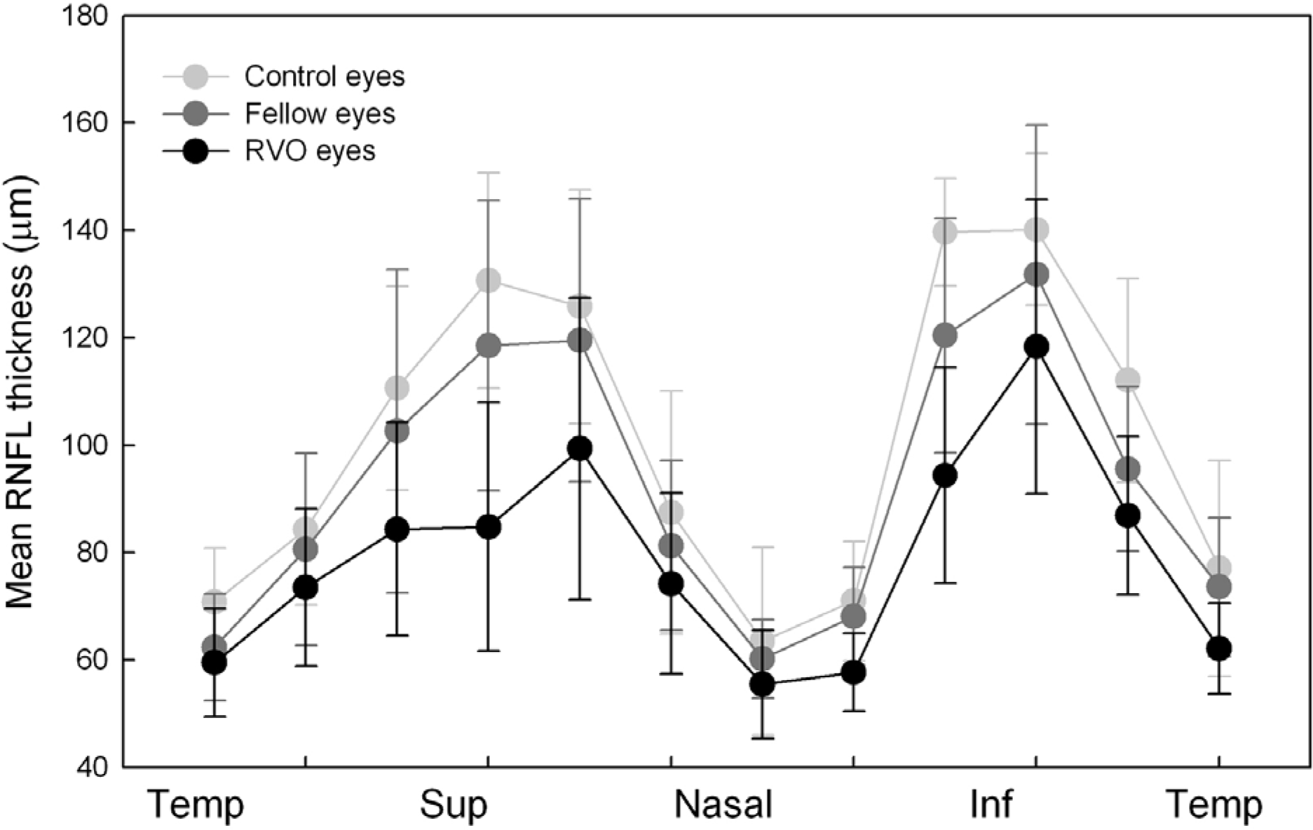
Figure 2.
Fluctuation of intraocular pressure (IOP) and ocular perfusion pressure (OPP) in healthy control eyes and fellow eyes of unilateral branched retinal vein occlusion. The mean diurnal variation of IOP, mean OPP (MOPP) and mean diastolic OPP (DOPP) at different time points are shown (left column). The mean IOP, MOPP, and DOPP shows no significant difference at any time point between groups. The absolute diurnal changes of MOPP and DOPP shows significant difference (*) between the control eyes versus fellow eyes (right column).
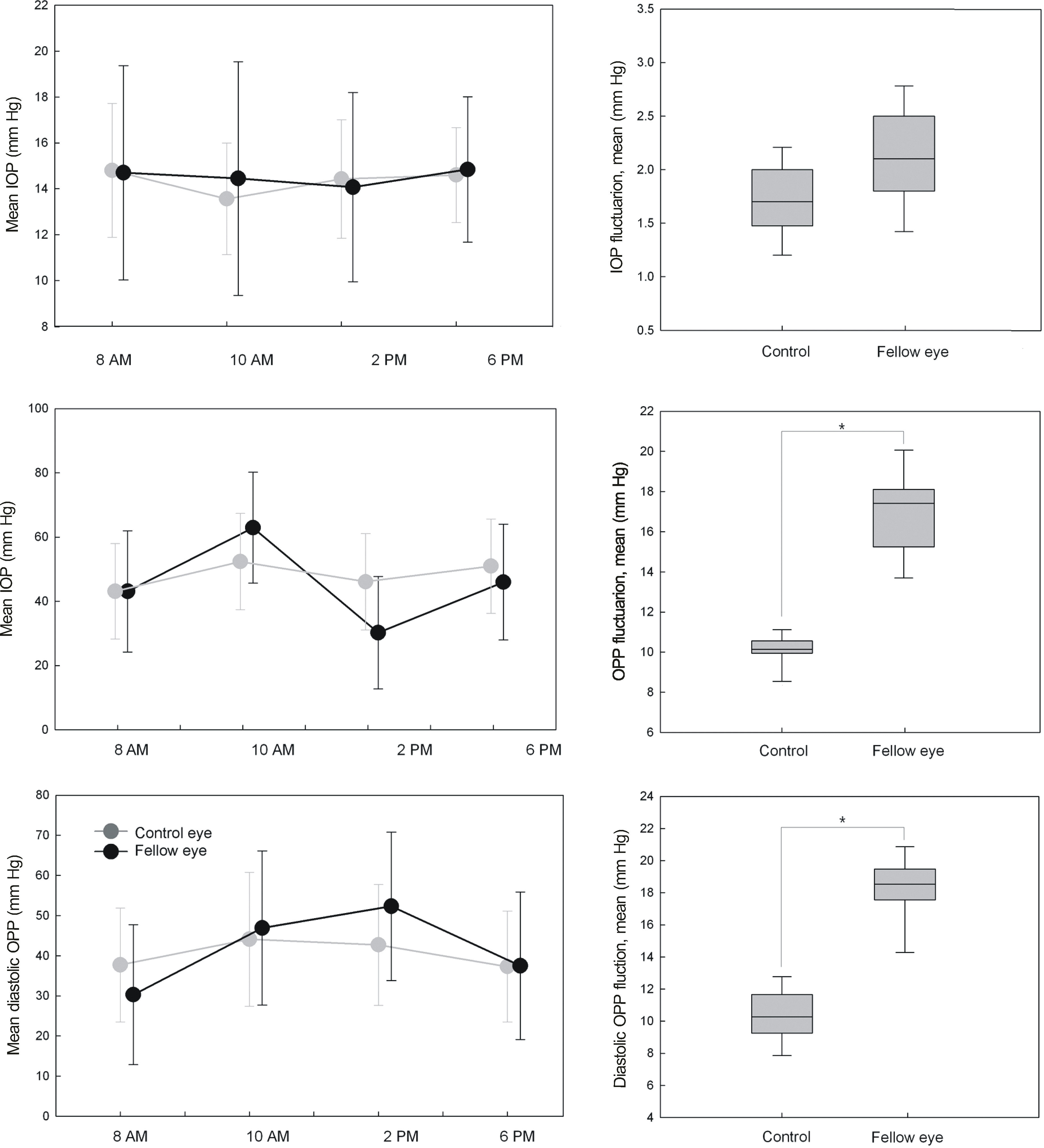
Figure 3.
Representative case 1: 52-year-old man with branched retinal vein occlusion (BRVO) in the left eye. Larger diurnal MOPP, and DOPP fluctuation are observed in the fellow eye of the patient, compared to the mean of control eyes. The localized retinal nerve fiber layer (RNFL) defect is found in the superotemporal area in the fellow eye and superotemporal RNFL is reduced on OCT measurements.
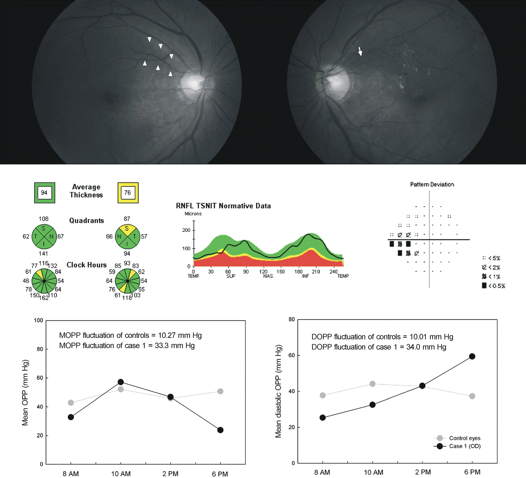
Table 1.
Mean retinal nerve fiber layer thickness measured by optical coherence tomography
| Control | Fellow eyes | BRVO eyes | p-value* | p-value† | p-value‡ | |
|---|---|---|---|---|---|---|
| Average | 110.52 ± 10.45 | 92.78 ± 13.35 | 69.79 ± 13.10 | <0.001 | <0.001 | <0.001 |
| Quadrant | ||||||
| Temporal | 76.05 ± 10.79 | 70.73 ± 8.81 | 62.25 ± 8.93 | 0.033 | 0.004 | 0.665 |
| Superior | 125.52 ± 11.84 | 111.06 ± 20.68 | 89.32 ± 18.47 | 0.011 | <0.001 | <0.001 |
| Nasal | 75.06 ± 7.77 | 74.49 ± 11.24 | 64.87 ± 8.74 | 0.257 | 0.024 | 0.152 |
| Inferior | 127.47 ± 11.41 | 111.83 ± 12.67 | 103.72 ± 16.26 | <0.001 | <0.001 | 0.193 |
| Clock hours | ||||||
| 1 | 125.77 ± 17.39 | 119.50 ± 26.37 | 99.31 ± 28.07 | 0.013 | <0.001 | <0.001 |
| 2 | 87.45 ± 19.30 | 81.30 ± 15.85 | 74.18 ± 16.83 | 0.378 | 0.214 | 0.360 |
| 3 | 63.45 ± 7.81 | 60.20 ± 7.34 | 55.48 ± 10.11 | 0.455 | 0.489 | 0.532 |
| 4 | 70.99 ± 14.19 | 68.09 ± 9.13 | 57.67 ± 7.29 | 0.314 | 0.303 | 0.157 |
| 5 | 139.67 ± 21.75 | 120.45 ± 21.81 | 94.35 ± 20.11 | <0.001 | <0.001 | <0.001 |
| 6 | 140.13 ± 22.57 | 131.74 ± 27.89 | 118.32 ± 27.42 | 0.040 | <0.001 | 0.017 |
| 7 | 112.03 ± 17.44 | 95.56 ± 15.31 | 86.89 ± 14.73 | <0.001 | <0.001 | 0.164 |
| 8 | 77.06 ± 11.06 | 73.60 ± 12.90 | 62.12 ± 8.48 | 0.212 | 0.004 | 0.054 |
| 9 | 70.83 ± 10.04 | 62.35 ± 9.92 | 59.48 ± 10.02 | 0.236 | 0.017 | 0.608 |
| 10 | 84.31 ± 14.13 | 80.55 ± 17.90 | 73.49 ± 14.61 | 0.172 | 0.048 | 0.366 |
| 11 | 110.59 ± 19.00 | 102.61 ± 30.09 | 84.34 ± 19.88 | 0.147 | <0.001 | <0.001 |
| 12 | 130.66 ± 20.05 | 118.43 ± 27.04 | 84.78 ± 23.11 | <0.001 | <0.001 | <0.001 |
Table 2.
Systemic and ocular measurements
| Normal controls | BRVO patients | p-value | |
|---|---|---|---|
| Number | 50 | 50 | |
| Age (years) | 63.63 ± 6.54 | 64.84 ± 5.67 | 0.721† |
| Sex (male:female) | 28:22 | 26:24 | 0.665‡ |
| Spherical equivalent (diopters)* | -0.63 ± 1.11 | -0.54 ± 1.27 | 0.713† |
| Intraocular pressure (mm Hg)* | |||
| Mean | 14.35 ± 2.33 | 14.54 ± 2.35 | 0.990† |
| Peak | 15.50 ± 2.39 | 16.27 ± 2.16 | 0.146† |
| Fluctuation | 1.59 ± 0.85 | 2.07 ± 0.96 | 0.112† |
| Visual field exam (dB)* | |||
| Mean deviation | -0.64 ± 2.46 | -1.47 ± 1.26 | 0.255† |
| Pattern standard deviation | 1.57 ± 2.14 | 1.94 ± 1.04 | 0.131† |
| Systemic hypertension (n) | 10 (20.0%) | 30 (60.0%) | <0.001‡ |
| Taking antihypertensive medication | 8 (80.0%) | 25 (83.3%) | 0.831‡ |
| Systolic BP (mm Hg) | |||
| Mean | 125.93 ± 12.66 | 136.48 ± 12.19 | 0.164 |
| Fluctuation | 18.8 ± 3.55 | 25.67 ± 3.53 | 0.045 |
| Diastolic BP (mm Hg) | |||
| Mean | 73.43 ± 11.94 | 80.60 ± 22.68 | 0.201 |
| Fluctuation | 19.0 ± 5.04 | 27.23 ± 4.86 | 0.037 |
| Mean arterial pressure (mm Hg) | |||
| Mean | 92.26 ± 9.82 | 92.56 ± 10.35 | 0.535 |
| Fluctuation | 10.22 ± 7.03 | 19.89 ± 9.85 | 0.011 |
Table 3.
Measurements in intraocular pressure and ocular perfusion pressure
| Control eyes | Fellow eyes | p-value* | |
|---|---|---|---|
| Ocular perfusion pressure | |||
| Mean | 47.16 ± 2.72 | 47.05 ± 2.08 | 0.791 |
| Fluctuation | 10.27 ± 2.31 | 17.71 ± 3.32 | <0.001 |
| Diastolic ocular perfusion pressure | |||
| Mean | 36.60 ± 2.85 | 35.45 ± 2.15 | 0.573 |
| Fluctuation | 10.01 ± 3.18 | 18.13 ± 3.36 | <0.001 |
Table 4.
Significance of independent variables in predicting outcomes of retinal nerve fiber layer thickness in the fellow eyes of unilateral BRVO by univariate models
| Variable | Beta [95% CI] | p-value |
|---|---|---|
| Age | 1.012 [0.721-4.433] | 0.601 |
| SE | 0.934 [0.315-1.216] | 0.522 |
| History of systemic hypertension | 1.167 [0.916-5.973] | 0.048* |
| Systemic antihypertensive medication | 1.033 [0.840-5.723] | 0.379 |
| VF MD | 1.046 [0.753-3.816] | 0.462 |
| VF PSD | 1.058 [0.801-3.573] | 0.411 |
| Mean IOP | 1.121 [1.014-4.168] | 0.130 |
| Peak IOP | 1.008 [0.912-3.410] | 0.286 |
| IOP fluctuation | 1.104 [0.830-3.533] | 0.187 |
| Mean SBP | 0.572 [0.313-0.914] | 0.518 |
| SBP fluctuation | 1.032 [0.852-4.260] | 0.386 |
| Mean DBP | 1.040 [0.971-1.114] | 0.247 |
| DBP fluctuation | 1.173 [1.015-2.016] | 0.016* |
| Mean MAP | 0.928 [0.716-1.097] | 0.540 |
| MAP fluctuation | 1.043 [0.917-1.180] | 0.382 |
| MOPP | 1.044 [0.985-1.116] | 0.251 |
| MOPP fluctuation | 1.028 [0.999-2.005] | 0.256 |
| Mean DOPP | 1.128 [1.087-2.025] | 0.200 |
| DOPP fluctuation | 0.163 [1.121-2.309] | 0.009* |
BRVO = branched retinal vein occlusion; SE = spherical equivalent; VF = visual field; MD = mean deviation; PSD = pattern standard deviation; IOP = intraocular pressure; SBP = systolic blood pressure; DBP = diastolic blood pressure; MAP = mean arterial pressure; MOPP = mean ocular perfusion pressure; DOPP = diastolic ocular perfusion pressure.
Table 5.
Significance of independent variables in predicting outcomes of retinal nerve fiber layer thickness in the fellow eyes of unilateral BRVO by multivariate models
| Variable | Beta [95% CI] | p-value |
|---|---|---|
| Age | 1.008 [0.578-3.267] | 0.720 |
| SE | 0.819 [0.310-1.082] | 0.618 |
| History of systemic hypertension | 1.964 [0.893-4.510] | 0.097 |
| Systemic antihypertensive medication | 0.617 [0.127-1.815] | 0.446 |
| DOPP fluctuation | 1.911 [1.184-4.630] | 0.005* |
 XML Download