

 PDF
PDF ePub
ePub Citation
 Print
Print


Abstract
Purpose
To evaluate the causes of secondary macular hole after vitrectomy and the possibility of their prevention.
Methods
27 patients (28 eyes) who experienced macular hole formation after vitrectomy were reviewed retrospectively. Age, sex, operation methods, duration between the vitrectomy and the secondary macular hole surgery and causes of the primary vitrectomy were recorded. Best-corrected visual acuity (BCVA) before and after primary vitrectomy; preoperative and postoperative macular findings with optical coherence tomography and fundus examination; and BCVA before and after macular hole surgery were analyzed.
Results
Of the 2945 eyes that had undergone vitrectomy, 28 eyes (0.96%) experienced macular hole formation. As causes of primary vitrectomy, 12 eyes had proliferative diabetic retinopathy, 6 eyes had rhegmatogenous retinal detachment, 2 eyes had branch retinal vein occlusion, 3 eyes had age-related macular degeneration and 5 eyes had trauma such as eyeball rupture or intraocular foreign body. The mean duration between primary vitrectomy and macular hole formation was 20.4 months (4 days-115 months). The estimated causes of macular hole formation included cystoid macular edema (CME) (n = 13), thinning of the macula (n = 6), thickening of internal limiting membrane or recurrence of preretinal membrane (PRM) (n = 7), recurrence of subretinal hemorrhage (n = 1) and macular damage during vitrectomy (n = 2). Final BCVA after macular hole surgery decreased in most cases compared to BCVA before macular hole formation except in 7 eyes (25%).
Conclusions
Close observation of the macula after primary vitrectomy especially in eyes with continuous CME, and recurrent PRM and proper management on them including timely removal of the tangential traction force are necessary for preventing macular hole formation. In addition, surgeons should make efforts not to exert excessive tractional force on the macula to avoid iatrogenic damage during removal of the preretinal membrane.
References
1. Gass JD.Idiopathic senile macular hole. Its early stages and pathogenesis. Arch Ophthalmol. 1988; 106:629–39.

2. Niwa H, Terasaki H, Ito Y, Miyake Y.Macular hole development in fellow eyes of patients with unilateral macular hole. Am J Ophthalmol. 2005; 140:370–5.
3. Puliafito CA, Hee MR, Lin CP. . Imaging of macular diseases with optical coherence tomography. Ophthalmology. 1995; 102:217–29.
4. Ruiz-Moreno JM, Staicu C, Piñero DP. . Optical coherence to-mography predictive factors for macular hole surgery outcome. Br J Ophthalmol. 2008; 92:640–4.
5. Smiddy WE.Atypical presentations of macular holes. Arch Ophthalmol. 1993; 111:626–31.
6. Lee SH, Park KH, Kim JH. . Secondary macular hole for-mation after vitrectomy. Retina. 2010; 30:1072–7.
7. Tsujikawa M, Saito Y, Lewis JM, Tano Y.Secondary vitrectomy for the treatment of macular holes occurring after vitrectomy. Ophthalmic Surg Lasers. 1997; 28:336–7.
8. Gass JD.Reappraisal of biomicroscopic classification of stages of development of a macular hole. Am J Ophthalmol. 1995; 119:752–9.
9. Lipham WJ, Smiddy WE.Idiopathic macular hole following vi-trectomy: implications for pathogenesis. Ophthalmic Surg Lasers. 1997; 28:633–9.
10. Lo WR, Hubbard GB.Macular hole formation, spontaneous clo-sure, and recurrence in a previously vitrectomized eye. Am J Ophthalmol. 2006; 141:962–4.
11. Melberg NS, Meredith TA.Success with macular hole surgery. Ophthalmology. 1996; 103:200–1.
12. Melberg NS, Williams DF.More on macular holes. Ophthalmology. 1994; 101:1764–5.
13. Shaikh S, Garretson B.Spontaneous closure of a recurrent macular hole following vitrecomy corroborated by optical coherence tomography. Ophthalmic Surg Lasers Imaging. 2003; 34:172–4.
14. Smiddy WE, Flynn HW Jr.Pathogenesis of macular holes and ther-apeutic implications. Am J Ophthalmol. 2004; 137:525–37.
15. Tsilimbaris MK, Gotzaridis S, Charisis SK. . Spontaneous clo-sure of macular holes developed after pars plana vitrectomy. Semin Ophthalmol. 2007; 22:39–42.
16. Akiba J, Quiroz MA, Trempe CL.Role of posterior vitreous detach-ment in idiopathic macular holes. Ophthalmology. 1990; 97:1610–3.
17. Frangieh GT, Green WR, Engel HM.A histopathologic study of macular cysts and holes. Retina. 1981; 1:311–36.
18. Trempe CL, Weiter JJ, Furukawa H.Fellow eyes in cases of mac-ular hole. Biomicroscopic study of the vitreous. Arch Ophthalmol. 1986; 104:93–5.
19. Flynn HW Jr.Macular hole surgery in patients with proliferative diabetic retinopathy. Arch Ophthalmol. 1994; 112:877–8.
20. Brazitikos PD, Stangos NT.Macular hole formation in diabetic ret-inopathy: the role of coexisting macular edema. Doc Ophthalmol. 1999; 97(3-4):273–8.
21. Ghoraba H.Types of macular holes encountered during diabetic vitrectomy. Retina. 2002; 22:176–82.
22. Krizova L, Kalousova M, Kubena A. . Increased uric acid and glucose concentrations in vitreous and serum of patients with dia-betic macular oedema. Ophthalmic Res. 2011; 46:73–9.
23. Karacorlu M, Karacorlu S, Ozdemir H.Iatrogenic punctate cho-rioretinopathy after internal limiting membrane peeling. Am J Ophthalmol. 2003; 135:178–82.
24. Steven P, Laqua H, Wong D, Hoerauf H.Secondary paracentral ret-inal holes following internal limiting membrane removal. Br J Ophthalmol. 2006; 90:293–5.
25. Choi YH, Park JW, Cho YW.Internal limiting membrane peeling with or without indocyanine green in macular hole surgery. J Korean Ophthalmol Soc. 2005; 46:1342–50.
Fiagure 1.
Serial optical coherence tomography of macular hole formation due to cystoid macular edema 3 months (A) and 36 months (B) after primary vitrectomy.
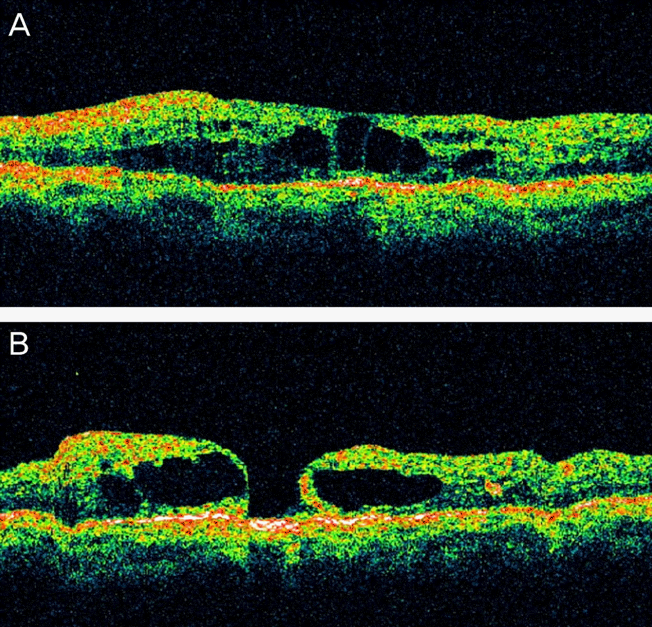
Figure 2.
Serial optical coherence tomography of macular hole formation due to macular thinning 1 months (A) and 6 months (B) after primary vitrectomy.
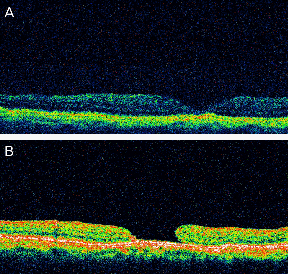
Figure 3.
Serial optical coherence tomography of macular hole formation due to internal limiting membrane thickening and/or epiretinal membrane recurrence after primary vitrectomy. Macular image before (A, B) and after (C, D) macular hole formation.
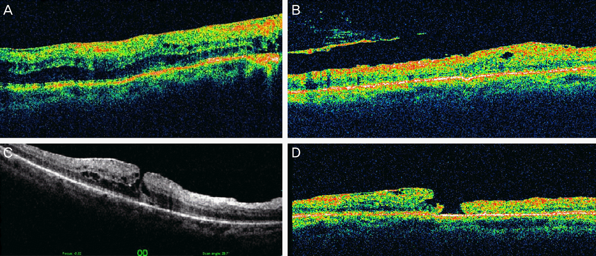
Table 1.
Causes of primary pars plana vitrectomy
Table 2.
Estimated causes of macular hole formation after primary vitrectomy
Table 3.
Clinical characteristics of primary vitrectomy groups with secondary macular hole formation
| Group1* | Group2† | Group3‡ | Group4§ | Group5∏ | p-value# | |
|---|---|---|---|---|---|---|
| No. of subjects | 6 | 12 | 2 | 3 | 5 | |
| Age at primary PPV (years) | 51.7 ± 14.5 | 52.9 ± 11.5 | 70.2 ± 2.8 | 77.7 ± 12.1 | 46.4 ± 13.1 | 0.025 |
| Duration between primary PPV and MH formation (months) | 22.1 ± 36.2 | 19.6 ± 34.1 | 8.5 ± 10.6 | 2.0 ± 2.0 | 36.1 ± 0.0 | 0.530 |
| MH size (DD) | 0.36 ± 0.36 | 0.53 ± 0.79 | 0.33 ± 0.11 | 0.20 ± 0.0 | 0.40 ± 0.13 | 0.446 |
| Spherical equivalent (diopter) | -6.10 ± 10.2 | -0.76 ± 1.7 | -1.93 ± 1.5 | -0.75 ± 0.9 | -3.92 ± 4.5 | 0.228 |
| Axial length (mm) | 25.90 ± 2.92 | 22.53 ± 0.72 | 21.87 ± 0.88 | 22.45 ± 1.69 | 23.50 ± 1.71 | 0.062 |
Table 4.
Clinical characteristics by estimated causes of macular hole formation
| CME | EMR and/or ILM thickening | Macular thinning | Iatrogenic macular damage | Recurrence of SMH | p-value* | |
|---|---|---|---|---|---|---|
| No. of subjects | 12 | 7 | 6 | 2 | 1 | |
| Sex distribution (male : female) | 3 : 9 | 5 : 2 | 2 : 4 | 2 : 0 | 0 : 1 | |
| Diabetes mellitus | 8 | 3 | 2 | 1 | 0 | |
| Age at primary PPV (years) | 55.2 ± 11.5 | 40.3 ± 10.4 | 62.5 ± 6.4 | 71.0 ± 11.3 | 89.0 ± 0.00 | 0.006 |
| Duration between primary PPV and MH formation (months) | 16.7 ± 31.1 | 31.4 ± 43.9 | 24.0 ± 35.8 | 3.0 ± 4.2 | 2.0 ± 0.00 | 0.790 |
| MH size (DD) | 0.61 ± 0.83 | 0.44 ± 0.14 | 0.22 ± 0.05 | 0.17 ± 0.02 | 0.2 ± 0.00 | 0.072 |
| Spherical equivalent (diopter) | -1.6 ± 2.8 | -3.2 ± 4.4 | -3.4 ± 9.7 | 0.4 ± 0.9 | -0.3 ± 0.0 | 0.545 |
| Axial length (mm) | 22.8 ± 1.82 | 23.8 ± 1.7 | 24.3 ± 3.5 | 23.4 ± 0.8 | 22.7 ± 0.0 | 0.568 |
Table 5.
Surgical outcome of primary vitrectomy and secondary macular hole surgery by primary vitrectomy groups
| Primary PPV groups |
BCVA at primary PPV (log MAR) |
BCVA at MH surgery (log MAR) |
||||
|---|---|---|---|---|---|---|
| Preoperative | Postoperative | p-value* | Preoperative | Postoperative | p-value* | |
| Group1† | -1.65 ± 0.85 | -0.85 ± 0.50 | 0.005 | -1.30 ± 0.76 | -0.87 ±0.95 | 0.087 |
| Group2‡ | -1.09 ± 0.64 | -1.08 ± 0.75 | 0.930 | -1.65 ± 0.82 | -1.61 ± 1.34 | 0.860 |
| Group3§ | -1.40 ± 0.00 | -1.05 ± 0.49 | 0.500 | -1.20 ± 0.28 | -1.40 ± 0.00 | 0.500 |
| Group4∏ | -1.83 ± 0.75 | -1.60 ± 1.01 | 0.317 | -1.40 ± 0.79 | -1.57 ± 1.05 | 0.655 |
| Group5# | -2.10 ± 1.34 | -1.10 ± 1.14 | 0.104 | -1.47 ± 0.79 | -0.83 ± 0.67 | 0.069 |
| Total | -1.49 ± 0.87 | -1.08 ± 0.77 | 0.007 | -1.49 ± 0.70 | -1.29 ± 1.08 | 0.176 |
| p-value** | 0.814 | 0.738 | ||||
Table 6.
Surgical outcome of primary vitrectomy and secondary macular hole surgery by the estimated causes of macular hole formation
| Primary PPV groups |
BCVA at primary PPV (log MAR) |
BCVA at MH surgery (log MAR) |
||||
|---|---|---|---|---|---|---|
| Preoperative | Postoperative | p-value* | Preoperative | Postoperative | p-value* | |
| CME | -1.24 ± 0.76 | -1.13 ± 0.69 | 0.544 | -1.64 ± 0.82 | -1.55 ± 1.06 | 0.589 |
| ERM and/or ILM thickening | -1.76 ± 1.26 | -1.11 ± 1.21 | 0.141 | -1.54 ± 0.87 | -1.28 ± 1.61 | 0.498 |
| Macular thinning | -1.70 ± 0.81 | -0.92 ± 0.55 | 0.042 | -1.21 ± 0.29 | -0.87 ± 0.48 | 0.109 |
| Iatrogenic macular damage | -1.40 ± 0.00 | -1.40 ± 0.00 | 1.000 | -1.40 ± 0.00 | -1.00 ± 0.57 | 0.655 |
| Recurrence of SMH | -1.40 ± 0.00 | -0.70 ± 0.00 | - | -1.40 ± 0.00 | -1.40 ± 0.00 | - |
| p-value† | 0.845 | 0.417 | ||||
 XML Download