

 PDF
PDF ePub
ePub Citation
 Print
Print


Abstract
Purpose
To evaluate the macular function by a multifocal electroretinogram (mfERG) in patients with diabetic retinopathy (DR), and to assess the correlation between responses of mfERG and the threshold of the visual field test (VF).
Methods
The records of patients with DR (16 eyes, 16 patients) and control subjects (14 eyes, 14 subjects) were retrospectively reviewed. mfERG and VF were divided into Ring 1, Ring 2 and Ring 3 at 6-degree intervals from the central macula. The correlation between the amplitude/peak time and the threshold of each ring was analyzed.
Results
In patients with DR, the amplitude was decreased in all areas, the peak time was delayed in Ring 2 and the threshold was decreased in Rings 2 and 3, compared to control subjects. The amplitude of mfERG and the threshold of VF showed statistically significant positive correlations in Rings 2 and 3 (p < 0.05). The peak time of mfERG and the threshold of VF showed statistically significant negative correlations in Ring 3 (p < 0.05).
Conclusions
The threshold of VF was more significantly correlated with the amplitude than with the peak time of mfERG in patients with DR. mfERG and VF were useful tests to assess the macular function, and alteration of macular function was early detected because two tests were conducted at the same time.
Go to : 
References
1. Ola MS, Nawaz MI, Siddiquei MM. . Recent advances in un-derstanding the biochemical and molecular mechanism of diabetic retinopathy. J Diabetes Complications. 2012; 26:56–64.

2. Frank RN.Etiologic mechanisms in diabetic retinopathy. Ryan SJ, Retina Schachat AP., editors4th ed.Los Angeles: Mosby;2006. v. 2. chap. 66.
3. Yoon JH, Kim MJ, Chin HS, Moon YS.Effect of PRP on visual acuity, visual field and subjective symptoms in very severe NPDR. J Korean Ophthalmol Soc. 2003; 44:2545–52.
4. Lee BH, Cho YW.Quantitative analysis of diabetic macular edema by optical coherence tomography. J Korean Ophthalmol Soc. 2004; 45:1858–64.
5. Ito H, Horii T, Nishijima K. . Association between fluorescein leakage and optical coherence tomographic characteristics of mi-croaneurysms in diabetic retinopathy. Retina. 2013; 33:732–9.
6. Hood DC.Assessing retinal function with the multifocal technique. Prog Retin Eye Res. 2000; 19:607–46.
7. Sutter EE, Dodsworth-Feldman B, Haegerstrom-Portnoy G.Simultaneous multifocal ERGs in diseased retina. Invest Ophthalmol Vis Sci. 1986; 27:301–2.
8. Lung JC, Swann PG, Wong DS, Chan HH.Global flash multifocal electroretinogram: early detection of local functional changes and its correlations with optical coherence tomography and visual field tests in diabetic eyes. Doc Ophthalmol. 2012; 125:123–35.
9. Hood DC, Zhang X.Multifocal ERG and VEP responses and visu-al fields: comparing disease-related changes. Doc Ophthalmol. 2000; 100(2-3):115–37.
10. Hood DC, Bach M, Brigell M. et al. International Society For Clinical Electrophysiology of Vision. ISCEV standard for clinical multifocal electroretinography (mfERG) (2011 edition). Doc Ophthalmol. 2012; 124:1–13.
11. Harris A, Bingaman D, Ciulla TA, Martin B.Retinal and choroidal blood flow in health and disease. Ryan SJ, Schachat AP, editors. Retina. 4th ed.Los Angeles: Mosby;2006. v. 1:chap. 5.
12. Hood DC, Frishman LJ, Saszik S, Viswanathan S.Retinal origins of the primate multifocal ERG: implications for the human response. Invest Ophthalmol Vis Sci. 2002; 43:1673–85.
13. Hood DC, Greenstein V, Frishman L. . Identifying inner retinal contributions to the human multifocal ERG. Vision Res. 1999; 39:2285–91.
14. Miller RF.The physiology and morphology of the vertebrate retina. Ryan SJ, Schachat AP, editors. Retina. 4th ed.Los Angeles: Mosby;2006. v. 1:chap. 9.
15. Kim SJ, Yu HG.The clinical applications of multifocal electro-retinogram in diabetic retinopathy. J Korean Ophthalmol Soc. 2004; 45:64–8.
16. Kondo M, Miyake Y, Horiguchi M. . Clinical evaluation of multifocal electroretinogram. Invest Ophthalmol Vis Sci. 1995; 36:2146–50.
17. Fine BS, Brucker AJ.Macular edema and cystoid macular edema. Am J Ophthalmol. 1981; 92:466–81.
18. Roth JA.Central visual field in diabetes. Br J Ophthalmol. 1969; 53:16–25.
19. Bengtsson B, Heijl A, Agardh E.Visual fields correlate better than visual acuity to severity of diabetic retinopathy. Diabetologia. 2005; 48:2494–500.
20. Federman JL, Lloyd J.Automated static perimetry to evaluate dia-betic retinopathy. Trans Am Ophthalmol Soc. 1984; 82:358–70.
21. Pahor D.Automated static perimetry as a screening method for evaluation of retinal perfusion in diabetic retinopathy. Int Ophthalmol. 1997-1998; 21:305–9.
22. Greenstein VC, Holopigian K, Hood DC. . The nature and ex-tent of retinal dysfunction associated with diabetic macular edema. Invest Ophthalmol Vis Sci. 2000; 41:3643–54.
Go to :
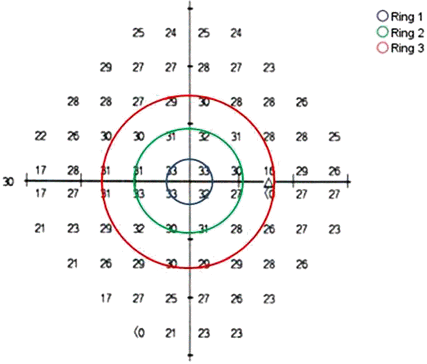 | Figure 1.Threshold plot of automated perimetry (dB) was divided into Ring 1, Ring 2, Ring 3 by every 6 degree. |
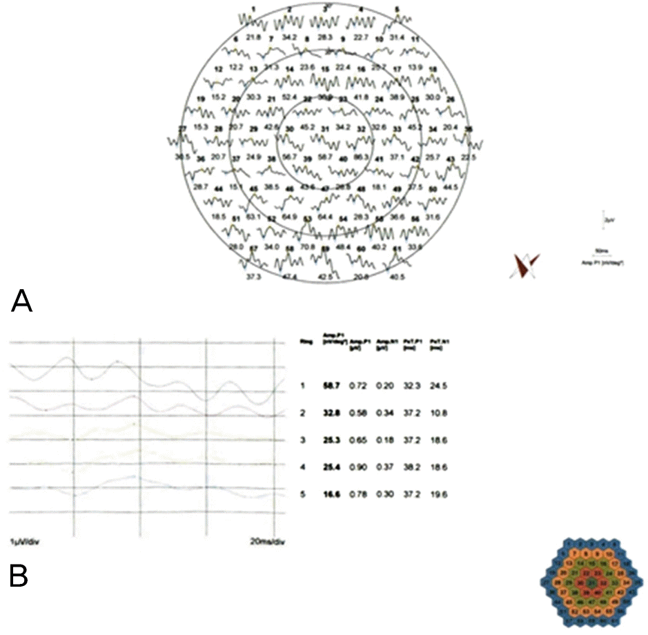 | Figure 2.Trace arrays of mfERG (A) show the result on 61 point of 30 degree on central visual field. Ring average of mfERG (B) show mean amplitude and peak time of concentric ring. |
 | Figure 3.Relationship between the threshold of AVF and log (amplitude) of mfERG in DR group (A) and normal group (C). Relationship between the threshold of AVF and log (peak time) of mfERG in DR group (B) and normal group (D). |
Table 1.
Demographic characteristics of the patients
| DR | Normal | p-value* | |
|---|---|---|---|
| Number (eyes) | 16 (16) | 14 (14) | |
| Sex (male/female) | 10/6 | 9/5 | 0.846 |
| Age (years) | 55.86 ± 12.36 | 56.14 ± 11.36 | 0.825 |
| BCVA (log MAR) | 0.13 ± 0.13 | 0.01 ± 0.04 | 0.201 |
| IOP (mm Hg) | 14.00 ± 2.34 | 13.64 ± 2.59 | 0.699 |
| SE (diopter) | -0.81 ± 1.43 | -0.48 ± 1.03 | 0.215 |
| CPT (um) | 226.63 ± 50.42 | 214.00 ± 24.25 | 0.094 |
Table 2.
Comparison of the threshold (dB) of AVF in diabetic retinopathy group and normal group
| Ring 1 | Ring 2 | Ring 3 | |
|---|---|---|---|
| DR | 26.50 ± 2.97 | 24.60 ± 4.48 | 20.19 ± 4.09 |
| Normal | 30.18 ± 1.79 | 28.84 ± 2.16 | 26.10 ± 1.90 |
| p-value | 0.268 | 0.039* | 0.003* |
Table 3.
Comparison of the amplitude (nV/deg2) of mfERG in diabetic retinopathy group and normal group
| Ring 1 | Ring 2 | Ring 3 | |
|---|---|---|---|
| DR (log unit) | 46.08 ± 11.55 (1.65 ± 0.12) | 30.64 ± 6.51 (1.48 ± 0.09) | 20.36 ± 3.46 (1.30 ± 0.08) |
| Noraml (log unit) | 92.11 ± 22.20 (1.98 ± 0.12) | 62.16 ± 20.16 (1.77 ± 0.15) | 45.72 ± 8.17 (1.65 ± 0.08) |
| p-value | 0.016* | 0.000* | 0.001* |
Table 4.
Comparison of the peak time (msec) of mfERG in diabetic retinopathy group and normal group
| Ring 1 | Ring 2 | Ring 3 | |
|---|---|---|---|
| DR (log unit) | 38.06 ± 4.98 (1.58 ± 0.06) | 39.45 ± 3.30 (1.59 ± 0.04) | 39.43 ± 2.62 (1.59 ± 0.03) |
| Noraml (log unit) | 37.17 ± 3.01 (1.57 ± 0.04) | 36.67 ± 1.92 (1.56 ± 0.02) | 37.84 ± 1.86 (1.58 ± 0.02) |
| p-value | 0.387 | 0.012* | 0.508 |
Table 5.
Relationship between the threshold of AVF and responses of mfERG in diabetic retinopathy group
| R1 |
mfERG |
R2 |
mfERG |
R3 |
mfERG |
|||
|---|---|---|---|---|---|---|---|---|
| Amp* | Pe.T† | Amp | Pe.T | Amp | Pe.T | |||
| AVF | r=0.401‡ | r=-0.219‡ | AVF | r=0.779 | r=-0.276 | AVF | r=0.578 | r=-0.638 |
| (p=0.124) | (p=0.416) | (p=0.001§) | (p=0.214) | (p=0.043§) | (p=0.008§) | |||
 XML Download