Abstract
Purpose
To report a case of central serous chorioretinopathy development after glaucoma filtering surgery and spontaneous resolution in a patient with a history of central serous chorioretinopathy in the contralateral eye.
Case summary
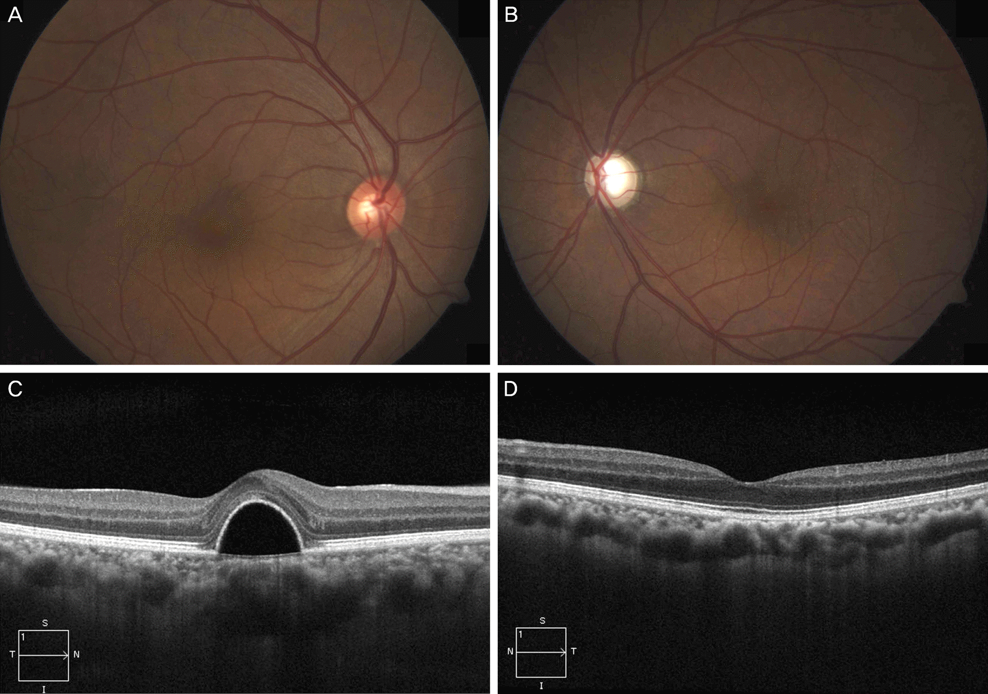
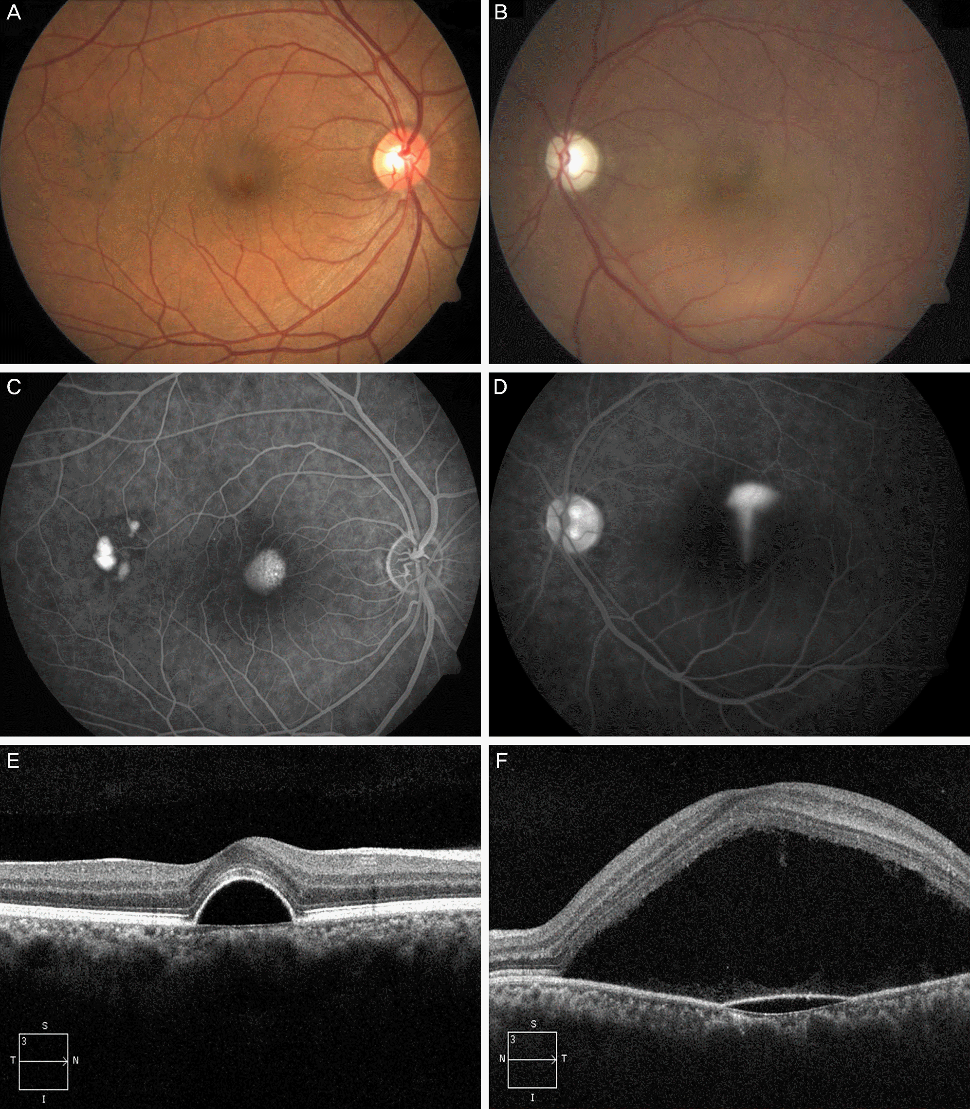
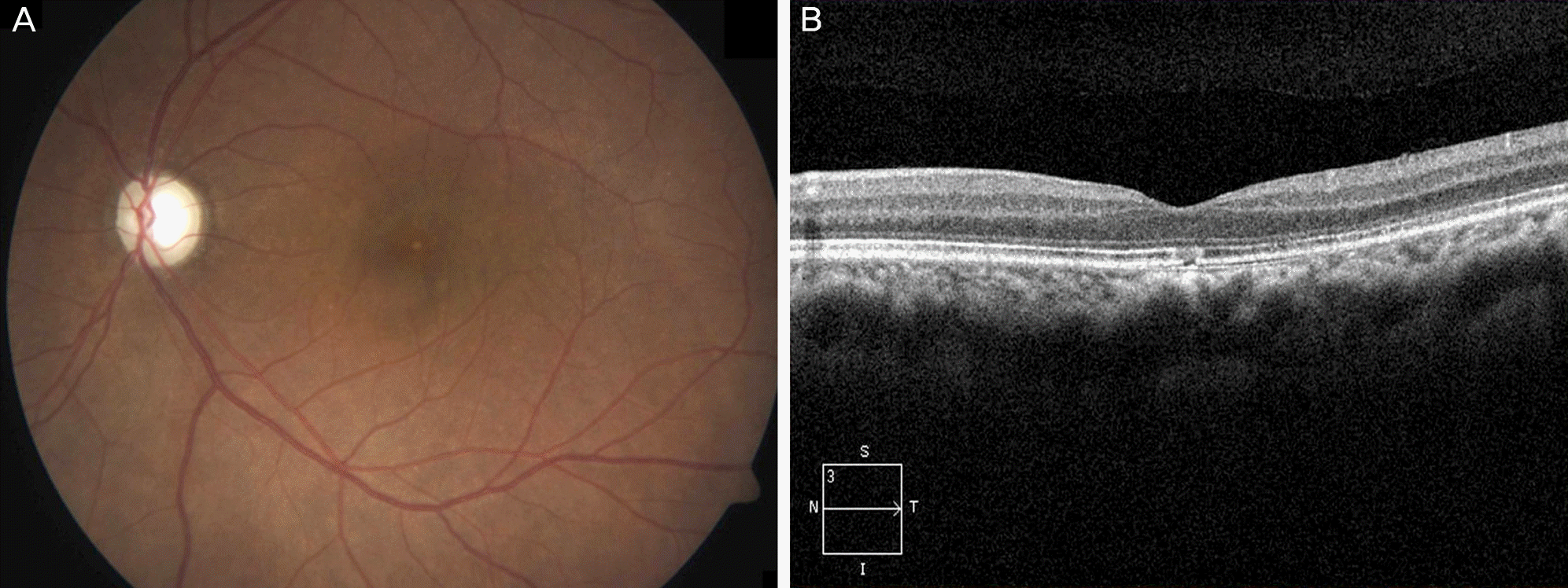
A 46-year-old male with a history of chronic uveitis in both eyes presented with uncontrolled intraocular pressure (IOP) in his left eye. Initial IOP was 34 mm Hg in his left eye. On preoperative evaluation, central serous chorioretinopathy, which was diagnosed in another clinic 1 month prior, was observed in his right eye. Slightly pale optic disc and retinal nerve fiber layer defects were noted in the left eye. However, macular abnormalities were not observed in the left eye. Trabeculectomy and peripheral iridectomy using mitomycin C were performed in the left eye. The patient was prescribed triamcinolone 8 mg daily for 4 days to reduce the post-surgical inflammation. On postoperative day 4, IOP in the left eye was 7 mm Hg and newly developed central serous chorioretinopathy was noted. On follow-up, IOP was maintained at 7-10 mm Hg and central serous chorioretinopathy disappeared 7 months postoperatively.
Go to : 
References
1. Yannuzzi LA. Type A behavior and central serous chorioretinopathy. Retina. 2012; 32(Suppl 1):709.

2. Marmor MF. New hypotheses on the pathogenesis and treatment of serous retinal detachment. Graefes Arch Clin Exp Ophthalmol. 1988; 226:548–52.

3. Simmons-Rear A, Yeh S, Chan-Kai BT, et al. Characterization of serous retinal detachments in uveitis patients with optical coherence tomography. J Ophthalmic Inflamm Infect. 2012; 2:191–7.

4. Lavin M, Franks W, Hitchings RA. Serous retinal detachment following glaucoma filtering surgery. Arch Ophthalmol. 1990; 108:1553–5.

5. Roe R, Branco BC, Cunningham ET Jr. Hypotony maculopathy in a patient with HLA-B27-associated uveitis. Ocul Immunol Inflamm. 2008; 16:107–8.

6. Roe RM, Donohue KV, Khalil SM, Sonenshine DE. Hormonal regulation of metamorphosis and reproduction in ticks. Front Biosci. 2008; 13:7250–68.

7. Lin JM, Tsai YY. Retinal pigment epithelial detachment in the fellow eye of a patient with unilateral central serous chorioretinopathy treated with steroid. Retina. 2001; 21:377–9.

8. Bouzas EA, Karadimas P, Pournaras CJ. Central serous chorioretinopathy and glucocorticoids. Surv Ophthalmol. 2002; 47:431–48.

9. Carvalho-Recchia CA, Yannuzzi LA, Negrão S, et al. Corticosteroids and central serous chorioretinopathy. Ophthalmology. 2002; 109:1834–7.

10. Haimovici R, Rumelt S, Melby J. Endocrine abnormalities in patients with central serous chorioretinopathy. Ophthalmology. 2003; 110:698–703.

11. Khairallah M, Kahloun R, Tugal-Tutkun I. Central serous chorioretinopathy, corticosteroids, and uveitis. Ocul Immunol Inflamm. 2012; 20:76–85.

12. Jampol LM, Weinreb R, Yannuzzi L. Involvement of corticosteroids and catecholamines in the pathogenesis of central serous chorioretinopathy: a rationale for new treatment strategies. Ophthalmology. 2002; 109:1765–6.

13. Arndt C, Sari A, Ferre M, et al. Electrophysiological effects of corticosteroids on the retinal pigment epithelium. Invest Ophthalmol Vis Sci. 2001; 42:472–5.
14. Sibayan SA, Kobuch K, Spiegel D, et al. Epinephrine, but not dexamethasone, induces apoptosis in retinal pigment epithelium cells in vitro: possible implications on the pathogenesis of central serous chorioretinopathy. Graefes Arch Clin Exp Ophthalmol. 2000; 238:515–9.

15. Hardy P, Nuyt AM, Abran D, et al. Nitric oxide in retinal and choroidal blood flow autoregulation in newborn pigs: interactions with prostaglandins. Pediatr Res. 1996; 39:487–93.

16. Mann RM, Riva CE, Stone RA, et al. Nitric oxide and choroidal blood flow regulation. Invest Ophthalmol Vis Sci. 1995; 36:925–30.
17. Hardy P, Abran D, Li DY, et al. Free radicals in retinal and choroidal blood flow autoregulation in the piglet: interaction with prostaglandins. Invest Ophthalmol Vis Sci. 1994; 35:580–91.
Go to : 
 | Figure 1.Preoperative fundus photograph reveals (A) a focal elevated lesion at macula in right eye (B) and slightly pale optic disc and no definite abnormal findings of macula in left eye. Preoperative optical coherence tomography (C) shows focal pigment epithelial detachment of macula in right eye (D) and no definite abnormal findings of macula in left eye. |
 | Figure 2.At postoperative fourth day, fundus photographs show (A) no definite change with preoperative examination of the right eye (B) and newly developed serous macular detachment of the left eye. (C) Late-phase fluorescein angiography of the right eye shows a hyperfluorescent spot without active leakage, suggesting the previous occurrence of central serous chorioretinopathy. (D) Fluorescein angiography of the left eye shows a smokestack ejection close to the fovea. Optical coherence tomography shows (E) no obvious change of small pigment epithelial detachment at macula of the right eye (F) and serous retinal detachment with pigment epithelial detachment of the left eye. |


 PDF
PDF ePub
ePub Citation
 Print
Print


 XML Download