Abstract
Purpose
To report a case of corrected residual refractive error after cataract surgery with sulcoflex piggyback intraouclar lens (IOL).
Case summary
A 77-year-old man was diagnosed as having hypermature cataract in the right eye and his corrected visual acuity in the same eye was hand-motion before surgery. Refractive error was +9.0 diopter (D) 6 months after conventional cataract surgery in the right eye. The authors performed additional cataract surgery using a piggyback method inserting a +10.0 D IOL in the ciliary sulcus. Four weeks after surgery, his refractive error was −1.25 D, visual acuity increased to 120/200 and 60/200 (with and without correction, respectively). No complication was observed during follow-up time and the patient was satisfied with his results.
Go to : 
References
1. West SK, Valmadrid CT. Epidemiology of risk factors for age-related cataract. Surv Ophthalmol. 1995; 39:323–34.
2. Ridley H. Intra-ocular acrylic lenses after cataract extraction. 1952. Bull World Health Organ. 2003; 81:758–61.
3. Ayala MJ, Pérez-Santonja JJ, Artola A, et al. Laser in situ keratomileusis to correct residual myopia after cataract surgery. J Refract Surg. 2001; 17:12–6.

4. Jin GJ, Crandall AS, Jones JJ. Changing indications for and improving outcomes of intraocular lens exchange. Am J Ophthalmol. 2005; 140:688–94.

5. Piñero DR, Ayala Espinosa MJ, Alió JL. LASIK outcomes following multifocal and monofocal intraocular lens implantation. J Refract Surg. 2010; 26:569–77.

6. Jin GJ, Crandall AS, Jones JJ. Intraocular lens exchange due to incorrect lens power. Ophthalmology. 2007; 114:417–24.

7. Fernández-Buenaga R, Alió JL, Pinilla-Cortés L, Barraquer RI. Perioperative complications and clinical outcomes of intraocular lens exchange in patients with opacified lenses. Graefes Arch Clin Exp Ophthalmol. 2013; 251:2141–6.

8. Gayton JL, Sanders VN. Implanting two posterior chamber intraocular lenses in a case of microphthalmos. J Cataract Refract Surg. 1993; 19:776–7.

9. Gayton JL, Sand, ers V, Van der Karr M, Raanan MG. Piggybacking intraocular implants to correct pseudophakic refractive error. Ophthalmology. 1999; 106:56–9.

10. Dagres E, Khan MA, Kyle GM, Clark D. Perioperative complications of intraocular lens exchange in patients with opacified Aqua-Sense lenses. J Cataract Refract Surg. 2004; 30:2569–73.

11. El Awady HE, Ghanem AA. Secondary piggyback implantation versus IOL exchange for symptomatic pseudophakic residual ametropia. Graefes Arch Clin Exp Ophthalmol. 2013; 251:1861–6.

12. Leysen I, Bartholomeeusen E, Coeckelbergh T, Tassignon MJ. Surgical outcomes of intraocular lens exchange: five-year study. J Cataract Refract Surg. 2009; 35:1013–8.
13. Hassan Z, Szalai E, Berta A, et al. Assessment of tear osmolarity and other dry eye parameters in post-LASIK eyes. Cornea. 2013; 32:e142–5. doi: 10.1097/ICO.0b013e318290496d.

14. Argento CJ, Cosentino MJ. Comparison of optical zones in hyperopic laser in situ keratomileusis: 5.9 mm versus smaller optical zones. J Cataract Refract Surg. 2000; 26:1137–46.

15. Llorente L, Barbero S, Merayo J, Marcos S. Total and corneal optical aberrations induced by laser in situ keratomileusis for hyperopia. J Refract Surg. 2004; 20:203–16.

16. Varley GA, Huang D, Rapuano CJ, et al. LASIK for hyperopia, hyperopic astigmatism, and mixed astigmatism: a report by the American Academy of Ophthalmology. Ophthalmology. 2004; 111:1604–17.
17. Fernández-Buenaga R, Alió JL, Pérez Ardoy AL, et al. Resolving refractive error after cataract surgery: IOL exchange, piggyback lens, or LASIK. J Refract Surg. 2013; 29:676–83.

18. Solebo LA, Eades Walker RJ, Dabbagh A. Intraocular lens exchange for pseudophakic refractive surprise due to incorrectly labeled intraocular lens. J Cataract Refract Surg. 2012; 38:2197–8.

19. Gayton JL, Apple DJ, Peng Q, et al. Interlenticular opacification: clinicopathological correlation of a complication of posterior chamber piggyback intraocular lenses. J Cataract Refract Surg. 2000; 26:330–6.

20. Iwase T, Tanaka N. Elevated intraocular pressure in secondary piggyback intraocular lens implantation. J Cataract Refract Surg. 2005; 31:1821–3.

22. Kodjikian L, Gain P, Donate D, et al. Malignant glaucoma induced by a phakic posterior chamber intraocular lens for myopia. J Cataract Refract Surg. 2002; 28:2217–21.

23. Chang DF, Masket S, Miller KM, et al. Complications of sulcus placement of single-piece acrylic intraocular lenses: recommendations for backup IOL implantation following posterior capsule rupture. J Cataract Refract Surg. 2009; 35:1445–58.
Go to : 
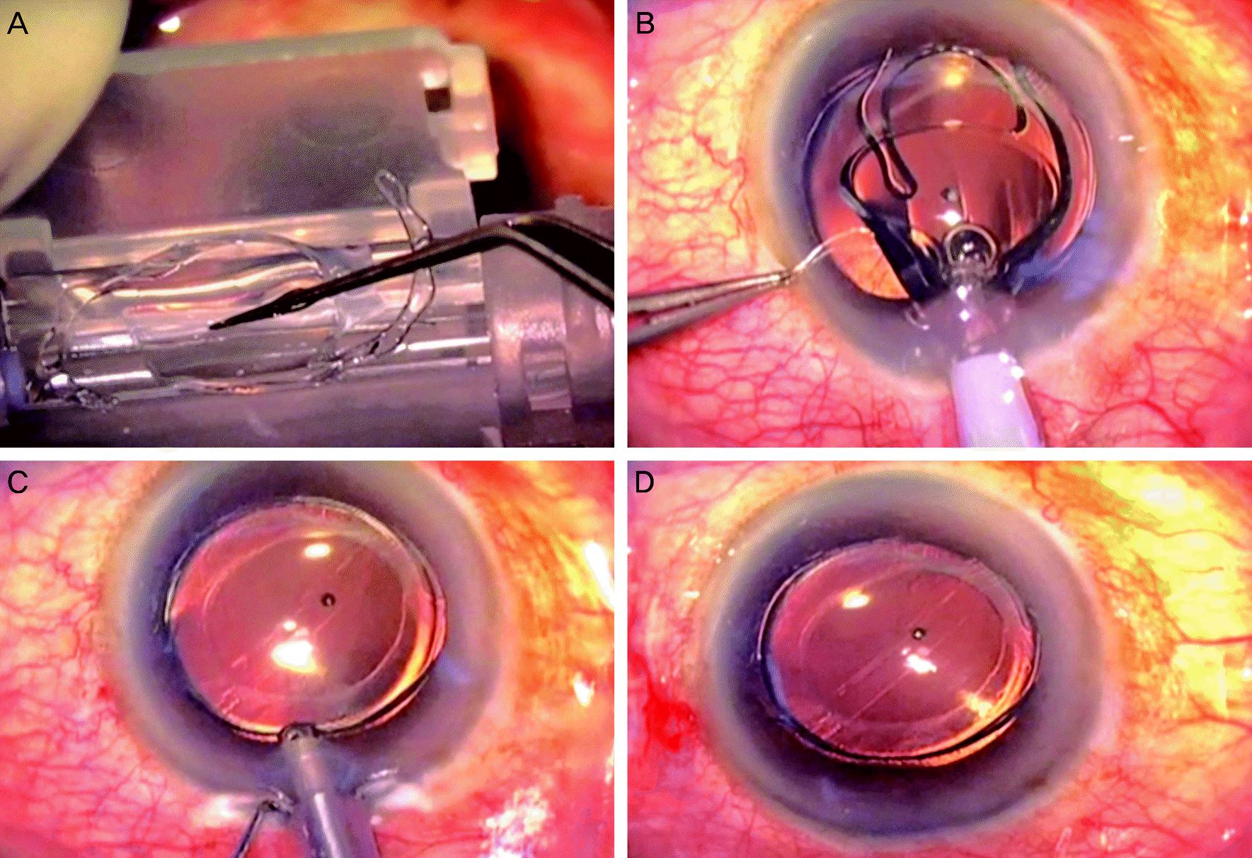
 | Figure 1.(A) The sulcoflex intraouclar lens (IOL) is placed on the central hinge of injector and pressed downward slightly. (B) The cartridge wings are closed and the secondary IOL is inserted into the ciliary sulcus. (C) Ophthalmic viscosurgical device is washed out. (D) The secondary IOL is well located in the ciliary sulcus. |
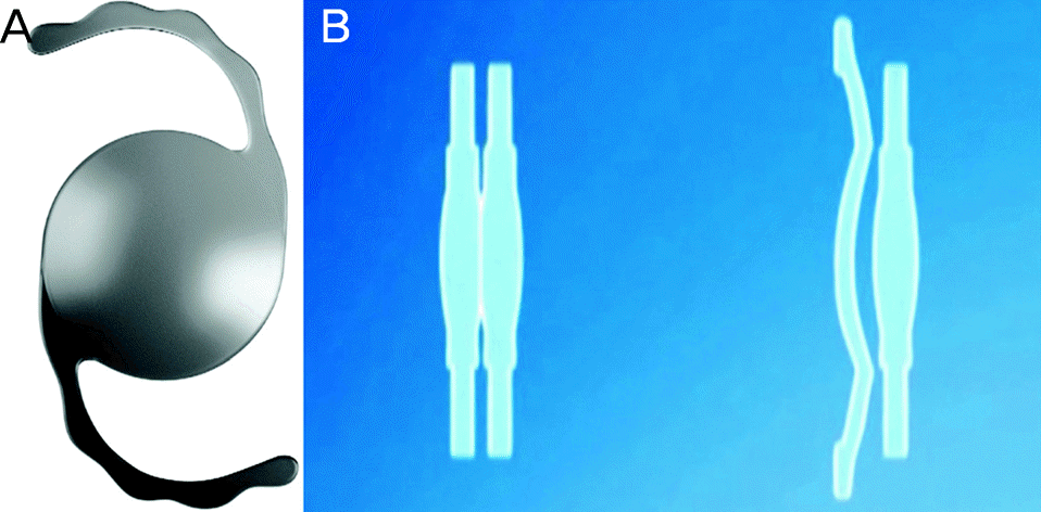
 | Figure 2.(A) Photograph of the sulcoflex intraouclar lens (IOL). The 6.5 mm optics reduce the risk of pupillary block and the 14 mm haptics maintain centration. Sulcoflex has a concave posterior surface to avoid contact with primary IOL. (B) Comparison between conventional piggyback IOL (left) and sulcoflex IOL (right). Specific design for sulcoflex IOL reduces physical contact between 2 IOLs. |


 PDF
PDF ePub
ePub Citation
 Print
Print


 XML Download