Journal List > J Korean Ophthalmol Soc > v.55(12) > 1009873

Cho and Lee: Correction of Hypertropia Coexisting with Intermittent Exotropia

Abstract

Purpose

To investigate the clinical features associated with hypertropia and report the surgical outcomes of hypertropia coexisting with exotropia.

Methods

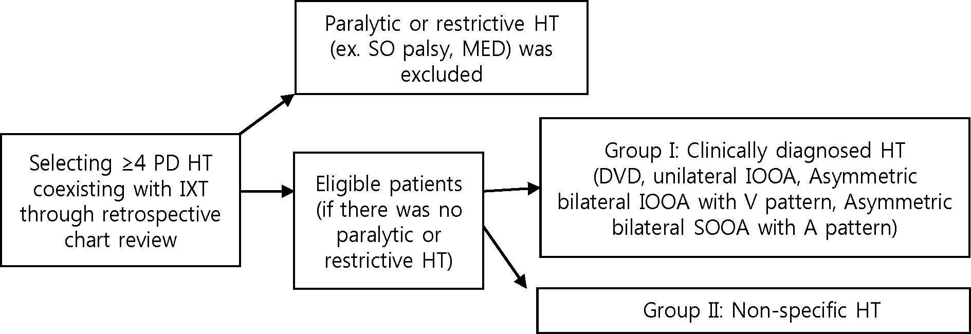
We reviewed the medical records of 148 patients with intermittent exotropia coexisting with hypertropia over 4 PD who received exotropia surgery. The cases accompanied by apparent paralytic strabismus such as superior oblique palsy were excluded. Patients were divided into group I (clinically diagnosed hypertropia) and group II (non-specific hypertropia) and the clinical features of coexisting hypertropia and surgical outcomes were analyzed.

Results

Among the 148 patients, group I consisted of 38 patients (26%) and group II of 110 patients (74%). The average amount of preoperative hypertropia angle in primary gaze was 9.58 ± 3.89 PD and 6.62 ± 2.69 PD in group I and II, respectively. Group I included 12 patients with dissociated vertical deviation (DVD), 10 patients with unilateral inferior oblique overaction, 13 patients with asymmetric bilateral inferior oblique overaction and 3 patients with superior oblique overaction. Group II included 19 patients with comitant hypertropia (17%), head tilt positive pattern (simulated superior oblique palsy) was found in 84 patients (76.3%) and variable incomitance was observed. In group I, 29 patients received simultaneous horizontal muscle with hypertropia surgery. Postoperative hypertropia angle in group I was 1.41 ± 2.93 PD and 4 cases were considered surgical failure. In group II, hypertropia was resolved with horizontal muscle surgery only and the amount of postoperative hypertropia was 0.45 ± 1.60 PD.

Conclusions

In this study, vertical deviations in intermittent exotropia with concomitant hypertropia related to obvious oblique muscle dysfunction or DVD were corrected effectively by oblique or vertical rectus muscle surgery. Nonspecific hypertropia can be resolved after horizontal muscle surgery alone, however, for precise differential diagnosis, careful examination for variable clinical features is necessary before determining surgery.

References

1. DUNLAP EA. VERTICAL DISPLACEMENT OF THE HORIZONTAL RECTI. Pac Med Surg. 1964; 72:360–2.
crossref
2. O'Neill JF. Surgical management of small-angle hypertropia by vertical displacement of the horizontal rectus muscles. Am Orthopt J. 1978; 28:32–42.
3. Paque JT, Mumma JV. Vertical offsets of the horizontal recti. J Pediatr Ophthalmol Strabismus. 1978; 15:205–9.
crossref
4. Metz HS. The use of vertical offsets with horizontal strabismus surgery. Ophthalmology. 1988; 95:1094–7.
crossref
5. Scott WE, Drummond GT, Keech RV. Vertical offsets of horizontal recti muscles in the management of A and V pattern strabismus. Aust N Z J Ophthalmol. 1989; 17:281–8.
crossref
6. Struck MC, Daley TJ. Resolution of hypertropia with correction of intermittent exotropia. Br J Ophthalmol. 2013; 97:1322–4.
crossref
7. Struck MC, Hariharan L, Kushner BJ, et al. Surgical management of clinically significant hypertropia associated with exotropia. J AAPOS. 2010; 14:216–20.
crossref
8. Lee JY, Kim SH, Yi ST, et al. Contemplation of the surgical normogram of lateral rectus recession for exotropia associated with superior oblique palsy. Korean J Ophthalmol. 2012; 26:195–8.
crossref
9. Shimko JF. Binocular Vision and Ocular Motility Theory and Management of Strabismus Gunter K. von Noorden, M.D.; Emilio C. Campos, M.D. Mosby Inc., Sixth Edition 2002, $149.00; 631 pages, 315 illustrations. Am Orthopt J. 2001; 51:161–2.
10. Moore S, Stockbridge L, Knapp P. A panoramic view of exotropias. Am Orthopt J. 1977; 27:70–9.
crossref
11. Pratt-Johnson JA, Tillson G. Management of the vertical associated with horizontal strabismus. Am Orthopt J. 1978; 28:24–7.
crossref
12. Cho YA, Kim SH. Surgical outcomes of intermittent exotropia associated with concomitant hypertropia including simulated superior oblique palsy after horizontal muscles surgery only. Eye (Lond). 2007; 21:1489–92.
crossref
13. Jampolsky A. Management of vertical strabismus. Trans New Orleans Acad Ophthalmol. 1986; 34:141–71.
14. Kushner BJ. Simulated superior oblique palsy. Ann Ophthalmol. 1981; 13:337–43.
15. Moore S, Stockbridge L. Fresnel prisms in the management of combined horizontal and vertical strabismus. Am Orthopt J. 1972; 22:14–21.
crossref
16. Wilson ME, Parks MM. Primary inferior oblique overaction in congenital esotropia, accommodative esotropia, and intermittent exotropia. Ophthalmology. 1989; 96:950–5. discussion 956-7.
crossref
17. Akar S, Gökyiğit B, Yilmaz OF. Graded anterior transposition of the inferior oblique muscle for V-pattern strabismus. J AAPOS. 2012; 16:286–90.
crossref
18. Bakunowicz-Lazarczyk A, Urban B, Lazarczyk J. [The surgical results of correcting strabismus with inferior oblique hyperfunction]. Klin Oczna. 2003; 105:398–400.
19. Kumar K, Prasad HN, Monga S, Bhola R. Hang-back recession of inferior oblique muscle in V-pattern strabismus with inferior oblique overaction. J AAPOS. 2008; 12:401–4.
crossref
20. Raab EL, Costenbader FD. Unilateral surgery for inferior oblique overaction. Arch Ophthalmol. 1973; 90:180–2.
crossref
21. Stein LA, Ellis FJ. Apparent contralateral inferior oblique muscle overaction after unilateral inferior oblique muscle weakening procedures. J AAPOS. 1997; 1:2–7.
crossref
22. Kim SH, Na JH, Cho YA. Inferior oblique transposition onto the equator: the role of the equator in development of contralateral inferior oblique overaction. J Pediatr Ophthalmol Strabismus. 2012; 49:98–102.
crossref
23. Lim HT, Smith DR, Kraft SP, Buncic JR. Dissociated vertical deviation in patients with intermittent exotropia. J AAPOS. 2008; 12:390–5.
crossref

Figure 1.
Flow diagram summarizing the stepwise categorization of the patients with hypertropia coexisting with intermittent exotropia. PD = prism diopter; HT = hypertropia; IXT = intermittent exotropia; SO = superior oblique; MED = monocular elevation deficiency; DVD = dissociated vertical deviation; IOOA = inferior oblique overaction; SOOA = superior oblique overaction.
jkos-55-1883f1.tif
Table 1.
Clinical characteristics of patients
Demographics Results
N 148
Sex (M:F) 70:78
Mean age at surgery (years) 12.47 ± 9.98
Mean amount of preoperative exodeviation (PD) 30.15 ± 1.76
Mean amount of postoperative exodeviation (PD) 7.17 ± 8.14
Mean amount of preoperative hypertropia (PD) 12.47 ± 9.89
Laterality of the coexisting hypertropia (n)
 RHT 78
 LHT 58
 DVD (both) 12
 Same as exotropic eye* 49
 Opposite to exotropic eye* 38

Values are presented as mean ± SD unless otherwise indicated.

PD = prism diopter; RHT = right hypertropia; LHT = left hypertropia; DVD = dissociated vertical deviation.

* Patients who had alternating fixation were excluded.

Table 2.
Distribution of surgery in clinically diagnosed hypertropia
Surgery No. of cases
DVD Bilateral IO anteriorization 2
SR recession 2
Oblique muscle dysfunction Bilateral IO recession 14
Unilateral IO recession 8
Bilateral IO myectomy 1
Bilateral SO recession 1
Bilateral SO tenotomy 1
Total 29

DVD = dissociated vertical deviation; IO = inferior oblique muscle; SR = superior rectus muscle; SO = superior oblique muscle.

Table 3.
Distribution of hypertropia coexisting with intermittent exotropia
Corrective surgery of HT N Preoperative HT (PD) Postoperative HT (PD)
Clinically diagnosed HT 38 9.58 ± 3.89 1.41 ± 2.93
 DVD + 4 10.0 ± 4.32 1.25 ± 2.5
- 8 6.75 ± 1.38 0.5 ± 1.41
 Unilateral IOOA + 10 11 ± 3.74 2.33 ± 4.47
- 0
 Asymmetric bilateral IOOA with V pattern + 13 9.0 ± 3.92 1.15 ± 1.91
- 0
 Asymmetric bilateral SOOA with A pattern + 2 9.87 ± 2.84 0
- 1 12 7
Non-specific HT + 0
- 110 6.62 ± 2.69 0.45 ± 1.60

Values are presented as mean ± SD.

HT = hypertropia; PD = prism diopter; DVD = dissociated vertical deviation; IOOA = inferior oblique overaction; SOOA = superior oblique overaction.

Table 4.
Surgical failure of hypertropia in clinically diagnosed hypertropia
Clinically diagnosed HT Surgical management
Overcorrection (>1 PD) 2 cases Unilateral IOOA Unilateral IO recession
Unilateral IOOA Unilateral IO recession
Undercorrection (>4 PD) 2 cases Unilateral IOOA Unilateral IO recession
Asymmetric bilateral IOOA Bilateral IO myectomy

HT = hypertropia; PD = prism diopter; IOOA = inferior oblique overaction; IO = inferior oblique muscle.

Table 5.
Classification of incomitance patterns in non-specific hypertropia
N HT in primary gaze (PD) HT in ipsilateral gaze (PD) HT in contralateral gaze (PD)
Pattern 1 13 6.07 ± 2.53 1.46 ± 3.01 5.76 ± 3.76
Pattern 2 19 6.52 ± 2.64 8.47 ± 3.43 1.84 ± 3.81

HT in primary gaze (PD) HT in ipsilateral HTT (PD) HT in contralateral HTT (PD)

Pattern 3 84 6.82 ± 2.85 11.80 ± 4.08 2.52 ± 3.91
Pattern 4 3 7.5 ± 2.51 3.75 ± 3.30 13.25 ± 2.21

Values are presented as mean ± SD; Pattern 1: increment of HT angle on contralateral gaze, contralateral side gaze PD-ipsilateral side PD >4 PD; Pattern 2: increment of HT angle on ipsilateral gaze, ipsilateral side gaze PD-contralateral side PD >4 PD; Pattern 3: Bielschowsky HTT positive (increment of HT angle on ipsilateral HTT), ipsilateral HTT PD contralateral HTT PD>8 PD; Pattern 4: reversed Bielschowsky HTT positive (increment of HT angle on contralateral HTT), contralateral HTT PD-ipsilateral HTT PD >8 PD. HT = hypertropia; PD = prism diopter; HTT = head tilt test.

Table 6.
Distribution of incomitance pattern in non-specific hypertropia
Distribution of incomitance patterns No. of patterns (%)
Pattern 1 only 2 (2)
Pattern 2 only 2 (2)
Pattern 3 only 57 (52)
Pattern 4 only 2 (2)
Pattern 1 & 3 11 (10)
Pattern 2 & 3 16 (14)
Pattern 1 & 4 0 (0)
Pattern 2 & 4 1 (1)
Comitant hypertropia (no pattern) 19 (17)
Total 110 (100)

Pattern 1: increment of hypertropia (HT) angle on contralateral gaze, contralateral side gaze prism diopter (PD)-ipsilateral side PD >4 PD; Pattern 2: increment of HT angle on ipsilateral gaze, ipsilateral side gaze PD-contralateral side PD >4 PD; Pattern 3: Bielschowsky head tilt test (HTT) positive (increment of HT angle on ipsilateral HTT), ipsilateral HTT PD-contralateral HTT PD >8 PD; Pattern 4: reversed Bielschowsky HTT positive (increment of HT angle on contralateral HTT), contralateral HTT PD-ipsilateral HTT PD >8 PD.

Table 7.
Outcomes of non-specific hypertropia after the intermittent exotropia correction
Distribution of preoperative hypertropia N Postoperative hypertropia (PD)
4-7 PD 67 0.22 ± 1.05
8-11 PD 34 0.76 ± 2.20
12-15 PD 9 1.0 ± 2.12

Values are presented as mean ± SD.

PD = prism diopter.

TOOLS
Similar articles