

 PDF
PDF ePub
ePub Citation
 Print
Print


Abstract
Purpose
To assess the changes of corneal astigmatism and higher order aberrations (HOAs) of the anterior and posterior corneal surface after cataract surgery with on-axis clear corneal incision in eyes with-the-rule (WTR) astigmatism and against-the-rule (ATR) astigmatism.
Methods
This study included 50 patients who underwent phacoemulsification and IOL insertion through a 2.8-mm on-axis clear corneal incision. The eyes were divided into two groups: (1) 26 eyes with WTR astigmatism with a superior incision and (2) 24 eyes with ATR astigmatism with a temporal incision. During the follow-up period, visual acuity was measured, and the surgically induced astigmatism (SIA) and HOAs of the anterior and posterior corneal surface were measured with Pentacam® (Occlus, Wetzlar, Germany) preoperatively and 1 week, 1 month, and 2 months postoperatively.
Results
There were no significant differences in UCVA and BCVA between the two groups. HOAs increased in both groups 1 week after surgery, but no significant differences were found between the groups (p > 0.05). Surgically induced astigmatism was larger in the WTR group than in the ATR group (p < 0.05). At postoperative 2 months, there were significant differences in HOAs between the two groups, and there were statistically significant differences in HOAs, oblique trefoil at front side, and in HOAs, horizontal coma at rear side (p < 0.05).
Conclusions
In conclusion, superior incision in eyes with WTR astigmatism resulted in higher SIA compared to temporal incision in eyes with ATR astigmatism. Moreover, HOAs was significantly decreased in eyes with WTR astigmatism with superior incision. Thus, superior incision could be more effective in reducing corneal astigmatism in eyes with WTR astigmatism.
Go to : 
References
1. Moon SJ, Lee DJ, Lee KH.Induced astigmatism and high-order aberrations after 1.8-mm, 2.2-mm and 3.0-mm coaxial phacoemulsi-fication incisions. J Korean Ophthalmol Soc. 2011; 52:407–13.

2. Artal P, Berrio E, Guirao A, Piers P.Contribution of the cornea and internal surfaces to the change of ocular aberrations with age. J Opt Soc Am A Opt Image Sci Vis. 2002; 19:137–43.
3. Borasio E, Mehta JS, Maurino V.Surgically induced astigmatism after phacoemulsification in eyes with mild to moderate corneal as-tigmatism: temporal versus on-axis clear corneal incisions. J Cataract Refract Surg. 2006; 32:565–72.
4. Alió J, Rodríguez-Prats JL, Galal A, Ramzy M.Outcomes of mi-croincision cataract surgery versus coaxial phacoemulsification. Ophthalmology. 2005; 112:1997–2003.
5. Artal P, Guirao A, Berrio E, Williams DR.Compensation of cor-neal aberrations by the internal optics in the human eye. J Vis. 2001; 1:1–8.
6. Artal P, Guirao A.Contributions of the cornea and the lens to the aberrations of the human eye. Opt Lett. 1998; 23:1713–5.
7. Guirao A, Redondo M, Geraghty E. . Corneal optical aberra-tions and retinal image quality in patients in whom monofocal in-traocular lenses were implanted. Arch Ophthalmol. 2002; 120:1143–51.
8. Oshika T, Kawana K, Hiraoka T. . Ocular higher-order wave-front aberration caused by major tilting of intraocular lens. Am J Ophthalmol. 2005; 140:744–6.
9. Fujikado T, Kuroda T, Maeda N. . Light scattering and optical aberrations as objective parameters to predict visual deterioration in eyes with cataracts. J Cataract Refract Surg. 2004; 30:1198–208.
10. Lee SY, Chung JL, Hong JP. . Comparative study of two aspheric, aberration-free intraocular lenses in cataract surgery. J Korean Ophthalmol Soc. 2009; 50:1520–6.
11. Holladay JT, Hill WE, Steinmueller A.Corneal power measure-ments using scheimpflug imaging in eyes with prior corneal re-fractive surgery. J Refract Surg. 2009; 25:862–8.
12. Yao K, Tang X, Ye P.Corneal astigmatism, high order aberrations, and optical quality after cataract surgery: microincision versus small incision. J Refract Surg. 2006; 22(9 Suppl):S1079–82.
13. Guirao A, Tejedor J, Artal P.Corneal aberrations before and after small-incision cataract surgery. Invest Ophthalmol Vis Sci. 2004; 45:4312–9.
14. Hwang SJ, Choi SK, Oh SH. . Surgically induced astigmatism and corneal higher order aberrations in microcoaxial and conven-tional cataract surgery. J Korean Ophthalmol Soc. 2008; 49:1597–602.
15. Meek KM, Newton RH.Organization of collagen fibrils in the cor-neal stroma in relation to mechanical properties and surgical practice. J Refract Surg. 1999; 15:695–9.
16. Oh HC, Lee DJ, Park WC.Changes of the corneal aberration following cataract surgery. J Korean Ophthalmol Soc. 2009; 50:518–22.
Go to :
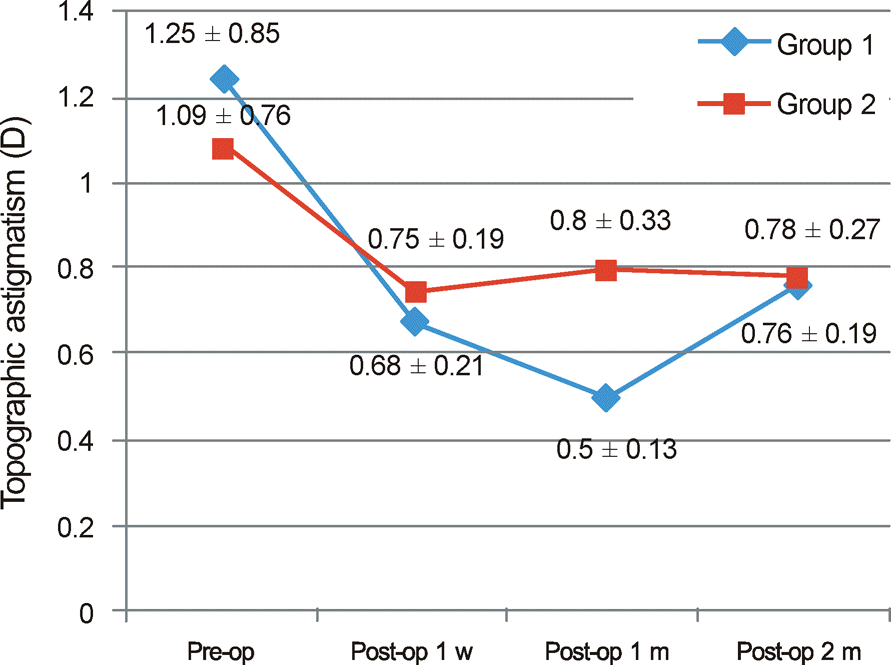 | Figure 1.Change of topographic astigmatism. Values are presented as mean ± SD. Pre-op = preoperative; Post-op 1 w = postoperative 1 week; Post-op 1 m = postoperative 1 month; Post-op 2 m = postoperative 2 months. p-value by Mann-Whitney U test. There is no statistically significant difference between two groups (p > 0.05). |
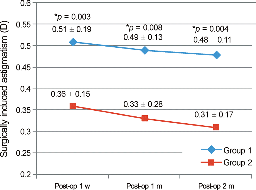 | Figure 2.Surgically-induced astigmatism calculated by vector analysis. Values are presented as mean ± SD. Pre-op = pre-operative; Post-op 1 w = postoperative 1 week; Post-op 1 m = postoperative 1 month; Post-op 2 m = postoperative 2 months. p-value by Mann-Whitney U test (* p < 0.05). |
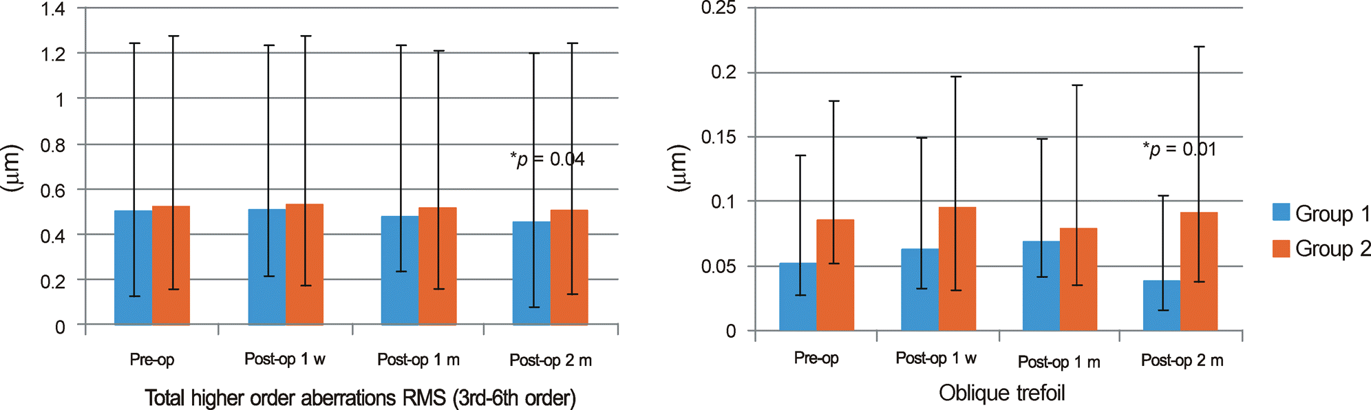 | Figure 3.Comparision of corneal total higher order aberrations RMS and oblique trefoil (Z(3,-3)) RMS on front side in two groups. Z = zernike coefficients; RMS = root mean square; Pre-op = preoperative; Post-op 1 w = postoperative 1 week; Post-op 1 m = postoperative 1 month; Post-op 2 m = postoperative 2 months. p-value by Mann-Whitney U test (* p < 0.05). |
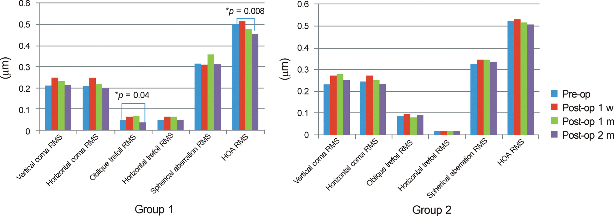 | Figure 4.Comparision of corneal total higher order aberations on front side in two groups. Z = zernike coefficients; RMS = root mean square; HOA = total higher order aberrations (3rd-6th order); Pre-op = preoperative; Post-op 1 w = postoperative 1 week; Post-op 1 m = postoperative 1 month; Post-op 2 m = postoperative 2 months. p-value by Wilcoxon rank test (* p < 0.05). |
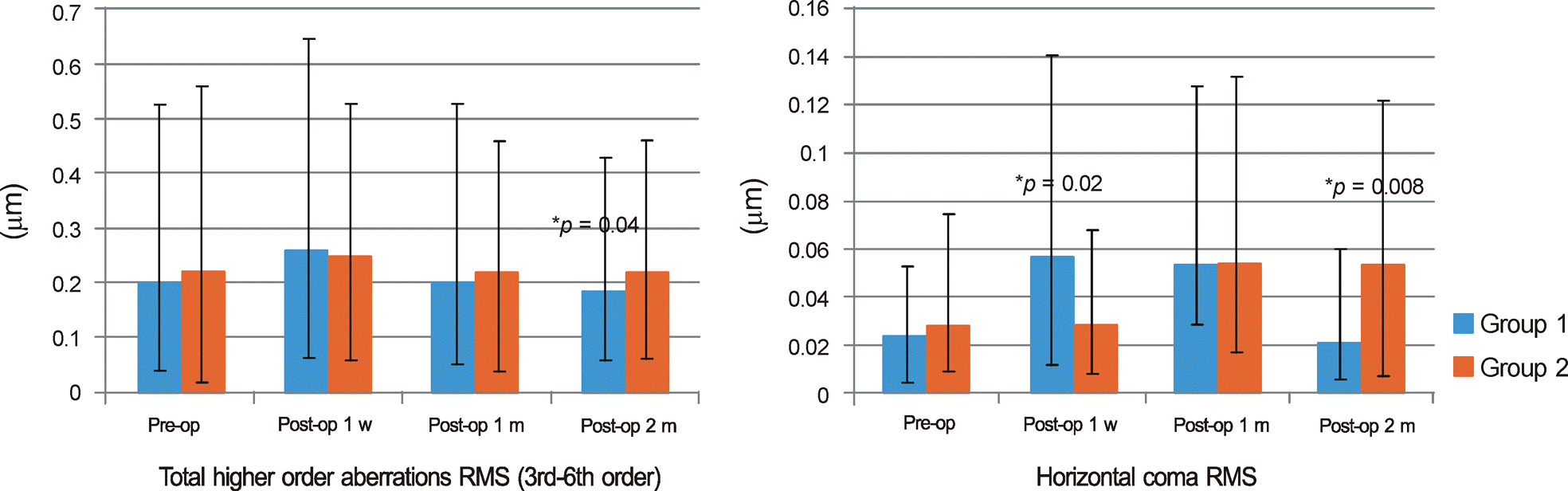 | Figure 5.Comparision of corneal total higher order aberations RMS and Horizontal coma (Z(3, 1)) RMS on rear side in two groups. Z = zernike coefficients; RMS = root mean square; HOA = total higher order aberrations (3rd-6th order); Pre-op = preoperative; Post-op 1 w = postoperative 1 week; Post-op 1 m = postoperative 1 month; Post-op 2 m = postoperative 2 months. p-value by Mann-Whitney U test (* p < 0.05). |
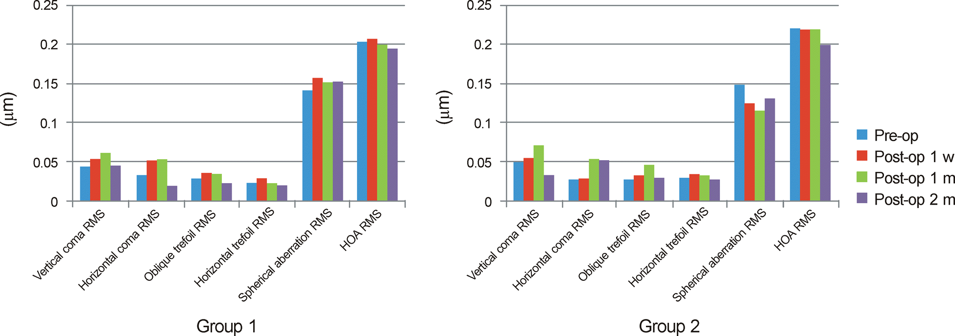 | Figure 6.Comparision of corneal total higher order aberrations on rear side in two groups. Z = zernike coefficients; RMS = root mean square; HOA = total higher order aberrations (3rd-6th order); Pre-op = preoperative; Post-op 1 w = postoperative 1 week; Post-op 1 m = postoperative 1 month; Post-op 2 m = postoperative 2 month. p-value by Wilcoxon rank test. There is no statistically significant difference among preoperative and postoperative 1 week, postoperative 1 month, postoperative 2 months in two groups, respectively (p > 0.05). |
Table 1.
Patient characteristics
| Variables | With the rule (Group 1) | Against the rule (Group 2) | p-value* |
|---|---|---|---|
| No. of eyes | 26 | 24 | |
| Laterality (OD:OS) | 14 : 12 | 12 : 12 | |
| Sex (M:F) | 13 : 13 | 11 : 13 | |
| Age (years) | 68.40 ± 6.87 | 73.90 ± 10.83 | 0.062 |
| Pachymetry (μm) | 563.64 ± 31.19 | 569.55 ± 33.75 | 0.558 |
| Endothelial cell count (cell/mm2) | 2703 ± 457.13 | 2825 ± 367.95 | 0.527 |
| Axial length (mm) | 23.95 ± 0.11 | 23.55 ± 0.53 | 0.424 |
Table 2.
Data summary of UCVA, BCVA, SE and topographic astigmatism
| With the rule (Group 1) | Against the rule (Group 2) | p-value* | ||
|---|---|---|---|---|
| Pre-op | UCVA (log MAR) | 0.78 ± 0.49 | 0.79 ± 0.43 | 0.25 |
| BCVA (log MAR) | 0.54 ± 0.37 | 0.59 ± 0.50 | 0.73 | |
| SE (diopter) | 2.29 ± 2.31 | 2.58 ± 1.90 | 0.829 | |
| Topographic astigmatism (diopter) | 1.25 ± 0.85 | 1.09 ± 0.76 | 0.23 | |
| Post-op 2 m | UCVA (log MAR) | 0.19 ± 0.32 | 0.18 ± 0.36 | 0.08 |
| BCVA (log MAR) | 0.05 ± 0.03 | 0.04 ± 0.07 | 0.08 | |
| SE (diopter) | -0.40 ± 0.42 | -0.39 ± 0.32 | 0.501 | |
| Topographic astigmatism (diopter) | 0.76 ± 0.19 | 0.78 ± 0.27 | 0.231 |
Table 3.
Postoperative changes of corneal aberrations on the front side
| Pre-op | Post-op 1 w | Post-op 1 m | Post-op 2 m | ||
|---|---|---|---|---|---|
| Z(3,-1) | Group 1* | 0.210 ± 0.024 | 0.251 ± 0.018 | 0.232 ± 0.025 | 0.216 ± 0.026 |
| Vertical coma | Group 2† | 0.231 ± 0.055 | 0.274 ± 0.014 | 0.280 ± 0.027 | 0.251 ± 0.057 |
| RMS (μm) | p-value | 0.649 | 0.287 | 0.468 | 0.14 |
| Z(3, 1) | Group 1* | 0.208 ± 0.221 | 0.249 ± 0.082 | 0.219 ± 0.022 | 0.197 ± 0.032 |
| Horizontal coma | Group 2† | 0.245 ± 0.055 | 0.272 ± 0.014 | 0.251 ± 0.05 | 0.235 ± 0.036 |
| RMS (μm) | p-value | 0.875 | 0.421 | 0.172 | 0.09 |
| Z(3, -3) | Group 1* | 0.052 ± 0.010 | 0.063 ± 0.012 | 0.068 ± 0.013 | 0.038 ± 0.012 |
| Oblique trefoil | Group 2† | 0.086 ± 0.019 | 0.096 ± 0.012 | 0.080 ± 0.079 | 0.092 ± 0.041 |
| RMS (μm) | p-value | 0.147 | 0.053 | 0.310 | 0.01‡ |
| Z(3, 3) | Group 1* | 0.052 ± 0.033 | 0.066 ± 0.015 | 0.063 ± 0.028 | 0.049 ± 0.013 |
| Horizontal trefoil | Group 2† | 0.051 ± 0.075 | 0.057 ± 0.047 | 0.059 ± 0.098 | 0.052 ± 0.042 |
| RMS (μm) | p-value | 0.482 | 0.785 | 0.612 | 0.745 |
| Z(4, 0) | Group 1* | 0.317 ± 0.028 | 0.308 ± 0.024 | 0.358 ± 0.039 | 0.311 ± 0.034 |
| Spherical aberration | Group 2† | 0.325 ± 0.047 | 0.347 ± 0.075 | 0.348 ± 0.049 | 0.336 ± 0.074 |
| RMS (μm) | p-value | 0.574 | 0.214 | 0.574 | 0.672 |
| HOA | Group 1* | 0.506 ± 0.034 | 0.512 ± 0.029 | 0.480 ± 0.036 | 0.455 ± 0.027 |
| Group 2† | 0.523 ± 0.078 | 0.531 ± 0.045 | 0.516 ± 0.062 | 0.507 ± 0.027 | |
| RMS (μm) | p-value | 0.613 | 0.06 | 0.186 | 0.04‡ |
Table 4.
Postoperative changes of corneal aberrations on the rear side
| Pre-op | Post-op 1 w | Post-op 1 m | Post-op 2 m | ||
|---|---|---|---|---|---|
| Z(3, -1) | Group 1* | 0.044 ± 0.006 | 0.054 ± 0.012 | 0.061 ± 0.007 | 0.037 ± 0.009 |
| Vertical coma | Group 2† | 0.050 ± 0.014 | 0.055 ± 0.021 | 0.071 ± 0.012 | 0.033 ± 0.015 |
| RMS (μm) | p-value | 0.281 | 0.253 | 0.749 | 0.924 |
| Z(3, 1) | Group 1* | 0.024 ± 0.003 | 0.063 ± 0.004 | 0.053 ± 0.004 | 0.021 ± 0.002 |
| Horizontal coma | Group 2† | 0.028 ± 0.012 | 0.029 ± 0.010 | 0.054 ± 0.006 | 0.053 ± 0.007 |
| RMS (μm) | p-value | 0.524 | 0.02‡ | 0.456 | 0.008‡ |
| Z(3, -3) | Group 1* | 0.029 ± 0.007 | 0.037 ± 0.058 | 0.035 ± 0.010 | 0.024 ± 0.008 |
| Oblique trefoil | Group 2† | 0.028 ± 0.011 | 0.034 ± 0.010 | 0.046 ± 0.009 | 0.031 ± 0.006 |
| RMS (μm) | p-value | 0.069 | 0.267 | 0.075 | 0.13 |
| Z(3, 3) | Group 1* | 0.024 ± 0.003 | 0.029 ± 0.005 | 0.024 ± 0.003 | 0.021 ± 0.004 |
| Horizontal trefoil | Group 2† | 0.031 ± 0.004 | 0.035 ± 0.110 | 0.034 ± 0.007 | 0.028 ± 0.006 |
| RMS (μm) | p-value | 0.07 | 0.09 | 0.144 | 0.161 |
| Z(4, 0) | Group 1* | 0.142 ± 0.007 | 0.167 ± 0.010 | 0.151 ± 0.008 | 0.163 ± 0.017 |
| Spherical aberration | Group 2† | 0.175 ± 0.010 | 0.125 ± 0.012 | 0.116 ± 0.009 | 0.131 ± 0.010 |
| RMS (μm) | p-value | 0.12 | 0.45 | 0.53 | 0.45 |
| HOA | Group 1* | 0.204 ± 0.011 | 0.266 ± 0.017 | 0.201 ± 0.010 | 0.185 ± 0.013 |
| Group 2† | 0.221 ± 0.019 | 0.240 ± 0.030 | 0.220 ± 0.011 | 0.200 ± 0.024 | |
| RMS (μm) | p-value | 0.4 | 0.214 | 0.186 | 0.04‡ |
 XML Download