Abstract
Purpose
We present a case of a patient with optic neuritis who had underlying suspicious idiopathic thrombocytopenic purpura.
Case summary
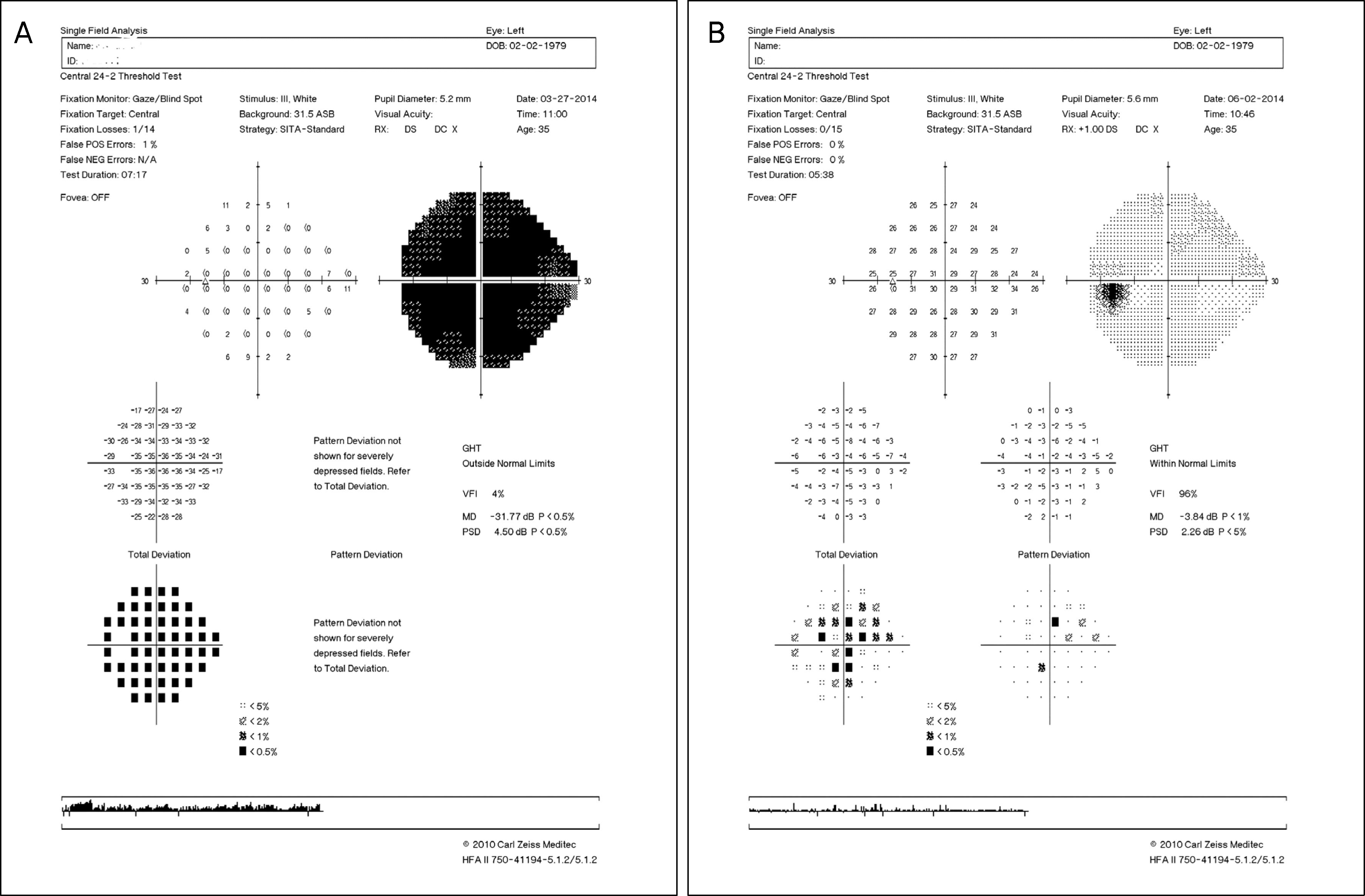
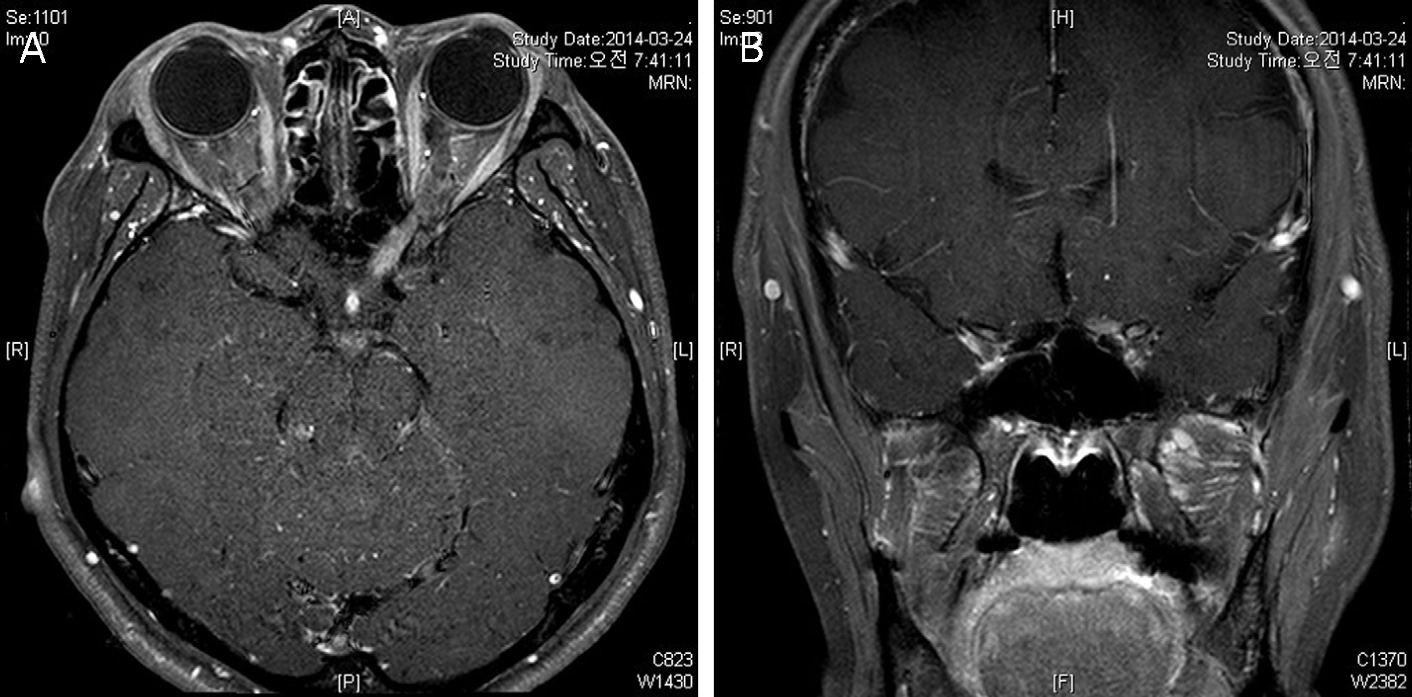
A 35-year-old female with no other systemic disease visited our clinic due to acutely decreased visual acuity in her left eye 10 days in duration. Relative afferent pupillary defect was observed, but without definite papilledema. Based on brain magnetic resonance imaging (MRI), optic neuritis was suspected. Laboratory tests showed increased red blood cells, hemoglobin and, hematocrit levels and decreased platelets. Peripheral blood smear test showed decreased platelets, relative lymphocytosis and atypical lymphocytes. Specific antibodies for autoimmune disease were not present. High-dose steroid pulse therapy (methyl prednisolone 1.0 g/d, 3 days) was started. One month after treatment her visual acuity and platelet count recovered and her visual field defect improved.
Go to : 
References
2. Swanton JK, Fernando K, Dalton CM, et al. Is the frequency of abnormalities on magnetic resonance imaging in isolated optic neuritis related to the prevalence of multiple sclerosis? A global comparison. J Neurol Neurosurg Psychiatry. 2006; 77:1070–2.

3. Petzold A, Plant GT. Diagnosis and classification of autoimmune optic neuropathy. Autoimmun Rev. 2014; 13:539–45.

4. Hui AC, Wong RS, Ma R, Kay R. Recurrent optic neuromyelitis with multiple endocrinopathies and autoimmune disorders. J Neurol. 2002; 249:784–5.

5. Yamout B, Usta J, Itani S, Yaghi S. Celiac disease, Behçet, and idiopathic thrombocytopenic purpura in siblings of a patient with multiple sclerosis. Mult Scler. 2009; 15:1368–71.

6. Sahraian MA, Eshaghi A. Concomitant multiple sclerosis and idiopathic thrombocytopenic purpura. Eur J Neurol. 2010; 17:e62–3.

7. Scott LM, Tong W, Levine RL, et al. JAK2 exon 12 mutations in polycythemia vera and idiopathic erythrocytosis. N Engl J Med. 2007; 356:459–68.
8. Alemany-Rodríguez MJ, Aladro Y, Amela-Peris R, et al. [Autoimmune diseases and multiple sclerosis]. Rev Neurol. 2005; 40:594–7.
9. Optic Neuritis Study Group. Multiple sclerosis risk after optic neuritis: final optic neuritis treatment trial follow-up. Arch Neurol. 2008; 65:727–32.
10. Ahn SM, Kim SS. A case of coexisting neuromyelitis optica in systemic lupus erythematosus. J Korean Ophthalmol Soc. 2013; 54:1469–74.

11. Adoni T, Lino AM, da Gama PD, et al. Recurrent neuromyelitis optica in Brazilian patients: clinical, immunological, and neuroimaging characteristics. Mult Scler. 2010; 16:81–6.

12. Sergio P, Mariana B, Alberto O, et al. Association of neuromyelitis optic (NMO) with autoimmune disorders: report of two cases and review of the literature. Clin Rheumatol. 2010; 29:1335–8.

13. Wingerchuk DM, Lennon VA, Lucchinetti CF, et al. The spectrum of neuromyelitis optica. Lancet Neurol. 2007; 6:805–15.

15. RABINOWITZ Y, DAMESHEK W. Systemic lupus erytematosus after “idiopathic” thrombocytopenic purpura: a review. Ann Intern Med. 1960; 52:1–28.
16. Hymes K, Blum M, Lackner H, Karpatkin S. Easy bruising, thrombocytopenia, and elevated platelet immunoglobulin G in Graves’ disease and Hashimoto's thyroiditis. Ann Intern Med. 1981; 94:27–30.

18. Sheremata WA, Jy W, Horstman LL, et al. Evidence of platelet activation in multiple sclerosis. J Neuroinflammation. 2008; 5:27.

19. Segal JB, Powe NR. Prevalence of immune thrombocytopenia: analyses of administrative data. J Thromb Haemost. 2006; 4:2377–83.

20. Killer HE, Huber A, Portman C, et al. Bilateral non-arteritic anterior ischemic optic neuropathy in a patient with autoimmune thrombocytopenia. Eur J Ophthalmol. 2000; 10:180–2.

21. Bermel RA, Balcer LJ. Optic neuritis and the evaluation of visual impairment in multiple sclerosis. Continuum (Minneap Minn). 2013; 19:1074–86.

22. Comi G, Filippi M, Barkhof F, et al. Effect of early interferon treatment on conversion to definite multiple sclerosis: a randomised study. Lancet. 2001; 357:1576–82.

23. Mandler RN, Ahmed W, Dencoff JE. Devic's neuromyelitis optica: a prospective study of seven patients treated with prednisone and azathioprine. Neurology. 1998; 51:1219–20.

24. Cusick MF, Libbey JE, Fujinami RS. Multiple sclerosis: autoimmunity and viruses. Curr Opin Rheumatol. 2013; 25:496–501.
Go to : 
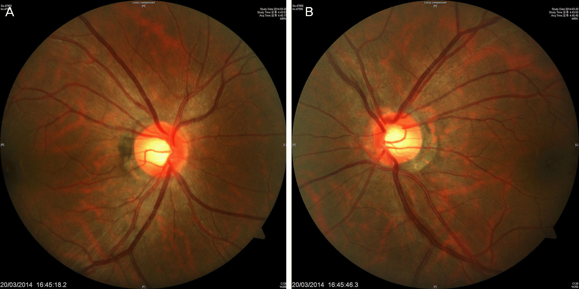
 | Figure 1.At the first visit, disc photography revealed no definite disc swelling in her both eyes. (A) Right eye, (B) left eye. |
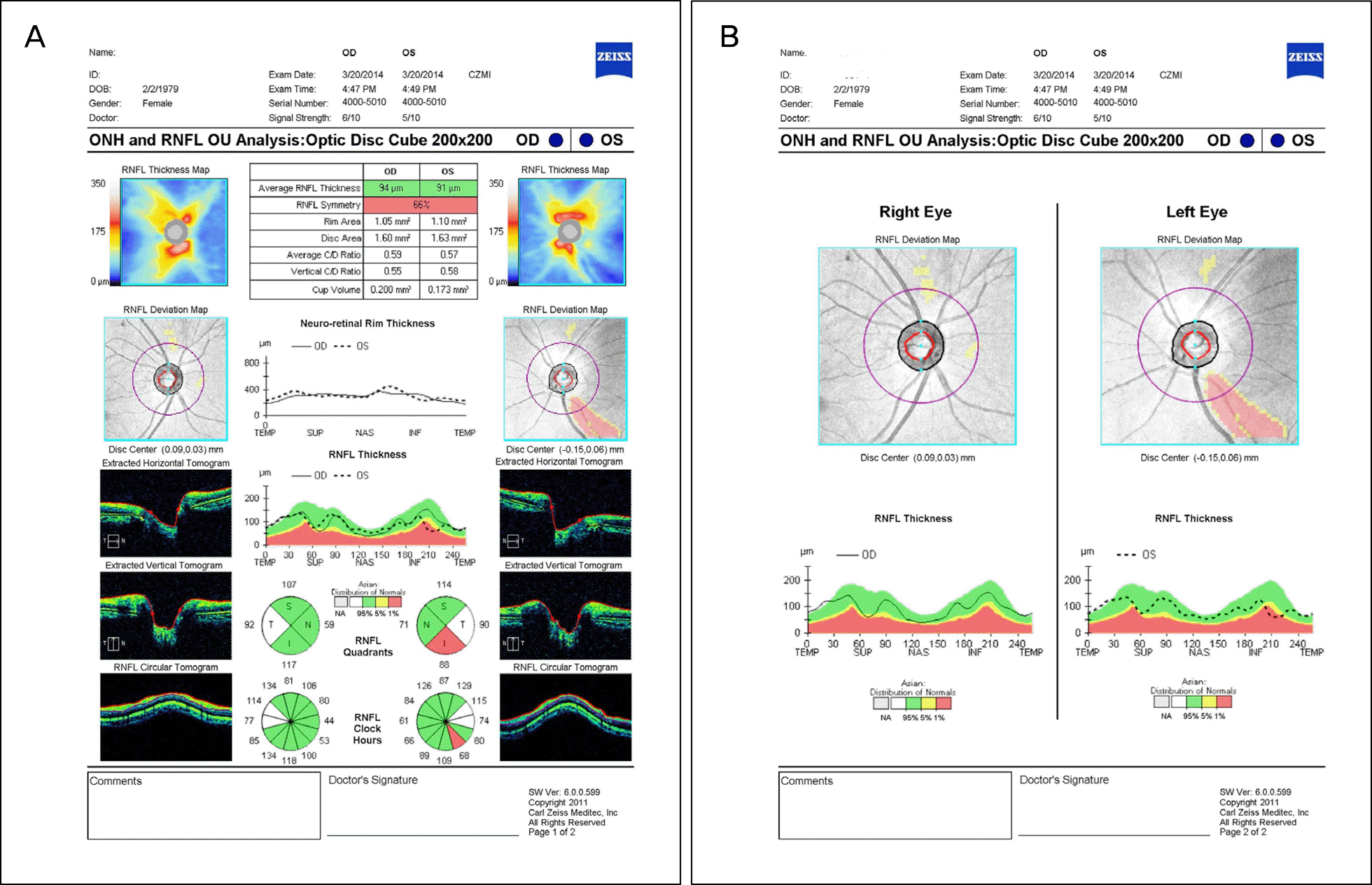
 | Figure 2.Decreased retinal nerve fiber layer (RNFL) thickness at the inferotemporal area of her left eye in optical coherence tomography. (A) RNFL quadrants and clock hours maps, (B) RNFL deviation map and thickness. OD = right eye; ONH = optic nerve head; OS = left eye; OU = both eyes; TEMP = temporal; SUP = superior, NAS = nasal, INF = inferior, C/D = cup to disc ratio. |
 | Figure 3.(A) Humphrey visual field test shows almost total scotoma in her left eye before high dose methylprednisolone treatment. (B) After treatment, improvement of visual field test in her left eye was noted. POS = positive; NEG = negative; GHT = glaucoma hemifield test; VFI = visual field index; MD = mean deviation; PSD = pattern standard deviation. |
 | Figure 4.Brain magentic resonance image (MRI) with enhance T1 weighted MRI. Diffuse enhancement of the left optic nerve, cisternal segment, suspicious of optic neuritis or neuropathy. No abnormal signal intensity or abnormal mass lesion in the brain parenchyma (A: axial view; B: coronal view). |


 PDF
PDF ePub
ePub Citation
 Print
Print


 XML Download