Abstract
Purpose
To report the characteristics and surgical outcome of macular holes (MHs) that develop after rhegmatogenous retinal detachment (RRD) repair.
Methods
A retrospective chart review was performed in patients who developed a new full-thickness macular hole after RRD repair between May 2010 and July 2013. For eyes that underwent pars plana vitrectomy with internal limiting membrane peeling and gas tamponade for MH repair, main outcomes included macular attachment status and postoperative visual acuity.
Results
Fourteen full-thickness MHs were detected in a series of 2,815 eyes (0.49% prevalence) that had undergone prior RRD surgery. Ten MHs developed after primary vitrectomy and four after scleral bucking surgery. The fovea was detached in eight of the 14 eyes at the time of RRD. Fourteen of 14 eyes were managed by pars plana vitrectomy, internal limiting membrane peeling, and intravitreal gas tamponade, and 12 of 14 eyes achieved MH closure. Mean preoperative Snellen best-corrected visual acuity (BCVA) was 20/63 (±0.25). Nine of 14 eyes had an improvement in visual acuity of at least two Snellen lines, and five eyes remained unchanged.
Conclusions
In this small retrospective study, the secondary MHs were found predominantly in foveal detachments after RRD repair, most commonly occurring after primary vitrectomy. In conclusion, the surgical outcome and postoperative visual acuity improvement were satisfactory, although the final BCVA depended on the macular status during the RRD.
Go to : 
References
1. Gass JD. Reappraisal of biomicroscopic classification of stages of development of a macular hole. Am J Ophthalmol. 1995; 119:752–9.
2. Gaudric A, Haouchine B, Massin P, et al. Macular hole formation: new data provided by optical coherence tomography. Arch Ophthalmol. 1999; 117:744–51.
3. Hee MR, Puliafito CA, Wong C, et al. Optical coherence tomography of macular holes. Ophthalmology. 1995; 102:748–56.

4. Smiddy WE. Macular hole formation without vitreofoveal traction. Arch Ophthalmol. 2008; 126:737–8.

5. Gordon LW, Glaser BM, Ie D, et al. Full-thickness macular hole formation in eyes with a pre-existing complete posterior vitreous detachment. Ophthalmology. 1995; 102:1702–5.

6. Brown GC. Macular hole following rhegmatogenous retinal detachment repair. Arch Ophthalmol. 1988; 106:765–6.

7. Ah Kiné D, Benson SE, Inglesby DV, Steel DH. The results of surgery on macular holes associated with rhegmatogenous retinal detachment. Retina. 2002; 22:429–34.

8. Boscia F, Recchimurzo N, Cardascia N, et al. Macular hole following conventional repair of bullous retinal detachment using air injection (D-ACE procedure). Eur J Ophthalmol. 2004; 14:572–4.

9. Moshfeghi AA, Salam GA, Deramo VA, et al. Management of macular holes that develop after retinal detachment repair. Am J Ophthalmol. 2003; 136:895–9.

10. Avins LR, Krummenacher TR. Macular holes after pneumatic retinopexy. Case reports. Arch Ophthalmol. 1988; 106:724–5.
11. Hejny C, Han DP. Vitrectomy for macular hole after pneumatic retinopexy. Retina. 1997; 17:356–7.

12. Garcia-Arumi J, Boixadera A, Martinez-Castillo V, et al. Macular holes after rhegmatogenous retinal detachment repair: surgical management and functional outcome. Retina. 2011; 31:1777–82.
13. Shibata M, Oshitari T, Kajita F, et al. Development of macular holes after rhegmatogenous retinal detachment repair in Japanese patients. J Ophthalmol. 2012; 2012:740591. Epub 2012 Jan 18.

14. Benzerroug M, Genevois O, Siahmed K, et al. Results of surgery on macular holes that develop after rhegmatogenous retinal detachment. Br J Ophthalmol. 2008; 92:217–9.

16. Kumagai K, Ogino N, Furukawa M, et al. Surgical outcomes for patients who develop macular holes after pars plana vitrectomy. Am J Ophthalmol. 2008; 145:1077–80.

17. Lee SH, Park KH, Kim JH, et al. Secondary macular hole formation after vitrectomy. Retina. 2010; 30:1072–7.

Go to : 
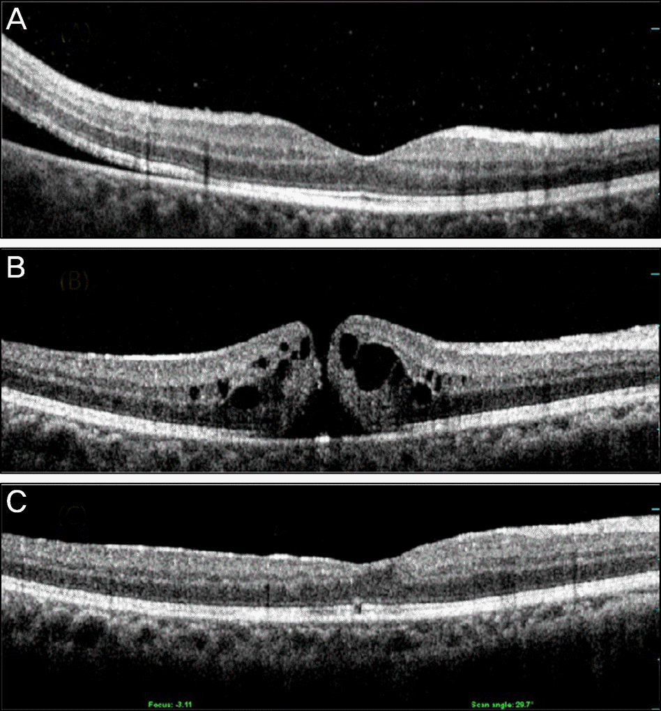
 | Figure 1.Optical coherence tomography images of patient number 12 that were taken at the diagnosis of retinal detachment (macular-spared) (A), 22 months after rhegmatogenous retinal detachment repair, macular hole had developed (B). 2 months after additional vitrectomy, internal limiting membrane peeling and fluid-gas exchange, macular hole had closed (C). The best-corrected visual acuity measured at each time point was 20/40, 20/50, 20/25, respectively. |
Table 1.
Characteristics of 14 patients who had macular holes that developed after prior rhegmatogenous retinal detachment repair


 PDF
PDF ePub
ePub Citation
 Print
Print


 XML Download