

 PDF
PDF ePub
ePub Citation
 Print
Print


Abstract
Purpose
To evaluate microvascular change (microaneurysm) in diabetic retinopathy patients who undergo intravitreal bevacizumab injection using fluorescein angiography (FAG).
Methods
Thirty one eyes of 31 diabetic retinopathy patients undergoing intravitreal bevacizumab injection (1.25 mg/0.05 mL) in only 1 eye were included in this study. The control group (31 eyes) consisted of the fellow eyes. We excluded cased with intra-vitreal bevacizumab injection in both eyes and medial opacity obscuring three fundus image due to vitreous hemorrhage. The microaneurysmal change was analyzed at the same site the circle with optic disc radius and macula using FAG 2 to 4 months after injection.
Results
The average number of microaneurysms was 42.58 ± 33.93 and significantly decreased to 28.74 ± 28.06 after intra-vitreal bevacizumab injection (p < 0.05). The decrease of 35.70 ± 24.79% in the treatment group was statistically higher than 13.95 ± 38.21% in the control group with the fellow eyes (p < 0.05).
Go to : 
References
1. Mun SJ, Kim IC, Lee DW, et al. The morphological study of muller cells with enzyme histochemical staining in retina of induced diabetic rats. J Korean Ophthalmol Soc. 2007; 48:708–14.
2. Kim HK, Oh TS, Lee SM, Lee JB. The initial fundus examination and severity of diabetic retinopathy at a primary eye clinic. J Korean Ophthalmol Soc. 2005; 46:982–8.
3. Tam J, Dhamdhere KP, Tiruveedhula P, et al. Subclinical capillary changes in non-proliferative diabetic retinopathy. Optom Vis Sci. 2012; 89:E692–703.

4. Wang H, Chhablani J, Freeman WR, et al. Characterization of diabetic microaneurysms by simultaneous fluorescein angiography and spectral-domain optical coherence tomography. Am J Ophthalmol. 2012; 153:861–7.e1.
5. Hellstedt T, Immonen I. Disappearance and formation rates of microaneurysms in early diabetic retinopathy. Br J Ophthalmol. 1996; 80:135–9.
6. Hoeben A, Landuyt B, Highley MS, et al. Vascular endothelial growth factor and angiogenesis. Pharmacol Rev. 2004; 56:549–80.
7. Lal BK, Varma S, Pappas PJ, et al. VEGF increases permeability of the endothelial cell monolayer by activation of PKB/akt, endothelial nitric-oxide synthase, and MAP kinase pathways. Microvasc Res. 2001; 62:252–62.
8. Liinamaa MJ, Savolainen MJ. High vitreous concentration of vascular endothelial growth factor in diabetic patients with proliferative retinopathy using statins. Ann Med. 2008; 40:209–14.
9. Funatsu H, Yamashita H, Noma H, et al. Aqueous humor levels of cytokines are related to vitreous levels and progression of diabetic retinopathy in diabetic patients. Graefes Arch Clin Exp Ophthalmol. 2005; 243:3–8.
10. Arevalo JF, Garcia-Amaris RA. Intravitreal bevacizumab for diabetic retinopathy. Curr Diabetes Rev. 2009; 5:39–46.
11. Aiello LP, Avery RL, Arrigg PG, et al. Vascular endothelial growth factor in ocular fluid of patients with diabetic retinopathy and other retinal disorders. N Engl J Med. 1994; 331:1480–7.
12. Crawford TN, Alfaro DV 3rd, Kerrison JB, Jablon EP. Diabetic retinopathy and angiogenesis. Curr Diabetes Rev. 2009; 5:8–13.
13. Ishida S, Usui T, Yamashiro K, et al. VEGF164 is proinflammatory in the diabetic retina. Invest Ophthalmol Vis Sci. 2003; 44:2155–62.
14. Roy S, Ha J, Trudeau K, Beglova E. Vascular basement membrane thickening in diabetic retinopathy. Curr Eye Res. 2010; 35:1045–56.
15. Hammes HP. Pericytes and the pathogenesis of diabetic retinopathy. Horm Metab Res. 2005; 37:39–43.
16. Stitt AW, Gardiner TA, Archer DB. Histological and ultrastructural investigation of retinal microaneurysm development in diabetic patients. Br J Ophthalmol. 1995; 79:362–7.
17. Ribeiro ML, Nunes SG, Cunha-Vaz JG. Microaneurysm turnover at the macula predicts risk of development of clinically significant macular edema in persons with mild nonproliferative diabetic retinopathy. Diabetes Care. 2013; 36:1254–9.
18. Haritoglou C, Kernt M, Neubauer A, et al. Microaneurysm formation rate as a predictive marker for progression to clinically significant macular edema in nonproliferative diabetic retinopathy. Retina. 2014; 34:157–64.
19. Sj⊘lie AK, Klein R, Porta M, et al. Retinal microaneurysm count predicts progression and regression of diabetic retinopathy. Post-hoc results from the DIRECT Programme. Diabet Med. 2011; 28:345–51.
20. Kohner EM, Sleightholm M. Does microaneurysm count reflect severity of early diabetic retinopathy? Ophthalmology. 1986; 93:586–9.
21. Nunes S, Pires I, Rosa A, et al. Microaneurysm turnover is a biomarker for diabetic retinopathy progression to clinically significant macular edema: findings for type 2 diabetics with nonproliferative retinopathy. Ophthalmologica. 2009; 223:292–7.
22. Photocoagulation treatment of proliferative diabetic retinopathy. Clinical application of Diabetic Retinopathy Study (DRS) findings, DRS Report Number 8. The Diabetic Retinopathy Study Research Group. Ophthalmology. 1981; 88:583–600.
23. Ferrara N, Hillan KJ, Gerber HP, Novotny W. Discovery and development of bevacizumab, an anti-VEGF antibody for treating cancer. Nat Rev Drug Discov. 2004; 3:391–400.
24. Spaide RF, Fisher YL. Intravitreal bevacizumab (Avastin) treatment of proliferative diabetic retinopathy complicated by vitreous hemorrhage. Retina. 2006; 26:275–8.
25. Han XX, Guo CM, Li Y, Hui YN. Effects of bevacizumab on the neovascular membrane of proliferative diabetic retinopathy: reduction of endothelial cells and expressions of VEGF and HIF-1α. Mol Vis. 2012; 18:1–9.
26. Leicht SF, Kernt M, Neubauer A, et al. Microaneurysm turnover in diabetic retinopathy assessed by automated RetmarkerDR image analysis–potential role as biomarker of response to ranibizumab treatment. Ophthalmologica. 2014; 231:198–203.
27. Kohner EM, Stratton IM, Aldington SJ, et al. Microaneurysms in the development of diabetic retinopathy (UKPDS 42). UK Prospective Diabetes Study Group. Diabetologia. 1999; 42:1107–12.
28. Horii T, Murakami T, Nishijima K, et al. Optical coherence tomographic characteristics of microaneurysms in diabetic retinopathy. Am J Ophthalmol. 2010; 150:840–8.
Go to :
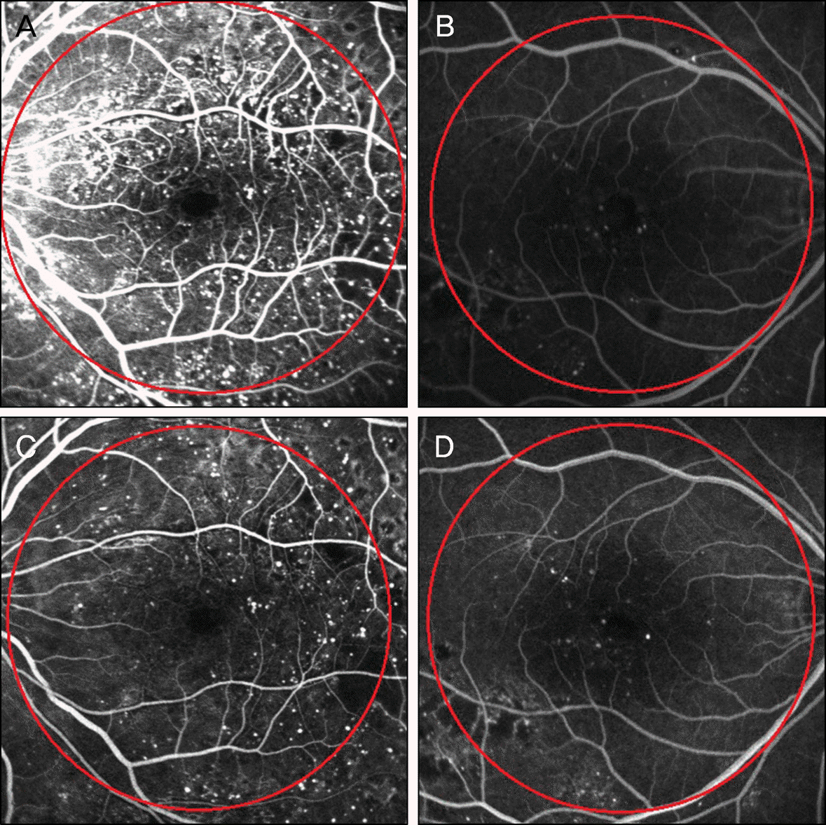 | Figure 1.Microaneurysm of preinjectional state (A, B) and at 8 week postinjectional state (C, D) after intravitreal bevacizumab injection. The circle with a radius of optic disc and macular (red line) (A, C = patients #24 in Fig. 2, 3) (B, D = patients #30 in Fig. 2, 3). |
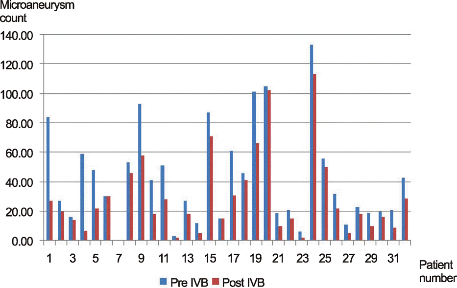 | Figure 2.The change of microaneurysm between pre intravitreous bevacizumab injection (IVB) and post IVB by fluorescein angiography (FAG). |
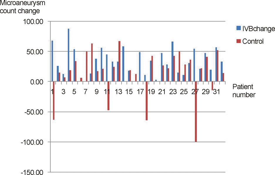 | Figure 3.Comparison between intravitreous bevacizumab injection (IVB) group and control group in microaneurysm changes. |
Table 1.
Characteristics of 31 patients who underwent intravitreal bevacizumab injection for diabetic retinopathy
 XML Download