Abstract
Purpose
To evaluate the clinical outcomes, complications and surgically induced astigmatism (SIA) after scleral fixation in patients with intraocular lens (IOL) or crystalline lens dislocation.
Methods
The present study retrospectively investigated the postoperative best corrected visual acuity (BCVA), refractory change, corneal astigmatism, clinical outcomes, and influencing factors of SIA in 57 eyes of 55 patients with a follow-up of 6 months after the IOL scleral fixation.
Results
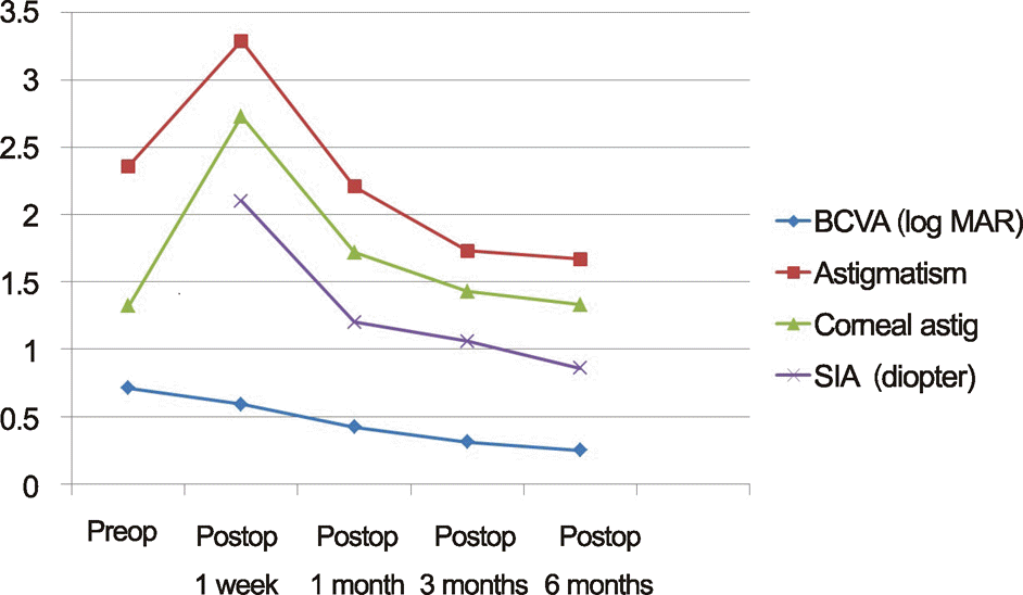
In comparison of preoperative and postoperative 6 months, BCVA, spherical equivalent and astigmatism were significantly improved but corneal astigmatism was not and SIA (diopter, D) improved from 2.10 ± 1.88 D to 0.86 ± 0.73 D (p = 0.002). 4 eyes having redislocation were repositioned and 4 eyes having tilted IOL, 6 eyes having elevated intraocular pressure, 3 eyes having exposure scleral knots, 1 eye having endophthalmitis, and 1 eye showing macular edema were observed. At postoperative 3 months, the SIA of a large incision size (>3 mm) and small incision size (≤3 mm) was significantly differented (p = 0.041). According to the location of scleral fixation, SIA at postoperative 1 month was significantly different but, was not different at postoperative 6 months.
Conclusions
Surgical management of dislocated IOLs or crystalline lens resulted in significant improvement of visual acuity and absence of influencing SIA factors. However, location of scleral fixation and small incision size influenced corneal astigmatism. J Korean Ophthalmol Soc 2014;55(10):1452-1459
Go to : 
References
1. Pallin SL, Walman GB. Posterior chamber intraocular lens implant centration: In or out of “the bag”. J Am Intraocular Implant Soc. 1982; 8:254–7.

2. Stark WJ Jr, Maumenee AE, Datiles M, et al. Intraocular lenses: complications and visual results. Trans Am Ophthalmol Soc. 1983; 81:280–309.
3. Smith SG, Lindstrom RL. Malpositioned posterior chamber lenses: etiology, prevention, and management. J Am Intraocul Implant Soc. 1985; 11:584–91.

4. Smiddy WE, Ibanez GV, Alfonso E, Flynn HW Jr. Surgical management of dislocated intraocular lenses. J Cataract Refract Surg. 1995; 21:64–9.

5. Mello MO Jr, Scott IU, Smiddy WE, et al. Surgical management and outcomes of dislocated intraocular lenses. Ophthalmology. 2000; 107:62–7.

6. Koh HJ, Kim CY, Lim SJ, Kwon OW. Scleral fixation technique using 2 corneal tunnels for a dislocated intraocular lens. J Cataract Refract Surg. 2000; 26:1439–41.

7. Cho SH, Kang SW, Jung MS. Four cases of modification of scleral fixation using 30 G needle for posterior chamber intraocular lens dislocation. J Korean Ophthalmol Soc. 2002; 43:917–21.
8. Chan CC, Crandall AS, Ahmed II. Ab externo scleral suture loop fixation for posterior chamber intraocular lens decentration: clinical results. J Cataract Refract Surg. 2006; 32:121–8.

9. Jehan FS, Mamalis N, Crandall AS. Spontaneous late dislocation of intraocular lens within the capsular bag in pseudoexfoliation patients. Ophthalmology. 2001; 108:1727–31.

10. Carlson AN, Stewart WC, Tso PC. Intraocular lens complications requiring removal or exchange. Surv Ophthalmol. 1998; 42:417–40.

11. Güell JL, Barrera A, Manero F. A review of suturing techniques for posterior chamber lenses. Curr Opin Ophthalmol. 2004; 15:44–50.
12. Ma DJ, Kim MK, Wee WR. Knotless external fixation technique for posterior chamber intraocular lens transscleral fixation: A 5-case analysis. J Korean Ophthalmol Soc. 2012; 53:1609–14.

13. Jung MO, Koh JW. Clinical results of modified Ab externo and one-knot technique. J Korean Ophthalmol Soc. 2012; 53:1783–8.

14. Lee DG, Nam KY, Kim JY. Modified scleral fixation of dislocated posterior chamber intraocular lenses. J Korean Ophthalmol Soc. 2009; 50:1071–5.

15. Naeser K, Hjortdal J. Polar value analysis of refractive data. J Cataract Refract Surg. 2001; 27:86–94.

16. Hill W. Expected effects of surgically induced astigmatism on AcrySof toric intraocular lens results. J Cataract Refract Surg. 2008; 34:364–7.

17. Michaeli A, Assia EI. Scleral and iris fixation of posterior chamber lenses in the absence of capsular support. Curr Opin Ophthalmol. 2005; 16:57–60.

18. Por YM, Lavin MJ. Techniques of intraocular lens suspension in the absence of capsular/zonular support. Surv Ophthalmol. 2005; 50:429–62.

19. Hayashi K, Hirata A, Hayashi H. Possible predisposing factors for in-the-bag and out-of-the-bag intraocular lens dislocation and outcomes of intraocular lens exchange surgery. Ophthalmology. 2007; 114:969–75.

20. Nikeghbali A, Falavarjani KG. Modified transscleral fixation technique for refixation of dislocated intraocular lenses. J Cataract Refract Surg. 2008; 34:743–8.

21. Grehn F, Sundmacher R. Fixation of posterior chamber lenses by transscleral sutures: technique and preliminary results. Arch Ophthalmol. 1989; 107:954–5.

22. Jang BH, Ahn M, Lee DW, Cho NC. The role of vitrectomy in transscleral fixation of posterior chamber intraocular lens. J Korean Ophthalmol Soc. 2005; 46:466–71.
23. Lee SJ, Kim KC, Hong YJ. Implantation of posterior chamber intraocular lens by trans - scleral fixation. J Korean Ophthalmol Soc. 1992; 33:704–10.
24. Roger FS. Cataract surgery technique, complications, management. 2nd ed.Saunders;2004. p. 413–6.
25. Taskapili M, Gulkilik G, Engin G, et al. Transscleral fixation of a single-piece hydrophilic foldable acrylic intraocular lens. Can J Ophthalmol. 2007; 42:256–61.

26. Merriam JC, Zheng L, Merriam JE, et al. The effect of incisions for cataract on corneal curvature. Ophthalmology. 2003; 110:1807–13.

27. Kammann J, Dornbach G, Allmers R. [Sutureless wound adaptation. Comparison between corneal and corneoscleral incision]. Ophthalmologe. 1994; 91:442–5.
Go to : 
 | Figure 1.Changes of best corrected visual acuity (BCVA), astigmatism, corneal astigmatism and surgically induced astigmatism (SIA) in scleral fixation of intraocular lens (IOL). |
Table 1.
Baseline characteristics of patient with scleral fixation of IOL
| Baseline characteristics | Number | p-value |
|---|---|---|
| Number of eyes (patients) | 57 (55) | |
| M:F (number of eyes) | 38 (39):17 (18) | |
| Age (years) (range) | 58.12 ± 14.00 (19-89) | |
| Preoperative UCVA (log MAR) | 1.03 ± 0.60 | |
| Preoperative BCVA (log MAR) | 0.71 ± 0.60 | 0.000* |
| Posteoperative BCVA (log MAR) | 0.25 ± 0.32 |
Table 2.
Comparision of BCVA, spherical equivalent, astigmatism, and corneal astigmatism before and after management
| Preop | 1 week | 1 month | 3 months | 6 months | |
|---|---|---|---|---|---|
| BCVA (log MAR) | 0.71 ± 0.60 | 0.59 ± 0.52 | 0.42 ± 0.46 | 0.31 ± 0.42 | 0.25 ± 0.32 |
| p-value | 0.188* | 0.001* | 0.000* | 0.000* | |
| Spherical equivalent (diopter) | 5.52 ± 7.13 | −2.05 ± 1.62 | −2.10 ± 1.46 | −1.87 ± 1.64 | −1.68 ± 1.50 |
| p-value | 0.000* | 0.000* | 0.000* | 0.000* | |
| Astigmatism (diopter) | 2.36 ± 2.23 | 3.29 ± 2.86 | 2.21 ± 1.73 | 1.73 ± 1.17 | 1.67 ± 1.04 |
| p-value | 0.028* | 0.650* | 0.028* | 0.008* | |
| Corneal astigmatism (diopter) | 1.32 ± 1.21 | 2.73 ± 2.38 | 1.72 ± 1.33 | 1.43 ± 1.07 | 1.33 ± 0.83 |
| p-value | 0.000* | 0.001* | 0.274* | 0.952* |
Table 3.
Comparision of surgically-induced corneal astigmatism in various factors
| Eyes (n) | 1 week | 1 month | 3 months | 6 months |
|---|---|---|---|---|
| Total SIA (diopter) | 2.10 ± 1.88 | 1.20 ± 1.33 | 1.06 ± 1.20 | 0.86 ± 0.73 |
| Trauma (15) | 2.09 ± 1.40 | 1.22 ± 1.19 | 0.87 ± 0.98 | 1.03 ± 0.97 |
| No trauma (42) | 2.10 ± 2.04 | 1.20 ± 1.40 | 1.12 ± 1.27 | 0.80 ± 0.62 |
| p-value | 0.520* | 0.758* | 0.289* | 0.189* |
| Vitrectomy (18) | 1.80 ± 1.59 | 1.26 ± 1.58 | 0.73 ± 0.44 | 0.77 ± 0.40 |
| No vitrectomy (39) | 2.23 ± 2.00 | 1.17 ± 1.23 | 1.20 ± 1.40 | 0.90 ± 0.84 |
| p-value | 0.460* | 0.817* | 0.542* | 0.655* |
| Pseudophakic (26) | 2.25 ± 1.86 | 1.33 ± 1.80 | 1.09 ± 1.37 | 0.93 ± 0.92 |
| Phakic (17) | 2.53 ± 2.26 | 1.14 ± 0.96 | 0.97 ± 1.11 | 0.89 ± 0.64 |
| Aphakic (14) | 1.26 ± 1.02 | 1.04 ± 0.62 | 1.11 ± 1.05 | 0.71 ± 0.41 |
| p-value | 0.109† | 0.611 | 0.621 | 0.584† |
| IOL exchange (9) | 2.56 ± 2.50 | 2.14 ± 2.61 | 1.38 ± 1.95 | 0.99 ± 0.69 |
| No IOL exchange (17) | 1.63 ± 1.41 | 0.93 ± 0.99 | 0.92 ± 0.93 | 0.84 ± 1.00 |
| p-value | 0.434* | 0.500* | 0.553* | 0.306* |
| Ab externo (26) | 1.88 ± 1.84 | 1.36 ± 1.37 | 1.06 ± 0.90 | 0.91 ± 0.62 |
| Ab interno (31) | 2.28 ± 1.92 | 1.07 ± 1.31 | 1.05 ± 1.42 | 0.82 ± 0.82 |
| p-value | 0.332* | 0.145* | 0.140* | 0.156* |
| Incision size > 3 mm (30) | 2.39 ± 1.95 | 1.47 ± 1.71 | 1.39 ± 1.54 | 1.02 ± 0.92 |
| Incision size ≤ 3 mm (27) | 1.77 ± 1.77 | 0.91 ± 0.61 | 0.68 ± 0.43 | 0.69 ± 0.37 |
| p-value | 0.141* | 1.581* | 0.041* | 0.366* |
| Fixation (o'clock) 3-9 (8) | 1.87 ± 1.32 | 1.82 ± 2.00 | 0.69 ± 0.33 | 0.57 ± 0.33 |
| 6-12 (22) | 1.65 ± 1.42 | 0.70 ± 0.55 | 1.07 ± 1.18 | 0.79 ± 0.46 |
| 5-11 (OD), 1-7 (OS) (14) | 2.67 ± 2.32 | 1.26 ± 1.29 | 0.94 ± 1.02 | 0.92 ± 1.04 |
| 1-7 (OD), 5-11 (OS) (13) | 1.37 ± 2.28 | 1.61 ± 1.65 | 1.38 ± 1.19 | 1.11 ± 0.87 |
| p-value | 0.522† | 0.033† | 0.692† | 0.612† |
Table 4.
Postoperative complications after management


 PDF
PDF ePub
ePub Citation
 Print
Print


 XML Download