Abstract
Purpose
To report a case of unilateral nasal hemianopsia caused by a large internal carotid artery aneurysm.
Case summary
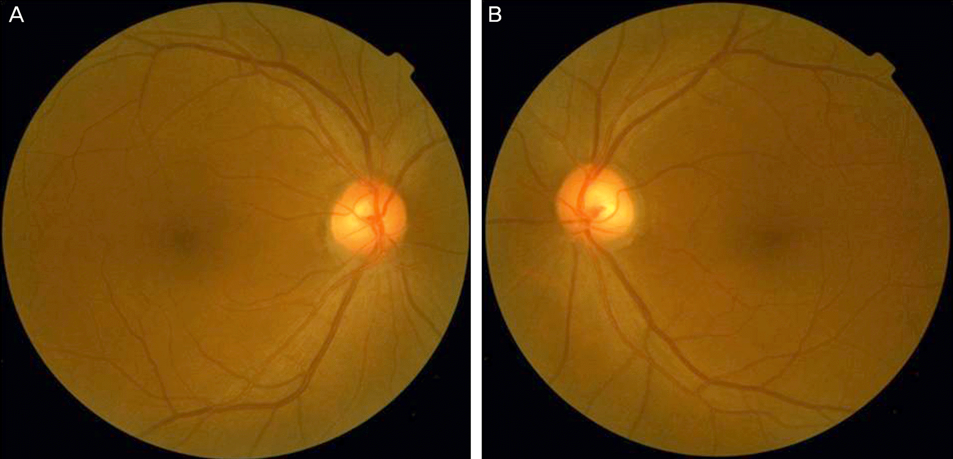
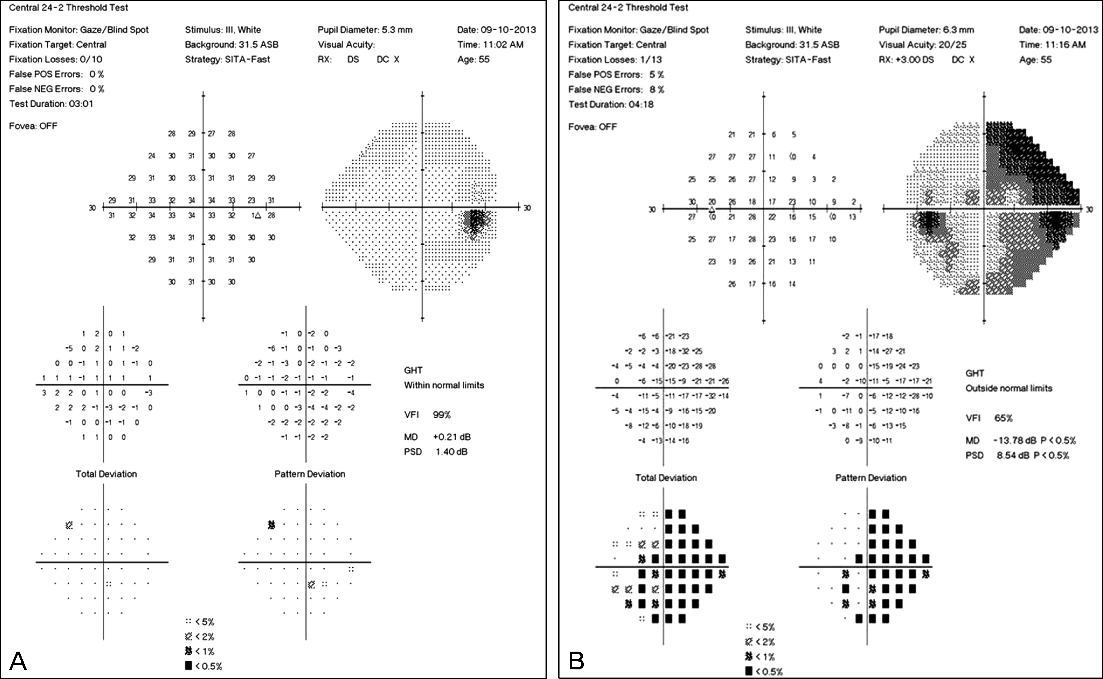
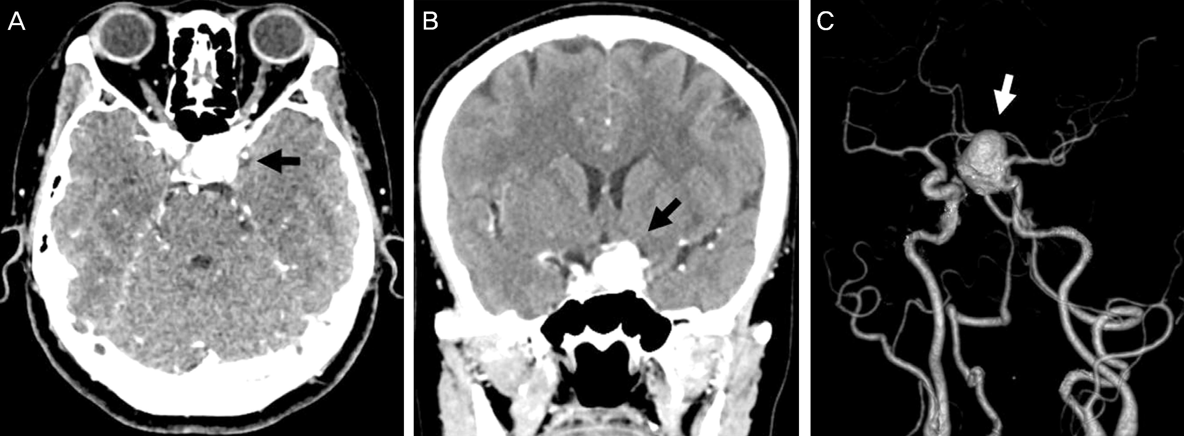
A 56-year-old female presented with large cupping in the left optic nerve head detected incidentally during a routine check-up. She had no underlying systemic disease except hypertension. The best corrected visual acuity was 20/20 in both eyes and a slit-lamp examination showed no abnormal findings. Ophthalmoscopy showed cup/disc ratios of 0.6 in the right eye and 0.75 in the left eye. Relative afferent papillary defect or color vision defect was not observed. A Humphrey visual-field test indicated unilateral nasal hemianopsia in the left eye. Brain CT and angiography revealed a large 2.2-cm aneurysm on the left internal carotid artery.
Go to : 
References
1. Cox TA, Corbett JJ, Thompson HS, Kassell NF. Unilateral nasal hemianopia as a sign of intracranial optic nerve compression. Am J Ophthalmol. 1981; 92:230–2.

2. Stacy RC, Jakobiec FA, Lessell S, Cestari DM. Monocular nasal hemianopia from atypical sphenoid wing meningioma. J Neuroophthalmol. 2010; 30:160–3.

3. AMYOT R. [Rupture of an aneurysm of the internal carotid; unilateral nasal hemianopsia; crossed hemiplegia caused by carotid spasm]. Union Med Can. 1959; 88:825–30.
4. Huber A. Eye signs and symptoms in brain tumors. St. Louis: C. V. Mosby Co.;1976. p. 266.
5. Meadows SP. Unusual clinical features and modes of presentation in pituitary adenoma, including pituitary apoplexy. Smith JL, editor. Neuroophthalmology. St. Louis: C.V. Mosby Co.;1968. 4:p. 178–89.
6. Rahman I, Nambiar A, Spencer AF. Unilateral nasal hemianopsia secondary to posterior subcapsular cataract. Br J Ophthalmol. 2003; 87:1045–6.

7. Karp CL, Fazio JR. Traumatic cataract presenting with unilateral nasal hemianopsia. J Cataract Refract Surg. 1999; 25:1302–3.

8. Chang BL. Neuroophthalmology. Seoul: Iljokak;2004. p. 128.
9. Levin PS, Newman SA, Quigley HA, Miller NR. A clinicopathologic study of optic neuropathies associated with intracranial mass lesions with quantification of remaining axons. Am J Ophthalmol. 1983; 95:295–306.
10. Chew SS, Cunnningham WJ, Gamble GD, Danesh-Meyer HV. Retinal nerve fiber layer loss in glaucoma patients with a relative afferent pupillary defect. Invest Ophthalmol Vis Sci. 2010; 51:5049–53.

11. Tatham AJ, Meira-Freitas D, Weinreb RN, et al. Estimation of retinal ganglion cell loss in glaucomatous eyes with a relative afferent pupillary defect. Invest Ophthalmol Vis Sci. 2014; 55:513–22.

12. Greenfield DS, Siatkowski RM, Glaser JS, et al. The cupped disc. Who needs neuroimaging? Ophthalmology. 1998; 105:1866–74.

13. Bianchi-Marzoli S, Rizzo JF 3rd, Brancato R, Lessell S. Quantitative analysis of optic disc cupping in compressive optic neuropathy. Ophthalmology. 1995; 102:436–40.
14. Alasil T, Wang K, Yu F, et al. Correlation of retinal nerve fiber layer thickness and visual fields in glaucoma: a broken stick model. Am J Ophthalmol. 2014; 157:953–9.

15. Wollstein G, Kagemann L, Bilonick RA, et al. Retinal nerve fibre layer and visual function loss in glaucoma: the tipping point. Br J Ophthalmol. 2012; 96:47–52.

16. Artmann H, Vonofakos D, Müller H, Grau H. Neuroradiologic and neuropathologic findings with growing giant intracranial aneurysm. Review of the literature. Surg Neurol. 1984; 21:391–401.

17. Horowitz MB, Yonas H, Jungreis C, Hung TK. Management of a giant middle cerebral artery fusiform serpentine aneurysm with distal clip application and retrograde thrombosis: case report and review of the literature. Surg Neurol. 1994; 41:221–5.

19. Dominick DiMaio, Vincent JM DiMaio. Forensic Pathology. 2nd ed.Florida: CRC Press;2001. p. 61.
20. Besada E, Fisher JP. Absent relative afferent pupillary defect in an asymptomatic case of lateral chiasmal syndrome from cerebral aneurysm. Optom Vis Sci. 2001; 78:195–205.

Go to : 
 | Figure 1.Fundus photographs show cup/disc ratios of 0.6 in the right eye (A) and 0.75 in the left eye (B). |
 | Figure 2.Humphrey visual field test demonstrating normal visual field in the right eye (A) and nasal hemianopsia on pattern-deviation plot in the left eye (B). |
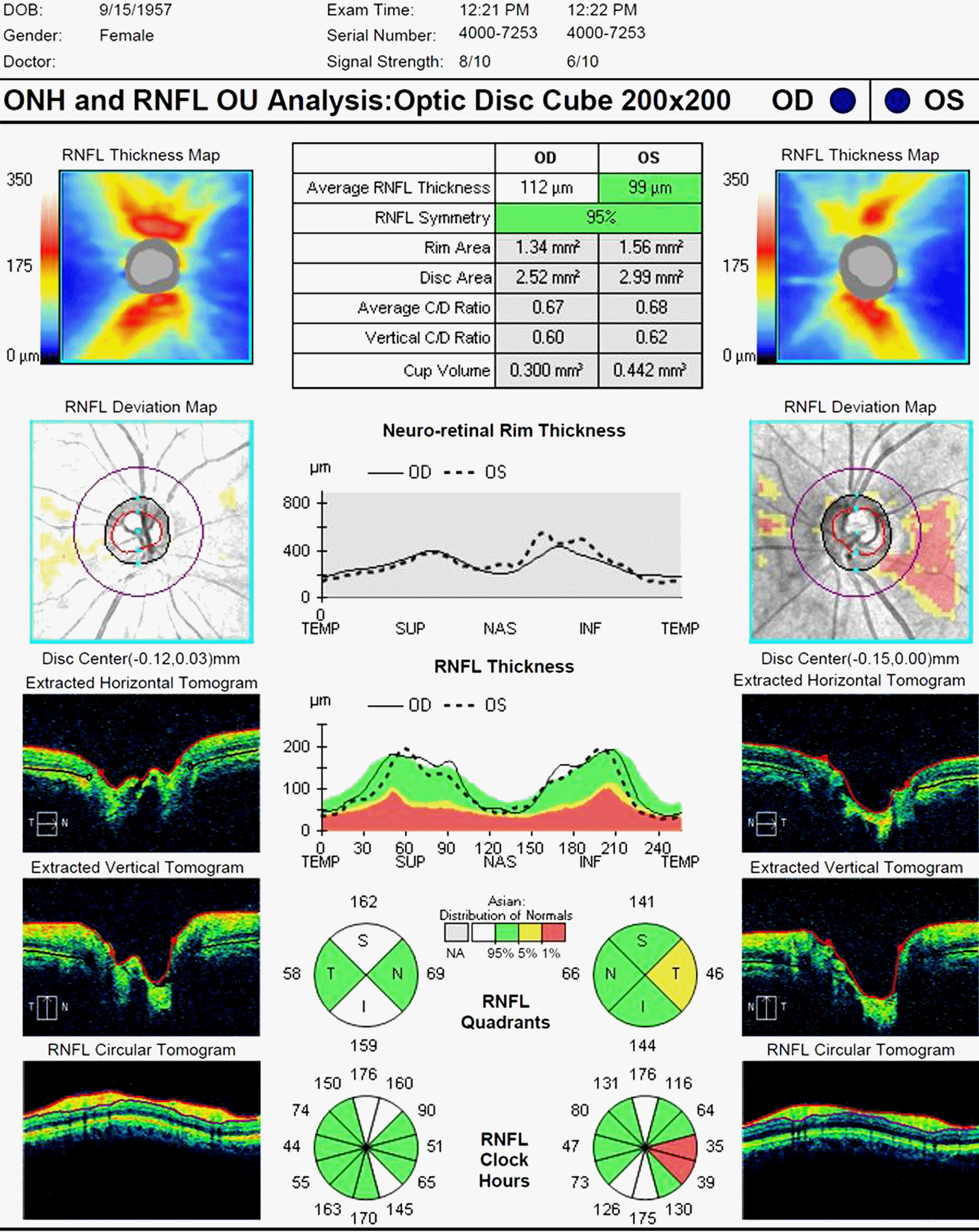
 | Figure 3.Optical coherence tomography demonstrating a slight but significant superotemporal and inferotemporal retinal nerve fiber layer thinning in the left eye relative to the right eye. ONH = optic nerve head; RNFL = retinal nerve fiber layer; OD = right; OS = left; C/D = cup/disc ratio; TEMP (T) = temporal; SUP (S) = superior; NAS (N) = nasal; INF (I) = inferior. |
 | Figure 4.Brain CT images of axial section (A) and coronal section (B) shows 2.2-cm large aneurysm on the left internal carotid artery (black arrow) compressing the lateral aspect of the left optic nerve in front of the optic chiasm. Brain CT angiography (C) demonstrates large aneurysm located on the paraclinoid portion of the left internal carotid artery (white arrow). |


 PDF
PDF ePub
ePub Citation
 Print
Print


 XML Download