

 PDF
PDF ePub
ePub Citation
 Print
Print


Abstract
Purpose
To report a case of anterior synechiolysis with lamellar corneal dissection in penetrating keratoplasty.
Case summary
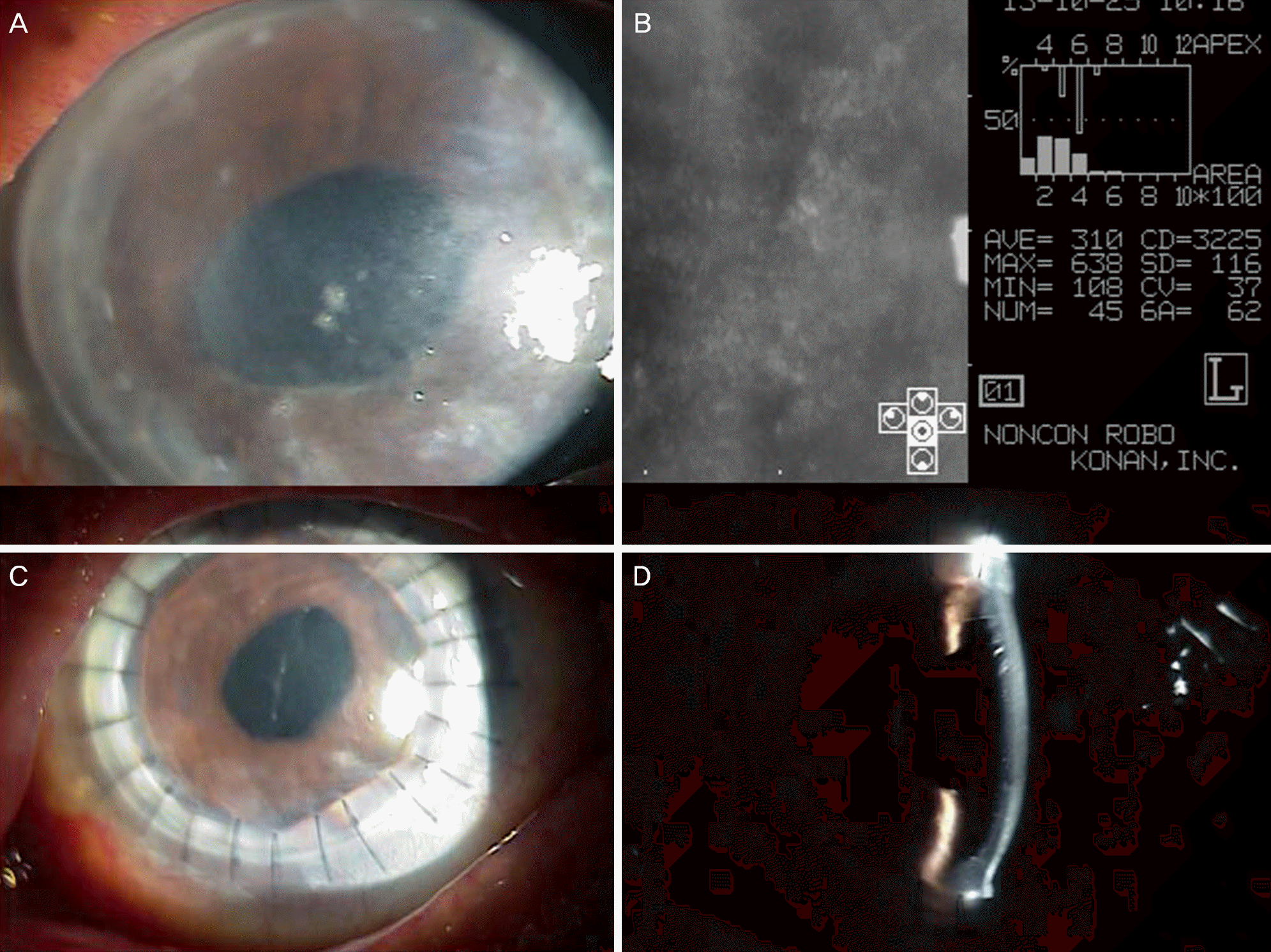
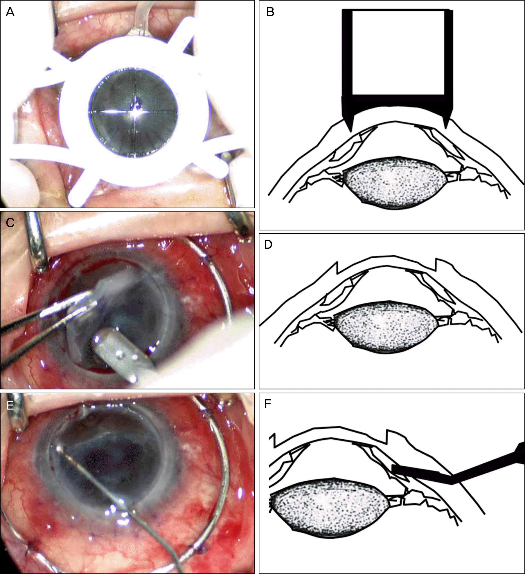
In an eye with graft failure and anterior synechiae, we performed anterior synechiolysis with a healon needle after lamellar dissection using vacuum trephine to visualize the anterior chamber during penetrating keratoplasty. Additionally, the remnant corneal layer was removed using corneal scissors. A donor cornea was harvested using a vacuum trephine and the corneal button was sewn in place with 10-0 nylon. We observed a well grafted cornea and well formed anterior chamber with no anterior synechiae observed on follow-up.
Go to : 
References
1. Chen WL, Hu FR, Wang IJ. Changing indications for penetrating keratoplasty in taiwan from 1987 to 1999. Cornea. 2001; 20:141–4.

2. Wilson SE, Kaufman HE. Graft failure after penetrating keratoplasty. Surv Ophthalmol. 1990; 34:325–56.
3. Castroviejo R. Surgical treatment of anterior synechiae before and after keratoplasty. Arch Ophthalmol. 1964; 72:240–5.
4. Stallard HB, Roper-Hall MJ. Stallard's eye surgery. 6th ed.Bristol [Eng.]: John Wiley & Sons;1980. p. 392–445.
5. Castroviejo R. Atlas of keratectomy and keratoplasty. 1st ed.Philadelphia: WB Saunders;1966. p. 350.
6. Ardjomand N, Fellner P, Moray M, et al. Lamellar corneal dissection for visualization of the anterior chamber before triple procedure. Eye (Lond). 2007; 21:1151–4.
Go to :
 XML Download