Abstract
Case summary
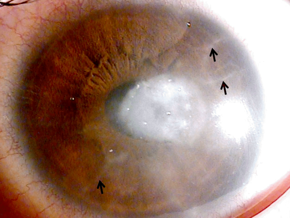
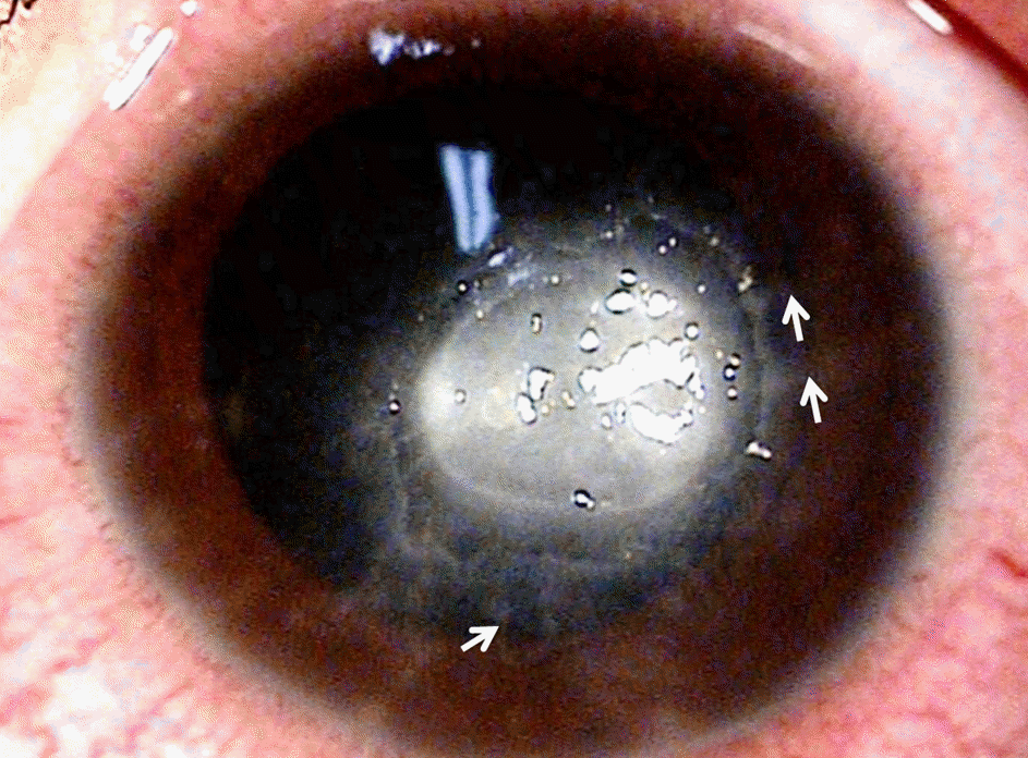
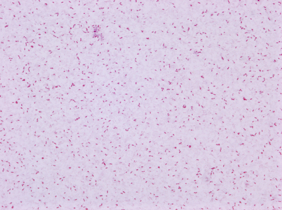
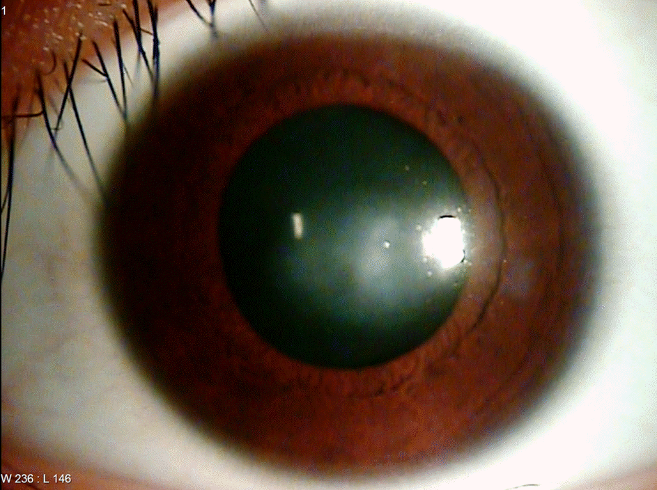
A 15-year-old female who wore cosmetic and orthokeratology contact lenses but performed inadequate lens care visited our clinic with severe pain and visual disturbance in her left eye. On slit lamp examination, central corneal epithelial defect and stromal infiltration with radial keratoneuritis were observed. Based on clinical findings and past history, Acanthamoeba keratitis was highly suspected. The patient was treated with topical chlorhexidine 0.02% (Sigma-Aldrich Co., MO, USA) and moxifloxacin 0.5% (Vigamox®, Alcon, TX, USA) per hour with 200 mg of oral itraconazol (Sporaone®, LG, Seoul, Korea) once a day. Symptoms and corneal lesions did not improve after three days. After Serratia marsenscens was isolated from her contact lenses and solution, topical chlorhexidine 0.02% was discontinued, and intravenous ceftazidime (Tazime®, Hanmi, Seoul, Korea) and fortified ceftazidime (50 mg/mL) eye drop was added. The corneal lesion dramatically improved, and after six months of follow-up, best-corrected visual acuity was 20/20 in the affected eye.
References
1. Joslin CE, Tu EY, Shoff ME, et al. The association of contact lens solution use and Acanthamoeba keratitis. Am J Ophthalmol. 2007; 144:169–80.

2. Park YM, Hahn TW, Choi SH, et al. Acanthamoeba keratitis related to cosmetic contact lenses. J Korean Ophthalmol Soc. 2007; 48:991–4.
3. Por YM, Mehta JS, Chua JL, et al. Acanthamoeba keratitis associated with contact lens wear in Singapore. Am J Ophthalmol. 2009; 148:7–12.e2.

4. Moore MB, McCulley JP, Kaufman HE, Robin JB. Radial keratoneuritis as a presenting sign in Acanthamoeba keratitis. Ophthalmology. 1986; 93:1310–5.
5. Joslin CE, Tu EY, McMahon TT, et al. Epidemiological characteristics of a Chicago-area Acanthamoeba keratitis outbreak. Am J Ophthalmol. 2006; 142:212–7.

6. Lindsay RG, Watters G, Johnson R, et al. Acanthamoeba keratitis and contact lens wear. Clin Exp Optom. 2007; 90:351–60.

7. Feist RM, Sugar J, Tessler H. Radial keratoneuritis in Pseudomonas keratitis. Arch Ophthalmol. 1991; 109:774–5.

8. Mutoh T, Matsumoto Y, Chikuda M. A case of radial keratoneuritis in non-Acanthamoeba keratitis. Clin Ophthalmol. 2012; 6:1535–8.

9. Rahimi F, Hashemian MN, Khodabande A. Serratia marcescens keratitis after photorefractive keratectomy. J Cataract Refract Surg. 2009; 35:1645–6.

10. Das S, Sheorey H, Taylor HR, Vajpayee RB. Association between cultures of contact lens and corneal scraping in contact lens related microbial keratitis. Arch Ophthalmol. 2007; 125:1182–5.
11. Alexandrakis G, Alfonso EC, Miller D. Shifting trends in bacterial keratitis in south Florida and emerging resistance to fluoroquinolones. Ophthalmology. 2000; 107:1497–502.

12. Mah-Sadorra JH, Najjar DM, Rapuano CJ, et al. Serratia corneal ulcers: a retrospective clinical study. Cornea. 2005; 24:793–800.
Figure 1.
Anterior segment photograph of the left eye at first visit shows corneal epithelial and stromal edema with Descemet's fold, circular corneal defect at the center, and stromal infiltration as radial keratoneuritis (black arrows).

Figure 2.
Anterior segment photograph of the left eye on day 3 reveals worsen corneal epithelial defect at center, and stromal infiltration with radial keratoneuritis (white arrows).


 PDF
PDF ePub
ePub Citation
 Print
Print


 XML Download