

 PDF
PDF ePub
ePub Citation
 Print
Print


Abstract
Purpose
To evaluate the long-term clinical outcomes of retinal pigment epithelium (RPE) tears after intravitreal injection of anti-vascular endothelial growth factor (VEGF) agent for the treatment of neovascular age-related macular degeneration (AMD).
Methods
The authors performed a retrospective chart review of 13 eyes of 13 patients who developed RPE tears after intravitreal anti-VEGF injection between February 2009 and June 2013. We investigated continuation of the treatment after tear, visual acuity, presence of cystoid macular edema, and central macular thickness (CMT) using optical coherence tomography (OCT) before and after treatment and visual outcomes depending on foveal sparing.
Results
After RPE tear, 12 of 13 patients continued injection of an anti-VEGF agent. The average number of injections was 6.08 ± 5.18. Mean visual acuity immediately after tear was 1.65 ± 0.8 log MAR, and that at the last visit was 1.82 ± 0.88 log MAR. Nine eyes with macular edema in OCT continued receiving injection, and improvement of macular edema was observed in four eyes at the final visit. The final visual acuity of patients with foveal involvement was 2.17 ± 0.49 log MAR, which was worse than the 1.51 ± 1.06 log MAR in patients without foveal involvement, although the difference was not significant (p = 0.295).
Conclusions
When anti-VEGF injections were continued after RPE tear, no improvement in visual acuity was observed, although better anatomical outcomes did result. Patients with foveal involvement had worse visual acuity than patients without foveal involvement, but the difference was not significant.
Go to : 
References
1. Chang LK, Sarraf D. Tears of the retinal pigment epithelium: an old problem in a new era. Retina. 2007; 27:523–34.
2. Gass JD. Pathogenesis of tears of the retinal pigment epithelium. Br J Ophthalmol. 1984; 68:513–9.

3. Pece A, Introini U, Bottoni F, Brancato R. Acute retinal pigment epithelial tear after photodynamic therapy. Retina. 2001; 21:661–5.
4. Carvounis PE, Kopel AC, Benz MS. Retinal pigment epithelium tears following ranibizumab for exudative age-related macular degeneration. Am J Ophthalmol. 2007; 143:504–5.
5. Dhalla MS, Blinder KJ, Tewari A, et al. Retinal pigment epithelial tear following intravitreal pegaptanib sodium. Am J Ophthalmol. 2006; 141:752–4.
6. Spandau UH, Jonas JB. Retinal pigment epithelium tear after intravitreal bevacizumab for exudative age-related macular degeneration. Am J Ophthalmol. 2006; 142:1068–70.
7. Pauleikhoff D, Löffert D, Spital G, et al. Pigment epithelial detachment in the elderly. Clinical differentiation, natural course and pathogenetic implications. Graefes Arch Clin Exp Ophthalmol. 2002; 240:533–8.
8. Coscas G, Koenig F, Soubrane G. The pretear characteristics of pigment epithelial detachments. A study of 40 eyes. Arch Ophthalmol. 1990; 108:1687–93.
9. Decker WL, Sanborn GE, Ridley M, et al. Retinal pigment epithelial tears. Ophthalmology. 1983; 90:507–12.
10. Casswell AG, Kohen D, Bird AC. Retinal pigment epithelial detachments in the elderly: classification and outcome. Br J Ophthalmol. 1985; 69:397–403.
11. Chan CK, Meyer CH, Gross JG, et al. Retinal pigment epithelial tears after intravitreal bevacizumab injection for neovascular age-related macular degeneration. Retina. 2007; 27:541–51.
12. Lommatzsch A, Heimes B, Gutfleisch M, et al. Serous pigment epithelial detachment in age-related macular degeneration: comparison of different treatments. Eye (Lond). 2009; 23:2163–8.
13. Chiang A, Chang LK, Yu F, Sarraf D. Predictors of anti-VEGF-associated retinal pigment epithelial tear using FA and OCT analysis. Retina. 2008; 28:1265–9.
14. Meyer CH, Mennel S, Schmidt JC, Kroll P. Acute retinal pigment epithelial tear following intravitreal bevacizumab (Avastin) injection for occult choroidal neovascularisation secondary to age related macular degeneration. Br J Ophthalmol. 2006; 90:1207–8.
15. Gamulescu MA, Framme C, Sachs H. RPE-rip after intravitreal bevacizumab (Avastin) treatment for vascularised PED secondary to AMD. Graefes Arch Clin Exp Ophthalmol. 2007; 245:1037–40.
16. Nicolò M, Ghiglione D, Calabria G. Retinal pigment epithelial tear following intravitreal injection of bevacizumab (Avastin). Eur J Ophthalmol. 2006; 16:770–3.
17. Gass JD. Serous retinal pigment epithelial detachment with a notch. A sign of occult choroidal neovascularization. Retina. 1984; 4:205–20.
18. Kiss C, Michels S, Prager F, et al. Retinal pigment epithelium tears following intravitreal ranibizumab therapy. Acta Ophthalmol Scand. 2007; 85:902–3.
19. Gutfleisch M, Heimes B, Schumacher M, et al. Long-term visual outcome of pigment epithelial tears in association with anti-VEGF therapy of pigment epithelial detachment in AMD. Eye (Lond). 2011; 25:1181–6.
20. Kook D, Wolf A, Neubauer AS, et al. [Retinal pigment epithelial tears after intravitreal injection of bevacizumab for AMD. Frequency and progress]. Ophthalmologe. 2008; 105:158–64.
21. Schoeppner G, Chuang EL, Bird AC. The risk of fellow eye visual loss with unilateral retinal pigment epithelial tears. Am J Ophthalmol. 1989; 108:683–5.
22. Chuang EL, Bird AC. Bilaterality of tears of the retinal pigment epithelium. Br J Ophthalmol. 1988; 72:918–20.
23. Garg S, Brod R, Kim D, et al. Retinal pigment epithelial tears after intravitreal bevacizumab injection for exudative age-related macular degeneration. Clin Experiment Ophthalmol. 2008; 36:252–6.
24. Sarraf D, Reddy S, Chiang A, et al. A new grading system for retinal pigment epithelial tears. Retina. 2010; 30:1039–45.
25. Lesniak SP, Fine HF, Prenner JL, Roth DB. Long-term follow-up of spontaneous retinal pigment epithelium tears in age-related macular degeneration treated with anti-VEGF therapy. Eur J Ophthalmol. 2011; 21:73–6.
26. Peiretti E, Iranmanesh R, Lee JJ, et al. Repopulation of the retinal pigment epithelium after pigment epithelial rip. Retina. 2006; 26:1097–9.
27. Chuang EL, Bird AC. Repair after tears of the retinal pigment epithelium. Eye (Lond). 1988; 2:106–13.
Go to :
 | Figure 1.The optical coherence tomography (OCT) images of 78-year-old male with retinal pigment epithelium (RPE) tear in wet age-related macular degeneration (AMD). The patients had discontinued the anti-VEGF injection after RPE tear. (A) There is no macular edema at the day of RPE tear. (B) Cystoid macular edema was detected on OCT image of the final follow-up day at 16 months after RPE tear. VEGF = vascular endothelial growth factor. |
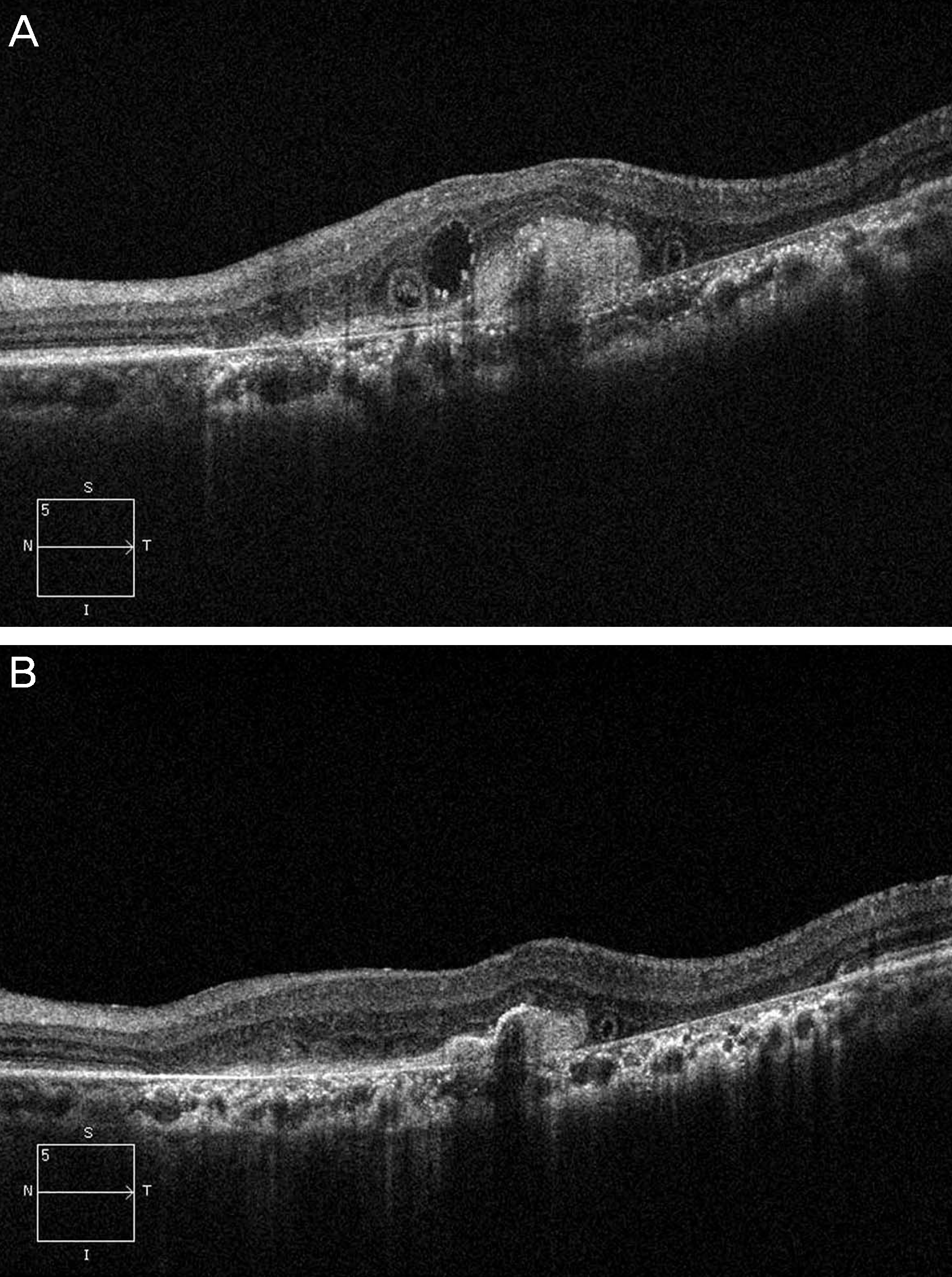 | Figure 2.The optical coherence tomography (OCT) images of 84-year-old male patient with continuing anti-VEGF treatment after retinal pigment epithelium (RPE) tear. The patient had been treated with bevacizumab on five times over period of 6 months. (A) The OCT shows cystic edema and a neurosensory detachment with reflectance that could represent the neovascularization complex. (B) The detachment resolved, and there is no persistent cystic change within the retina. VEGF = vascular endothelial growth factor. |
Table 1.
Summary of cases of retinal pigment epithelium tears after anti-VEGF therapy
Table 2.
OCT measurements of cases of retinal pigment epithelium tears after anti-VEGF therapy
 XML Download