

 PDF
PDF ePub
ePub Citation
 Print
Print


Abstract
Case summary
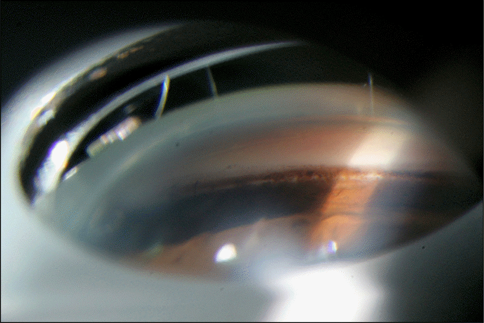
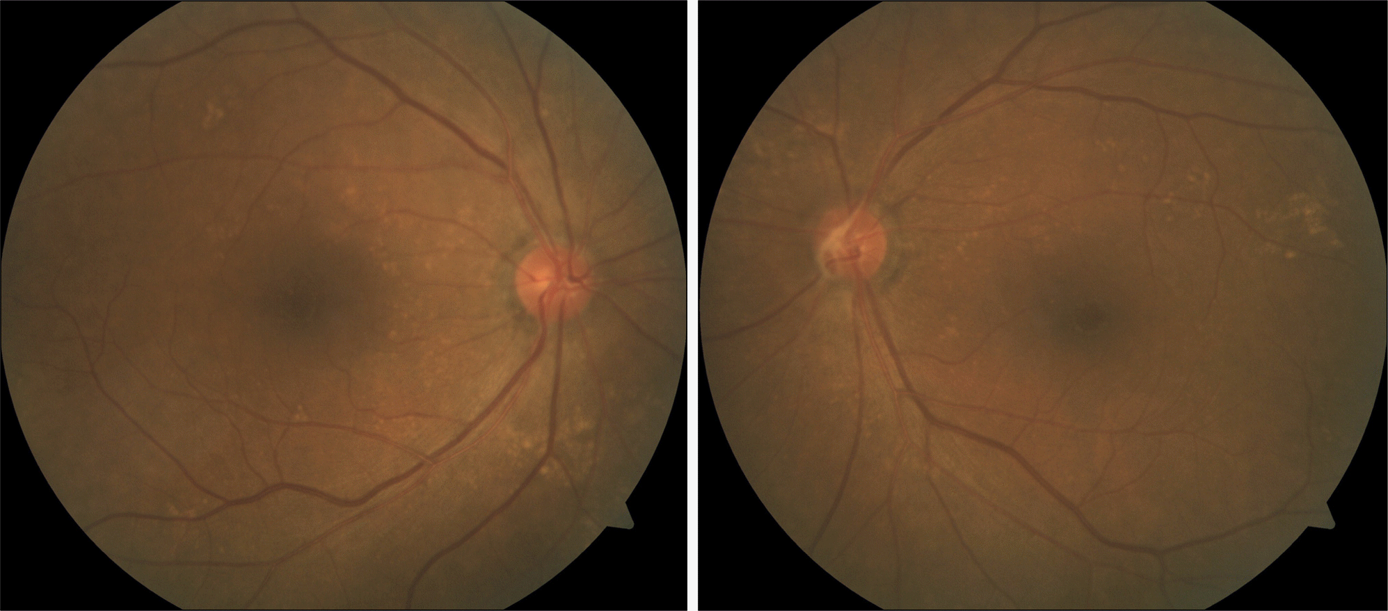
A 67-year-old female visited our clinic with complaint of visual disturbance in the left eye. She had no history of nyctalopia. Visual acuity was 0.6 in the right eye and 0.4 in the left eye. Intraocular pressure was 12 mm Hg in the right eye and 16 mm Hg in the left eye. Nuclear sclerosis was observed in the left lens. There was no pseudoexfoliative ma-terial observed. In the left eye, long anterior zonules with brown pigmented lens striae were spotted irregularly in every di-rection of the anterior lens. On gonioscopy, the angle was open, and dense, uniform, trabecular meshwork pigmentations were observed at the interior 120 degrees. On fundus examination, cup-to-disc ratio was 0.4 in the right eye, 0.3 in the left eye, and multiple hard exudates were observed in both retinas. Axial length was 22.03 mm in the right eye and 21.84 in the left eye. Anterior chamber depth was 2.71 mm in the right eye and 2.47 mm in the left eye. Defects in the retinal nerve fiber or visual field examination were not observed and pigment dispersion syndrome was diagnosed. The patient showed no significant change at the 9-month follow-up.
Go to : 
References
1. Sturrock GD, Tripathi RC.Pigmented lens striae. Br J Ophthalmol. 1976; 60:287–93.

2. Niyadurupola N, Broadway DC.Pigment dispersion syndrome and pigmentary glaucoma–a major review. Clin Experiment Ophthalmol. 2008; 36:868–82.
3. Roberts DK, Winters JE, Castells DD. . Pigmented striae of the anterior lens capsule and age-associated pigment dispersion of var-iable degree in a group of older African-Americans: an age, race, and gender matched study. Int Ophthalmol. 2001; 24:313–22.
4. Roberts DK, Winters JE, Castells DD. . A cross-sectional study of Krukenberg spindles and pigmented lens striae in a predom-inately black population: two highly associated clinical signs of an-terior segment pigment dispersal. J Glaucoma. 2005; 14:57–63.
5. Roberts DK, Wilensky J.Long anterior lens zonules. Clin Experiment Ophthalmol. 2012; 40:764–6.
6. Moroi SE, Lark KK, Sieving PA. . Long anterior zonules and pigment dispersion. Am J Ophthalmol. 2003; 136:1176–8.
7. Qing G, Wang N, Tang X. . Clinical characteristics of pigment dispersion syndrome in Chinese patients. Eye (Lond). 2009; 23:1641–6.
8. Ritch R.Exfoliation syndrome. Curr Opin Ophthalmol. 2001; 12:124–30.
9. Ritch R, Schlötzer-Schrehardt U.Exfoliation syndrome. Surv Ophthalmol. 2001; 45:265–315.
10. Hong C, Han SH, Sohn YH.A case of pigmentary glaucoma. J Korean Ophthalmol Soc. 1983; 24:435–9.
11. Drolsum L, Ringvold A, Nicolaissen B.Cataract and glaucoma surgery in pseudoexfoliation syndrome: a review. Acta Ophthalmol Scand. 2007; 85:810–21.
12. Choi J, Park KH.Clinical characteristics of Korean patients with pseudoexfoliation syndrome. J Korean Ophthalmol Soc. 2006; 47:577–86.
13. Subrayan V, Morris B, Armbrecht AM. . Long anterior lens zonules in late-onset retinal degeneration (L-ORD). Am J Ophthalmol. 2005; 140:1127–9.
14. Milam AH, Curcio CA, Cideciyan AV. . Dominant late-onset retinal degeneration with regional variation of sub-retinal pigment epithelium deposits, retinal function, and photoreceptor degeneration. Ophthalmology. 2000; 107:2256–66.
Go to :
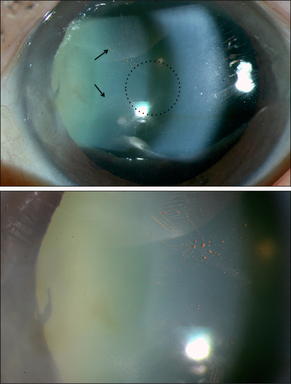 | Figure 1.Anterior segment photos of the left eye shows prom-inent long anterior lens zonules (black arrows) causing a zon-ule-free zone (dotted circle) only 2.5 mm in diameter. At the proximal insertion area of long anterior zonular fibers, several heavily pigmented granules are clustered in the lower picture. |
 XML Download