

 PDF
PDF ePub
ePub Citation
 Print
Print


Abstract
Purpose
Using the spectral domain optical coherence tomography (SD-OCT), we studied the difference in the choroidal morphology between the choroidal neovascularization (CNV) area and the area surrounding CNV.
Methods
This retrospective study consisted of 19 patients with myopic CNV lesion in eye; fellow eyes were used as controls. All eyes were analyzed by measuring the choroidal thickness and large choroidal vessel size using SD-OCT. Eyes with CNV were divided into groups; the neovascular lesion was defined as group 1, the surrounding area as group 2. Subfovea of the fellow eye was defined as group 3.
Results
The choroidal thickness was 80.00 ± 68.31 in group 1, 63.44 ± 67.75 in group 2 and 71.11 ± 65.69 µm in group 3. There was a significant difference between group 1 and group 2 (p = 0.038). There were no significant differences between group 1 and 3 or between group 2 and 3 (p = 0.365, p = 0.314). The large choroidal vessel size was 57.47 ± 39.78 in group 1, 40.45 ± 34.69 in group 2 and 45.63 ± 37.00 µm in group 3. There was a significant difference between group 1 and group 2 (p = 0.025). There were no significant differences between group 1 and 3 or between group 2 and 3 (p = 0.123, p = 0.325).
Conclusions
Choroidal thickness and large choroidal vessel size at the center of the CNV were greater than in the area surrounding CNV. The results suggest that although the CNVs were due to a thinned choroid caused by severe choroidal ischemia, the development of CNV requires maintenance of choriocapillaris and large choroid vessels.
References
1. Ikuno Y, Sayanagi K, Soga K, et al. Lacquer crack formation and choroidal neovascularization in pathologic myopia. Retina. 2008; 28:1124–31.

2. Keane PA, Liakopoulos S, Chang KT, et al. Comparison of the optical coherence tomographic features of choroidal neovascular membranes in pathological myopia versus age-related macular degeneration, using quantitative subanalysis. Br J Ophthalmol. 2008; 92:1081–5.
3. Yoshida T, Ohno-Matsui K, Yasuzumi K, et al. Myopic choroidal neovascularization: a 10-year follow-up. Ophthalmology. 2003; 110:1297–305.
4. Hayashi K, Ohno-Matsui K, Shimada N, et al. Long-term pattern of progression of myopic maculopathy: a natural history study. Ophthalmology. 2010; 117:1595–611. 1611.e1-4.
5. Curtin BJ. The posterior staphyloma of pathologic myopia. Trans Am Ophthalmol Soc. 1977; 75:67–86.
6. Ohno-Matsui K, Yoshida T, Futagami S, et al. Patchy atrophy and lacquer cracks predispose to the development of choroidal neovascularisation in pathological myopia. Br J Ophthalmol. 2003; 87:570–3.
7. Ohsugi H, Ikuno Y, Oshima K, Tabuchi H. 3-D choroidal thickness maps from EDI-OCT in highly myopic eyes. Optom Vis Sci. 2013; 90:599–606.
8. Hayashi M, Ito Y, Takahashi A, et al. Scleral thickness in highly myopic eyes measured by enhanced depth imaging optical coherence tomography. Eye (Lond). 2013; 27:410–7.
9. Farinha CL, Baltar AS, Nunes SG, et al. Choroidal thickness after treatment for myopic choroidal neovascularization. Eur J Ophthalmol. 2013; Jun 13:0. [Epub ahead of print].
10. Spaide RF, Koizumi H, Pozzoni MC. Enhanced depth imaging spectral-domain optical coherence tomography. Am J Ophthalmol. 2008; 146:496–500.
11. Margolis R, Spaide RF. A pilot study of enhanced depth imaging optical coherence tomography of the choroid in normal eyes. Am J Ophthalmol. 2009; 147:811–5.
12. Fujiwara T, Imamura Y, Margolis R, et al. Enhanced depth imaging optical coherence tomography of the choroid in highly myopic eyes. Am J Ophthalmol. 2009; 148:445–50.
13. El Matri L, Bouladi M, Chebil A, et al. [Macular choroidal thickness assessment with SD-OCT in high myopia with or without choroidal neovascularization]. J Fr Ophtalmol. 2013; 36:687–92.
14. Leveziel N, Caillaux V, Bastuji-Garin S, et al. Angiographic and optical coherence tomography characteristics of recent myopic choroidal neovascularization. Am J Ophthalmol. 2013; 155:913–9.
15. Wakabayashi T, Ikuno Y. Choroidal filling delay in choroidal neovascularisation due to pathological myopia. Br J Ophthalmol. 2010; 94:611–5.
16. Flores-Moreno I, Lugo F, Duker JS, Ruiz-Moreno JM. The relationship between axial length and choroidal thickness in eyes with high myopia. Am J Ophthalmol. 2013; 155:314–9.e1.
17. Kim EJ, Kim JH, Koo SH, et al. Choroidal thickness changes according to the refractive errors and axial length in Korean myopia patients. J Korean Ophthalmol Soc. 2012; 53:1814–22.
18. Kim KH, Kim DG. The relationship among refractive power, axial length and choroidal thickness measured by SD-OCT in Myopia. J Korean Ophthalmol Soc. 2012; 53:626–31.
Figure 1.
The enhanced depth imaging optical coherence tomography (EDI-OCT) horizontal scan passing through at the level of the superior margin of choroidal neovascularization (CNV) (A), center of CNV (B) and inferior margin of CNV (C). At each cross-sectional imaging, red dots are marked on the temporal and nasal end of the CNV. And then by connecting all the red dots, CNV area (green line) is obtained. Next a yellow box is drawn by tangent lines to the CNV area (D). Group 1 is defined as the center of the yellow box. Group 2 is defined as the four points, which are 250 μm apart from the superior, inferior, nasal and temporal boundaries of the CNV.
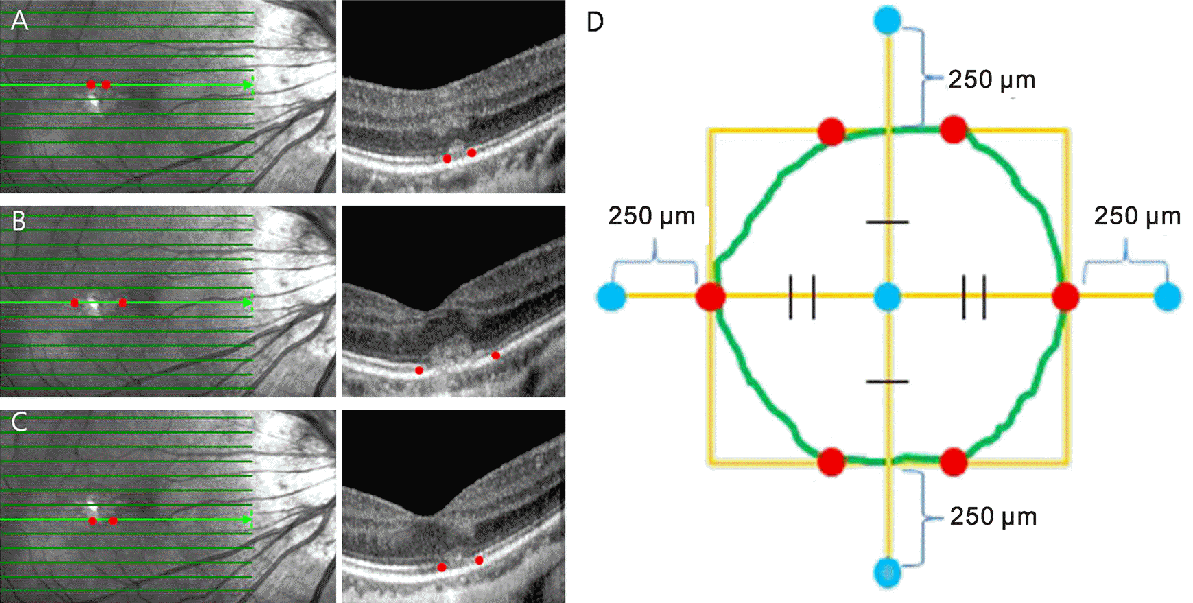
Figure 2.
A 59-year-old patient case with myopic choroidal nevoasucalization (CNV) in the left eye. The refractive error is −10.00 diopters and best-corrected visual acuity is 20/200. The enhanced depth imaging optical coherence tomography (EDI-OCT) horizontal scan passing through at the level of 250 um superior to the superior margin of the CNV (A), center of the CNV (B), 250 um inferior to the inferior margin of the CNV (C) and at the level of the subfovea of the fellow eye (D). Choroidal thickness (yellow arrows) and the large choroidal vessel size (red line) are measured at the center of the CNV lesion, at the four points which are 250 μm apart from the boundaries of the CNV and at the fovea of the fellow eye.
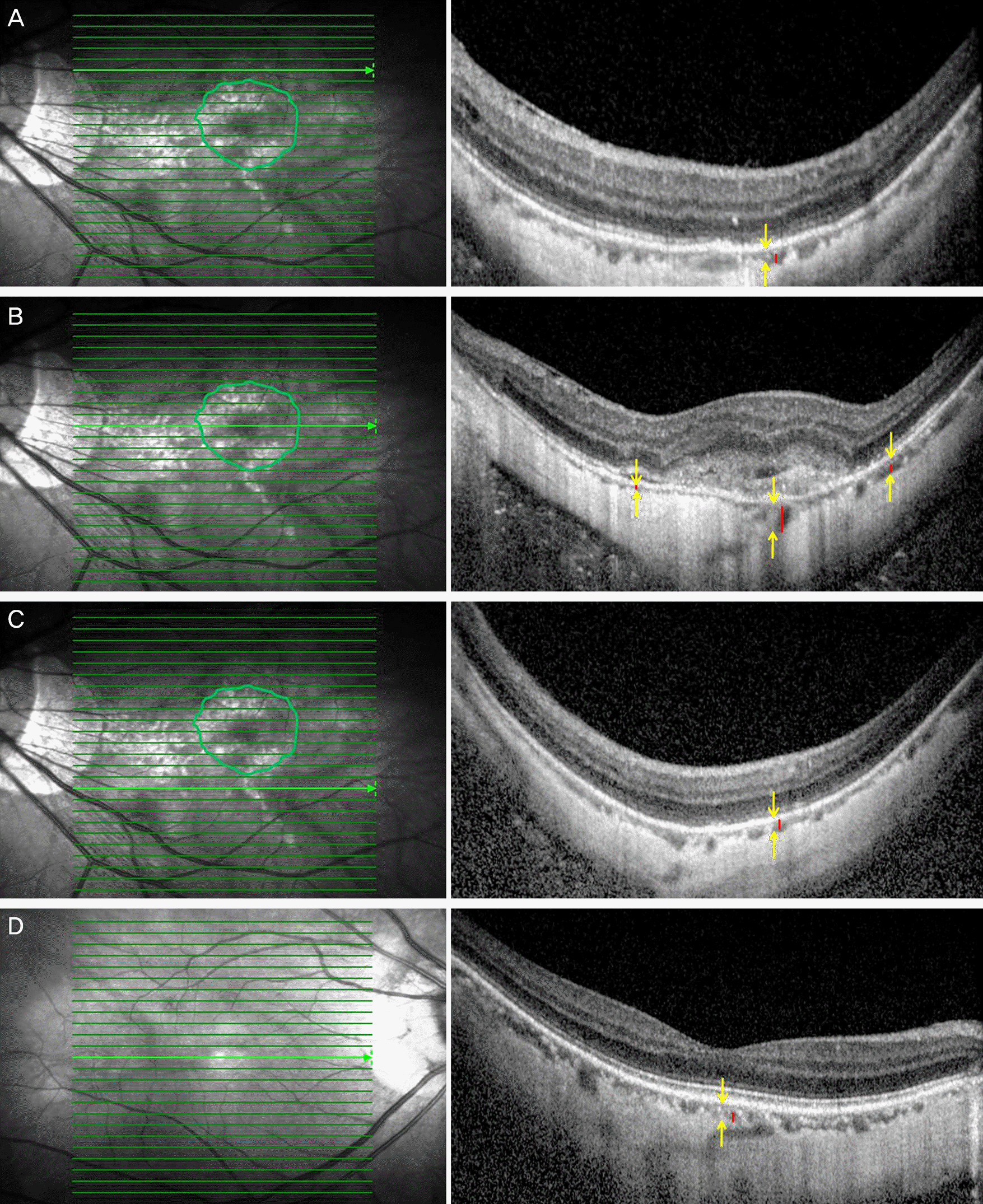
Table 1.
Demographics and characteristics of eyes with CNV
Table 2.
Comparison of eyes with CNV and fellow eyes
| Eyes with CNV | Fellow eyes | p-value* | |
|---|---|---|---|
| BCVA (log MAR) | 0.86 ± 0.40 | 0.33 ± 0.30 | 0.025 |
| Axial length (mm) | 28.65 ± 0.87 | 27.57 ± 1.82 | 0.005 |
| Spherical equivalent (diopter) | −14.44 ± 5.21 | −12.55 ± 6.88 | 0.212 |
| Subfoveal RT (μm) | 250.89 ± 85.30 | 202.89 ± 61.45 | 0.034 |
| Subfoveal CT (μm) | 57.32 ± 69.07 | 71.11 ± 65.69 | 0.070 |
Table 3.
Comparison of the retinal and choroidal thickness in three groups
| Group 1 | Group 2 | Group 3 | p* | p† | p‡ | |
|---|---|---|---|---|---|---|
| CT (μm) | 80.00 ± 68.31 | 63.44 ± 67.75 | 71.11 ± 65.69 | 0.038 | 0.365 | 0.314 |
| LCVS (μm) | 57.47 ± 39.78 | 40.45 ± 34.69 | 45.63 ± 37.00 | 0.025 | 0.123 | 0.325 |
 XML Download