

 PDF
PDF ePub
ePub Citation
 Print
Print


Abstract
Purpose
To compare the response of dry eye treatment in patients divided by the degree of lower lid laxity.
Methods
Thirty patients were classified into three groups - normal, moderate and severe, according to the degree of lower lid laxity. Tear break-up time (TBUT), Schirmer test (ST), ocular surface disease index (OSDI) scores and changes in OSDI score in each group were compared before and at 3 months after the treatment.
Results
TBUT, ST and OSDI scores were not different among the three groups at baseline. TBUT improved to 6.60 ± 1.43, 6.0 ± 2.54 and 6.0 ± 1.45 sec in normal, moderate and severe lower lid laxity group, respectively at 3 months after the treatment and no difference among the groups was found. ST scores did not increase after the treatment, while OSDI scores improved to 12.20 ± 1.40, 16.10 ± 4.63 and 20.80 ± 4.52 in each group, respectively and they were significantly different (p = 0.029, 0.029, <0.001, respectively). The response to the dry eye treatment as assessed by changes in OSDI scores was poorer in patients in the severe lower lid laxity group (p = 0.019 vs. moderate laxity group, <0.01 vs. normal group).
Go to : 
References
1. The definition and classification of dry eye disease: report of the Definition and Classification Subcommittee of the International Dry Eye WorkShop (2007). Ocul Surf. 2007; 5:75–92.
2. Shimazaki-Den S, Iseda H, Dogru M, Shimazaki J. Effects of diquafosol sodium eye drops on tear film stability in short BUT type of dry eye. Cornea. 2013; 32:1120–5.

3. Cho BJ, Lee JH, Shim OJ. The relation between clinical manifestations of dry eye patients and their BUTs. J Korean Ophthalmol Soc. 1992; 33:297–302.
4. Nichols KK, Nichols JJ, Mitchell GL. The lack of association between signs and symptoms in patients with dry eye disease. Cornea. 2004; 23:762–70.
5. Rees TD, Jelks GW. Blepharoplasty and the dry eye syndrome: guidelines for surgery? Plast Reconstr Surg. 1981; 68:249–52.
6. Mastrota KM. Impact of floppy eyelid syndrome in ocular surface and dry eye disease. Optom Vis Sci. 2008; 85:814–6.
7. Amano S. MGD Working Group: Definition and diagnostic criteria for meibomian gland dysfunction. J Eye (Atarashii Ganka). 2010; 27:627–31.
8. Dana MR, Hamrah P. Role of immunity and inflammation in corneal and ocular surface disease associated with dry eye. Adv Exp Med Biol. 2002; 506:729–38.
9. Stern ME, Gao J, Siemasko KF, et al. The role of the lacrimal functional unit in the pathophysiology of dry eye. Exp Eye Res. 2004; 78:409–16.
10. Le Q, Ge L, Li M, et al. Comparison on the vision-related quality of life between outpatients and general population with dry eye syndrome. Acta Ophthalmol. 2014; 92:e124–32.
11. Li M, Gong L, Chapin WJ, Zhu M. Assessment of vision-related quality of life in dry eye patients. Invest Ophthalmol Vis Sci. 2012; 53:5722–7.
12. Gonnering RS, Sonneland PR. Meibomian gland dysfunction in floppy eyelid syndrome. Ophthal Plast Reconstr Surg. 1987; 3:99–103.
13. Bron AJ, Tiffany JM, Gouveia SM, et al. Functional aspects of the tear film lipid layer. Exp Eye Res. 2004; 78:347–60.
14. Liu DT, Di Pascuale MA, Sawai J, et al. Tear film dynamics in floppy eyelid syndrome. Invest Ophthalmol Vis Sci. 2005; 46:1188–94.
15. Toda I, Fujishima H, Tsubota K. Ocular fatigue is the major symptom of dry eye. Acta Ophthalmol (Copenh). 1993; 71:347–52.
16. Jeong HS, Lim JS, Oh DK, et al. Prevalence and risk factors of dry eye syndrome in the Incheon area. J Korean Ophthalmol Soc. 2011; 52:1135–41.
Go to :
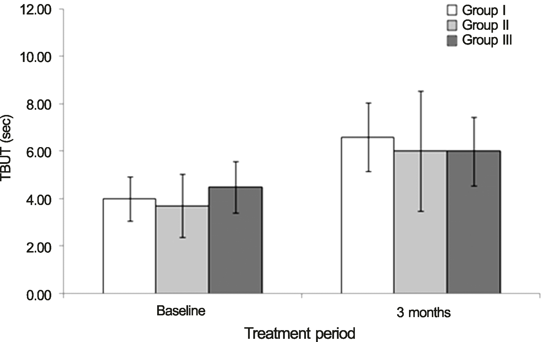 | Figure 1.Changes in tear break-up time (TBUT), before and at 3 months after the treatment in eyes grouped by the degree of lower lid laxity. |
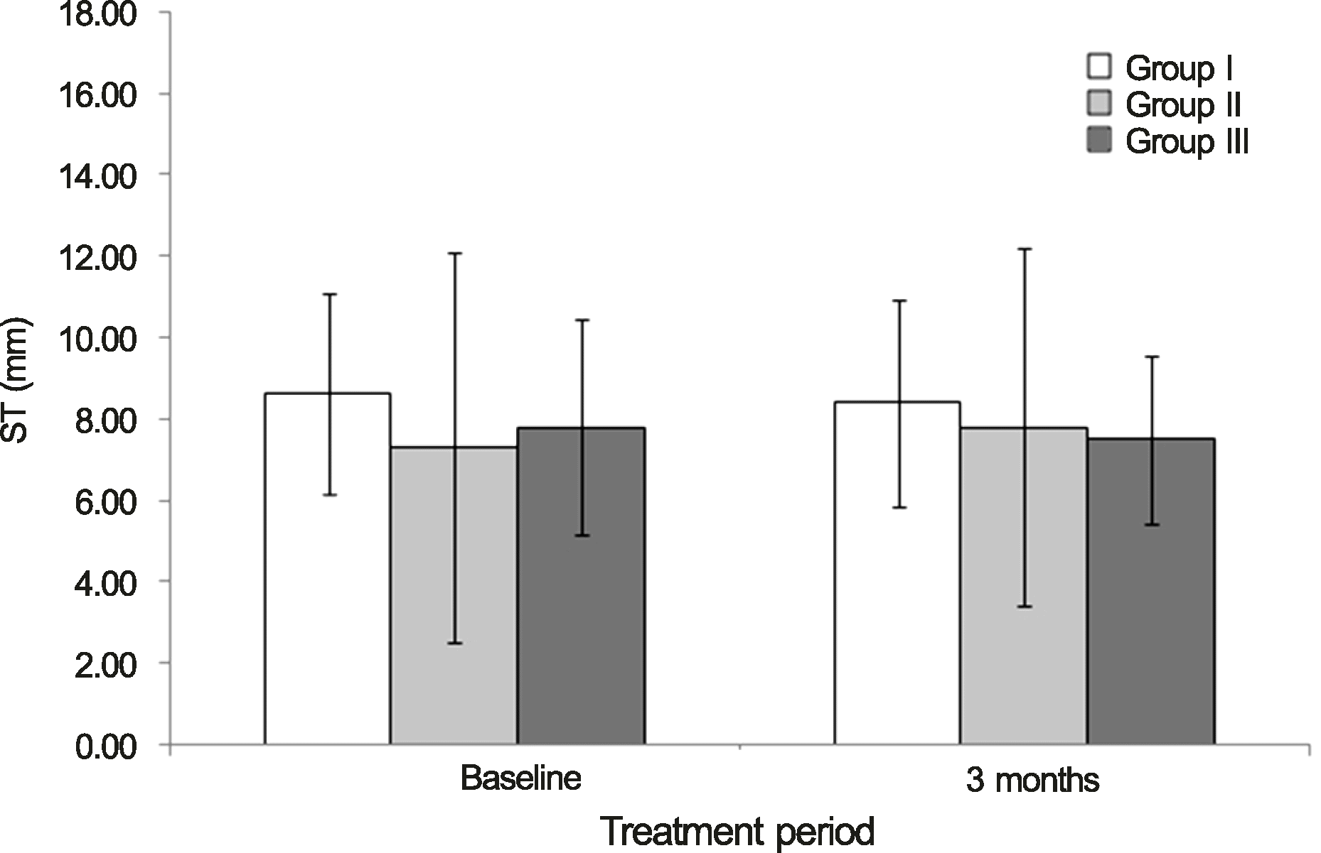 | Figure 2.Changes in Schirmer test (ST) before and at 3 months after the treatment in eyes grouped by the degree of lower lid laxity. |
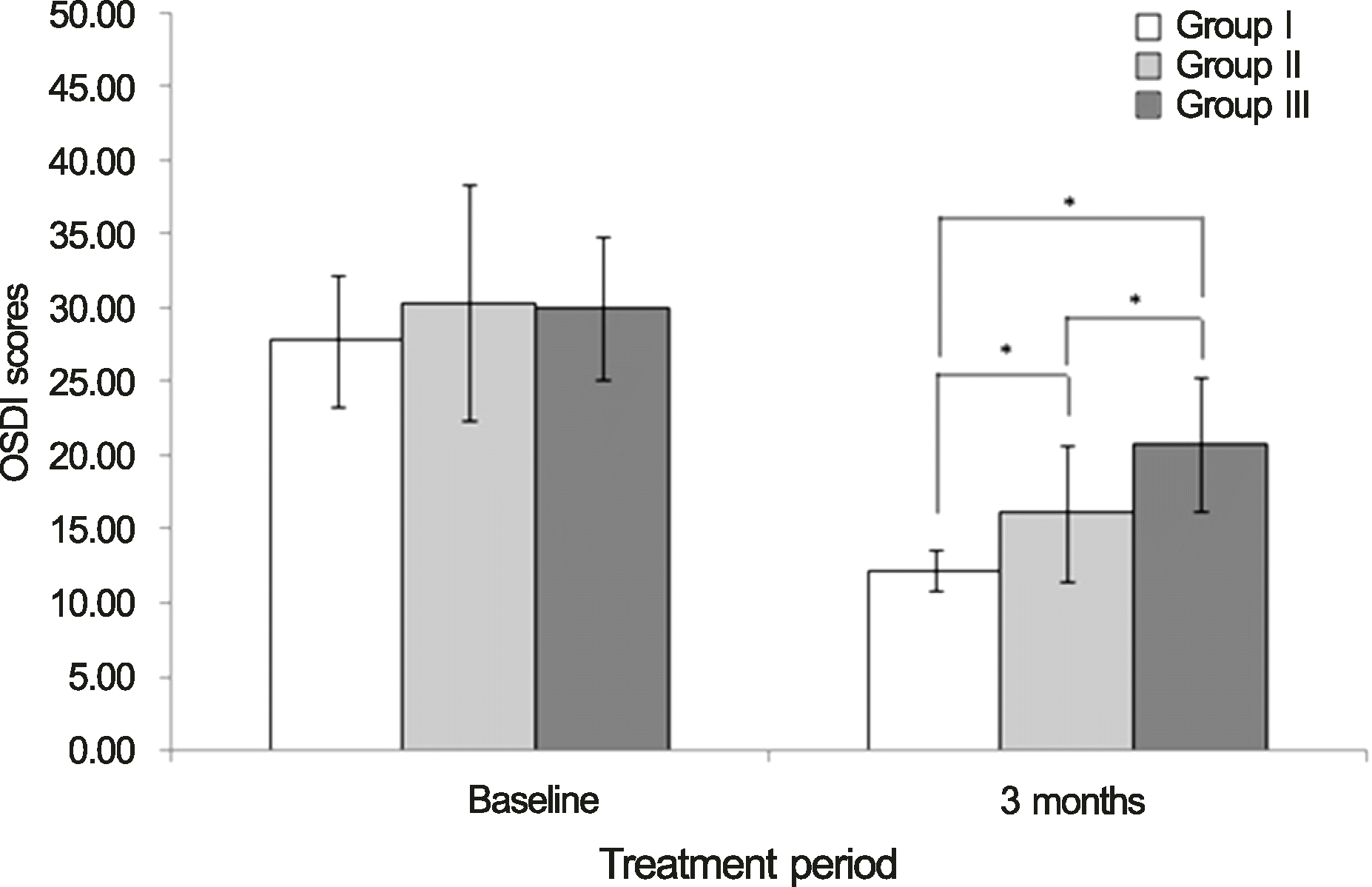 | Figure 3.Changes in ocular surface disease index (OSDI) scores before and at 3 months after the treatment in eyes grouped by the degree of lower lid laxity. *Mann Whitney test (significance level is p < 0.05). |
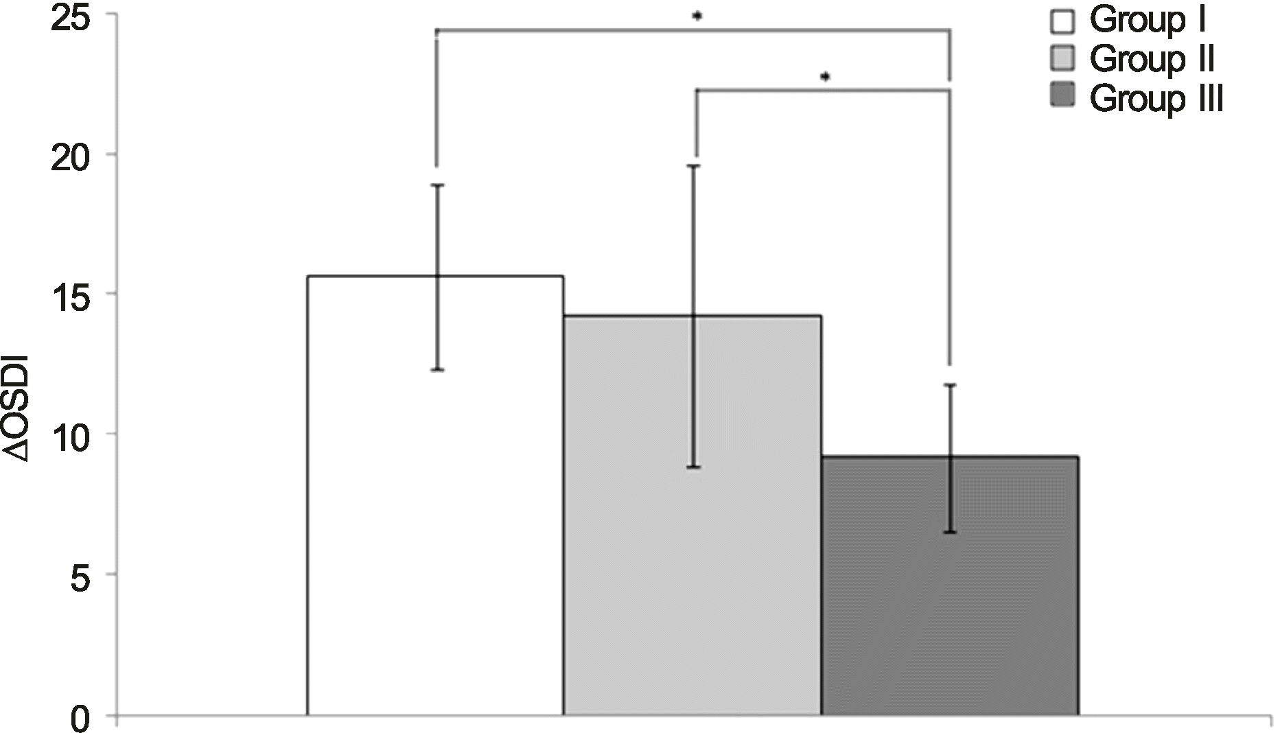 | Figure 4.Comparison of changes in ocular surface disease index (OSDI) scores between pretreatment and at 3 months after the treatment in eyes grouped by the degree of low er lid laxity. *Mann Whitney test (significance lev el is p < 0.05). |
Table 1.
Demographics and changes in tear break-up time (TBUT), Schirmer test (ST) and ocular surface diseases index (OSDI) scores before and at 3 months after the treatment in eyes grouped by the degree of lower lid laxity
| Parameters |
Groups |
p-value |
||||
|---|---|---|---|---|---|---|
| I (n = 10) | II (n = 10) | III (n = 10) | I & II | II & III | I & III | |
| Age (years) | 55.00 ± 5.29 | 56.70 ± 5.36 | 56.10 ±4.80 | 0.436 | 0.912 | 0.631 |
| Sex (M:F) | 0:10 | 1:9 | 2:8 | 1.000 | 1.000 | 0.474 |
| Treatments (%) | ||||||
| 0.1% Fluorometholon | 50 | 60 | 60 | 1.000 | 1.000 | 1.000 |
| 0.05% Cyclosporine A | 10 | 20 | 10 | 1.000 | 1.000 | 1.000 |
| Tetracyclin oint + Blephasol® | 0 | 20 | 40 | 0.474 | 0.628 | 0.087 |
| MGD | 1.20 ± 0.42 | 1.90 ± 0.74 | 2.00 ± 0.00 | 0.043† | 0.739 | 0.002† |
| TBUT (sec) | ||||||
| Baseline | 4.00 ± 0.94 | 3.70 ± 1.34 | 4.50 ± 1.08 | 0.739 | 0.218 | 0.315 |
| 3 months | 6.60 ± 1.43 | 6.00 ± 2.54 | 6.00 ± 1.45 | 0.631 | 0.684 | 0.436 |
| p-value | 0.004* | 0.011* | 0.010* | |||
| ΔTBUT | 2.60 ± 0.84 | 2.30 ± 1.57 | 1.50 ± 1.20 | 0.739 | 0.218 | 0.051 |
| ST (mm) | ||||||
| Baseline | 8.60 ± 2.46 | 7.30 ± 4.79 | 7.80 ± 2.66 | 0.105 | 0.247 | 0.529 |
| 3 months | 8.40 ± 2.55 | 7.80 ± 4.39 | 7.50 ± 2.07 | 0.353 | 0.631 | 0.481 |
| p-value | 0.557 | 0.496 | 0.457 | |||
| ΔST | −0.20 ± 2.15 | 0.50 ± 2.32 | −0.30 ± 1.83 | 0.631 | 0.579 | 0.853 |
| OSDI | ||||||
| Baseline | 27.80 ± 4.44 | 30.30 ± 7.99 | 30.00 ± 4.85 | 0.796 | 1.000 | 0.579 |
| 3 months | 12.20 ± 1.40 | 16.10 ± 4.63 | 20.80 ± 4.52 | 0.029† | 0.029† | <0.001† |
| p-value | 0.005* | 0.005* | 0.005* | |||
| ΔOSDI | 15.6 ± 3.31 | 14.2 ± 5.33 | 9.2 ± 2.62 | 0.529 | 0.019† | <0.001† |
 XML Download