Abstract
Purpose
To investigate clinical outcomes, response to treatment, and the related factors of recurrence and complication, following treatment of primary conjunctival malignant lymphoma.
Methods
The medical records of 39 patients diagnosed with primary conjunctival malignant lymphoma between January 2005 and June 2013 were retrospectively reviewed.
Results
The mean age of patients was 51.1 years old. The most common presenting symptom was hyperemia (33.3%). The most common anatomical location of the mass was the fornix (38.5%) and 25.6% patients had bilateral involvement. Histopathologically, mucosa-associated lymphoid tissue (MALT) lymphoma (92.3%) was the most common subtype. Every patient underwent radiotherapy (92.3%) or chemotherapy (7.7%) after surgical excision and had 100% complete remission. Local or systemic recurrence was observed in 15.4% of patients after treatment (mean 8.0 ± 3.3 months), but was completely remitted after additional radiation or chemotherapy. International prognostic index and location of tumor were significantly related factors for predicting tumor recurrence (p < 0.01, p = 0.02, respectively). Dry eye disease (DED) was the most common ocular complication (44.4%) after radiotherapy. Total radiation dosage and location of tumor were significantly associated factors for developing DED after radiotherapy (both p = 0.04).
Go to : 
References
1. Freeman C, Berg JW, Cutler SJ. Occurrence and prognosis of extranodal lymphomas. Cancer. 1972; 29:252–60.

2. Knowles DM, Jakobiec FA, McNally L, Burke JS. Lymphoid hyperplasia and malignant lymphoma occurring in the ocular adnexa (orbit, conjunctiva, and eyelids): a prospective multiparametric analysis of 108 cases during 1977 to 1987. Hum Pathol. 1990; 21:959–73.

3. Grossniklaus HE, Green WR, Luckenbach M, Chan CC. Conjunctival lesions in adults. A clinical and histopathologic review. Cornea. 1987; 6:78–116.
4. Shields CL, Shields JA, Carvalho C, et al. Conjunctival lymphoid tumors: clinical analysis of 117 cases and relationship to systemic lymphoma. Ophthalmology. 2001; 108:979–84.
6. Jenkins C, Rose GE, Bunce C, et al. Clinical features associated with survival of patients with lymphoma of the ocular adnexa. Eye (Lond). 2003; 17:809–20.

7. Coupland SE, Hummel M, Stein H. Ocular adnexal lymphomas: five case presentations and a review of the literature. Surv Ophthalmol. 2002; 47:470–90.
8. Matsuo T, Yoshino T. Long-term follow-up results of observation or radiation for conjunctival malignant lymphoma. Ophthalmology. 2004; 111:1233–7.
9. Liao SL, Kao SC, Hou PK, Chen MS. Results of radiotherapy for orbital and adnexal lymphoma. Orbit. 2002; 21:117–23.

10. Goda JS, Le LW, Lapperriere NJ, et al. Localized orbital mucosa-associated lymphoma tissue lymphoma managed with primary radiation therapy: efficacy and toxicity. Int J Radiat Oncol Biol Phys. 2011; 81:e659–66.

11. Lee SJ, Jung JH, Choi HY. Analysis of clinical features and prognostic factor analysis of orbital and adnexal lymphoma. J Korean Ophthalmol Soc. 2013; 54:12–8.

12. Lim DW, Im SK, Yoon KC. Clinical features and treatment results of conjunctival lymphoproliferative lesions. J Korean Ophthalmol Soc. 2004; 45:1820–6.
13. Swerdlow SH, Campo E, Harris NL, et al. WHO classification of tumours of haematopoietic and lymphoid tissues. 4th ed.2. Lyon, France: IARC Press;2008.
14. Carbone PP, Kaplan HS, Musshoff K, et al. Report of the Committee on Hodgkin's Disease Staging Classification. Cancer Res. 1971; 31:1860–1.
15. The definition and classification of dry eye disease: report of the Definition and Classification Subcommittee of the International Dry Eye WorkShop (2007). Ocul Surf. 2007; 5:75–92.
16. White WL, Ferry JA, Harris NL, Grove AS Jr. Ocular adnexal lymphoma. A clinicopathologic study with identification of lymphomas of mucosa-associated lymphoid tissue type. Ophthalmology. 1995; 102:1994–2006.
17. Johnson TE, Tse DT, Byrne GE Jr, et al. Ocular-adnexal lymphoid tumors: a clinicopathologic and molecular genetic study of 77 patients. Ophthal Plast Reconstr Surg. 1999; 15:171–9.
18. Coupland SE, Krause L, Delecluse HJ, et al. Lymphoproliferative lesions of the ocular adnexa. Analysis of 112 cases. Ophthalmology. 1998; 105:1430–41.
19. Abd Al-Kader L, Sato Y, Takata K, et al. A case of conjunctival follicular lymphoma mimicking mucosa-associated lymphoid tissue lymphoma. J Clin Exp Hematop. 2013; 53:49–52.
20. Jakobiec FA. Ocular adnexal lymphoid tumors: progress in need of clarification. Am J Ophthalmol. 2008; 145:941–50.

21. Ji JY, Ahn YC, Kim YD. Radiotherapy for malignant lymphoma of orbit and ocular adnexa. J Korean Ophthalmol Soc. 2005; 46:201–14.
22. Woo JM, Tang CK, Rho MS, et al. The clinical characteristics and treatment results of ocular adnexal lymphoma. Korean J Ophthalmol. 2006; 20:7–12.

23. Cho EY, Han JJ, Ree HJ, et al. Clinicopathologic analysis of ocular adnexal lymphomas: extranodal marginal zone b-cell lymphoma constitutes the vast majority of ocular lymphomas among Koreans and affects younger patients. Am J Hematol. 2003; 73:87–96.

24. Uno T, Isobe K, Shikama N, et al. Radiotherapy for extranodal, marginal zone, B-cell lymphoma of mucosa-associated lymphoid tissue originating in the ocular adnexa: a multiinstitutional, retrospective review of 50 patients. Cancer. 2003; 98:865–71.

25. Woolf DK, Ahmed M, Plowman PN. Primary lymphoma of the ocular adnexa (orbital lymphoma) and primary intraocular lymphoma. Clin Oncol (R Coll Radiol). 2012; 24:339–44.

26. Park SJ, Lee WS, Yang JW. The effect of rituximab, cyclophosphamide, vincristine, and prednisolone (R-CVP) chemotherapy in patients with ocular adnexal extranodal marginal zone B cell lymphoma of the mucosa-associated lymphoid tissue (MALT) lymphoma. J Korean Ophthalmol Soc. 2013; 54:1157–64.

27. Nückel H, Meller D, Steuhl KP, Dührsen U. Anti-CD20 monoclonal antibody therapy in relapsed MALT lymphoma of the conjunctiva. Eur J Haematol. 2004; 73:258–62.

28. Zinzani PL, Alinari L, Stefoni V, et al. Rituximab in primary conjunctiva lymphoma. Leuk Res. 2005; 29:107–8.

29. Kang HJ, Kim WS, Kim SJ, et al. Phase II trial of rituximab plus CVP combination chemotherapy for advanced stage marginal zone lymphoma as a first-line therapy: Consortium for Improving Survival of Lymphoma (CISL) study. Ann Hematol. 2012; 91:543–51.

30. Jaffe ES, Harris NL, Diebold J, Muller-Hermelink HK. World Health Organization classification of neoplastic diseases of the hematopoietic and lymphoid tissues. A progress report. Am J Clin Pathol. 1999; 111:S8–12.
31. Suh MH, Kim JH, Yu HG. Conjunctival MALToma patient with intraocular manifestation: a case report. J Korean Ophthalmol Soc. 2007; 48:460–4.
32. López-Guillermo A, Montserrat E, Bosch F, et al. Applicability of the International Index for aggressive lymphomas to patients with low-grade lymphoma. J Clin Oncol. 1994; 12:1343–8.

33. Troch M, Wöhrer S, Raderer M. Assessment of the prognostic indices IPI and FLIPI in patients with mucosa-associated lymphoid tissue lymphoma. Anticancer Res. 2010; 30:635–9.
34. Sjö LD, Heegaard S, Prause JU, et al. Extranodal marginal zone lymphoma in the ocular region: clinical, immunophenotypical, and cytogenetical characteristics. Invest Ophthalmol Vis Sci. 2009; 50:516–22.
35. Demirci H, Shields CL, Karatza EC, Shields JA. Orbital lymphoproliferative tumors: analysis of clinical features and systemic involvement in 160 cases. Ophthalmology. 2008; 115:1626–31. 1631.e1-3.

36. Ellis JH, Banks PM, Campbell RJ, Liesegang TJ. Lymphoid tumors of the ocular adnexa. Clinical correlation with the working formulation classification and immunoperoxidase staining of paraffin sections. Ophthalmology. 1985; 92:1311–24.
37. Avilés A, Neri N, Calva A, et al. Addition of a short course of chemotherapy did not improve outcome in patients with localized marginal B-cell lymphoma of the orbit. Oncology. 2006; 70:173–6.

38. Bhandare N, Moiseenko V, Song WY, et al. Severe dry eye syndrome after radiotherapy for head-and-neck tumors. Int J Radiat Oncol Biol Phys. 2012; 82:1501–8.

39. Bessell EM, Henk JM, Wright JE, Whitelocke RA. Orbital and conjunctival lymphoma treatment and prognosis. Radiother Oncol. 1988; 13:237–44.

40. Smitt MC, Donaldson SS. Radiotherapy is successful treatment for orbital lymphoma. Int J Radiat Oncol Biol Phys. 1993; 26:59–66.

41. Letschert JG, González González D, Oskam J, et al. Results of radiotherapy in patients with stage I orbital non-Hodgkin's lymphoma. Radiother Oncol. 1991; 22:36–44.

42. Erickson BA, Harris GJ, Enke CA, et al. Periocular lymphoproliferative diseases: natural history, prognostic factors, and treatment. Radiology. 1992; 185:63–70.

43. Claus F, Boterberg T, Ost P, De Neve W. Short term toxicity profile for 32 sinonasal cancer patients treated with IMRT. Can we avoid dry eye syndrome? Radiother Oncol. 2002; 64:205–8.
Go to : 
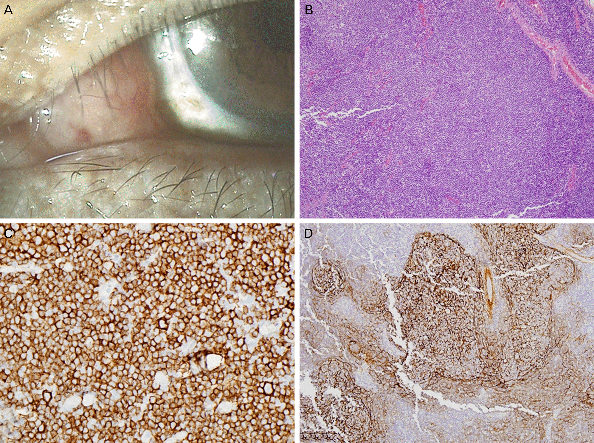
 | Figure 1.The representive case of the eye with extranodal marginal zone lymphoma of mucosa-associated lymphoid tissue (MALT lymphoma). (A) Slit-lamp exmanination reveals a ‘salmon-colored’ conjunctival mass in caruncle. (B) Histological examination demonstrates the diffuse proliferation of lymphoid cells in the conjunctiva (Hematoxylin-eosin stain, ×100). Immunohistochemical staining shows positivity of the tumor cells for (C) CD20 (×400) which is a marker for B-cells and (D) CD21 (×100) which reflects follicular dendritic cell meshwork (×100). |
 | Figure 2.The representive case of the eye with follicular lymphoma. (A) Clinical photograph of the follicular lymphoma presenting classical ‘salmon-colored’ conjunctival mass located in the fornix. Immunohistochemical stain of (B) CD10 and (C) Bcl-6 which are markers of follicular center B-cell differentiation (× 200). (D) Bcl-2 expression by the tumor cells (× 100). |
Table 1.
Anatomic distribution of primary conjunctival malignant lymphoma
| Conjunctival location | No. of patients (%) |
|---|---|
| Fornix | 15 (38.5) |
| Bulbar | 12 (30.8) |
| Limbus | 5 (12.8) |
| Caruncle/plica semilunaris | 4 (10.3) |
| Tarsus | 3 (7.7) |
Table 2.
Histopathologic diagnosis of primary conjunctival malignant lymphoma according to WHO classification
| Type | No. of patients (%) |
|---|---|
| MALT lymphoma | 36 (92.3) |
| Follcular lymphoma | 2 (5.1) |
| Small lymphocytic lymphoma | 1 (2.6) |
| Total | 39 (100.0) |
Table 3.
Analysis of predisposing factors of recurrence in patients with primary conjunctival malignant lymphoma
Table 4.
Comparison between patients with dry eye disease and patients without dry eye disease after radiotherapy
| Non-DED (n = 20) | DED (n = 16) | p-value | |
|---|---|---|---|
| Age (years) | 49.55 ± 15.79 | 53.19 ± 16.14 | 0.54* |
| Sex (male/female) | 8/12 | 5/11 | 0.59† |
| Total radiation dose (Gy) | 30.24 ± 2.59 | 32.24 ± 2.12 | 0.03* |
| BCVA (log MAR) | 0.02 ± 0.05 | 0.02 ± 0.05 | 0.54* |
| Tear parameters | |||
| TBUT (sec) | 10.10 ± 1.48 | 4.31 ± 1.01 | <0.01* |
| Basal tear secretion (mm) | 12.25 ± 1.86 | 5.94 ± 1.29 | <0.01* |
| Tear clearance rate ([log2]-1) | 5.40 ± 0.60 | 2.94 ± 0.68 | <0.01* |
| Keratoepitheliopathy score | 0.25 ± 0.44 | 2.19 ± 1.11 | <0.01* |
Table 5.
Analysis of risk factors for developing dry eye disease after radiotherapy in patients with primary conjunctival malignant lymphoma


 PDF
PDF ePub
ePub Citation
 Print
Print


 XML Download