

 PDF
PDF ePub
ePub Citation
 Print
Print


Abstract
Purpose
In this study we evaluated the location and shape of the optic canal using computed tomography (CT) for diagnosis and treatment of posterior orbital diseases.
Methods
Fifty patients, who had received a facial bone CT between November 2012 and June 2013 at Korea University Hospital were included in the present study. The location and shape of the optic canal was evaluated using 9 parameters on CT (P1: nasal bone tip; P2: middle point of tuberculum sellae; P3: root of columella nasi; P4: orbit end of the optic canal; P5: cranium end or the optic canal; P6: P1's projection on L2; L1: line that links P1 and P2; L2: goes through P3 and parallel to L1; L3: bisector of right and left and goes through P1).
Results
The distance between P3 and P4 was 81.5 mm and 75.6 mm in males and females, respectively (p = 0.001). The distance between P3 and P5 was 88.5 mm and 82.1 mm in, males and females, respectively (p = 0.001). The width of the orbital end and cranium end of the optic canal, the length of the optic canal was 2.4 mm, 4.1 mm, 10.9 mm in males and 2.3 mm, 3.6 mm, 10.2 mm, in females, respectively.
References
1. Hart CK, Theodosopoulos PV, Zimmer LA. Anatomy of the optic canal: a computed tomography study of endoscopic nerve decompression. Ann Otol Rhinol Laryngol. 2009; 118:839–44.

2. Hart CK, Zimmer LA. Computed tomography anatomy of the optic canal. Otolaryngology Head and Neck Surgery. 2008; 139:74.
3. Zhang J, Liao J, Yang Y, et al. [Applied anatomy study of optic canal by transnasal endoscopy]. Lin Chung Er Bi Yan Hou Tou Jing Wai Ke Za Zhi. 2009; 23:346–8.
4. Metson R, Pletcher SD. Endoscopic orbital and optic nerve decompression. Otolaryngol Clin North Am. 2006; 39:551–61. ix.
5. Akdemir G, Tekdemir I, Altin L. Transethmoidal approach to the optic canal: surgical and radiological microanatomy. Surg Neurol. 2004; 62:268–74. discussion 274.
6. Locatelli M, Caroli M, Pluderi M, et al. Endoscopic transsphenoidal optic nerve decompression: an anatomical study. Surg Radiol Anat. 2011; 33:257–62.
7. Liu S, Chen Y, Song J, et al. Optic canal location by computed tomography. J Craniofac Surg. 2013; 24:284–6.
8. Liu X, Zhou C, Zhang G, et al. [CT anatomic measurement of the optic canal and its clinical significance]. Zhonghua Er Bi Yan Hou Ke Za Zhi. 2000; 35:275–7.
Figure 1.
Center sagittal plane. P1: nasal bone tip; P2: middle point of tuberculum sellae; P3: root of columella nasi; P6: P1's projection on L2; L1: line links P1 and P2; L2: goes through P3 and parallel to L1; D1: the distance between L1 and L2; D2: the distance between P3 and P1.
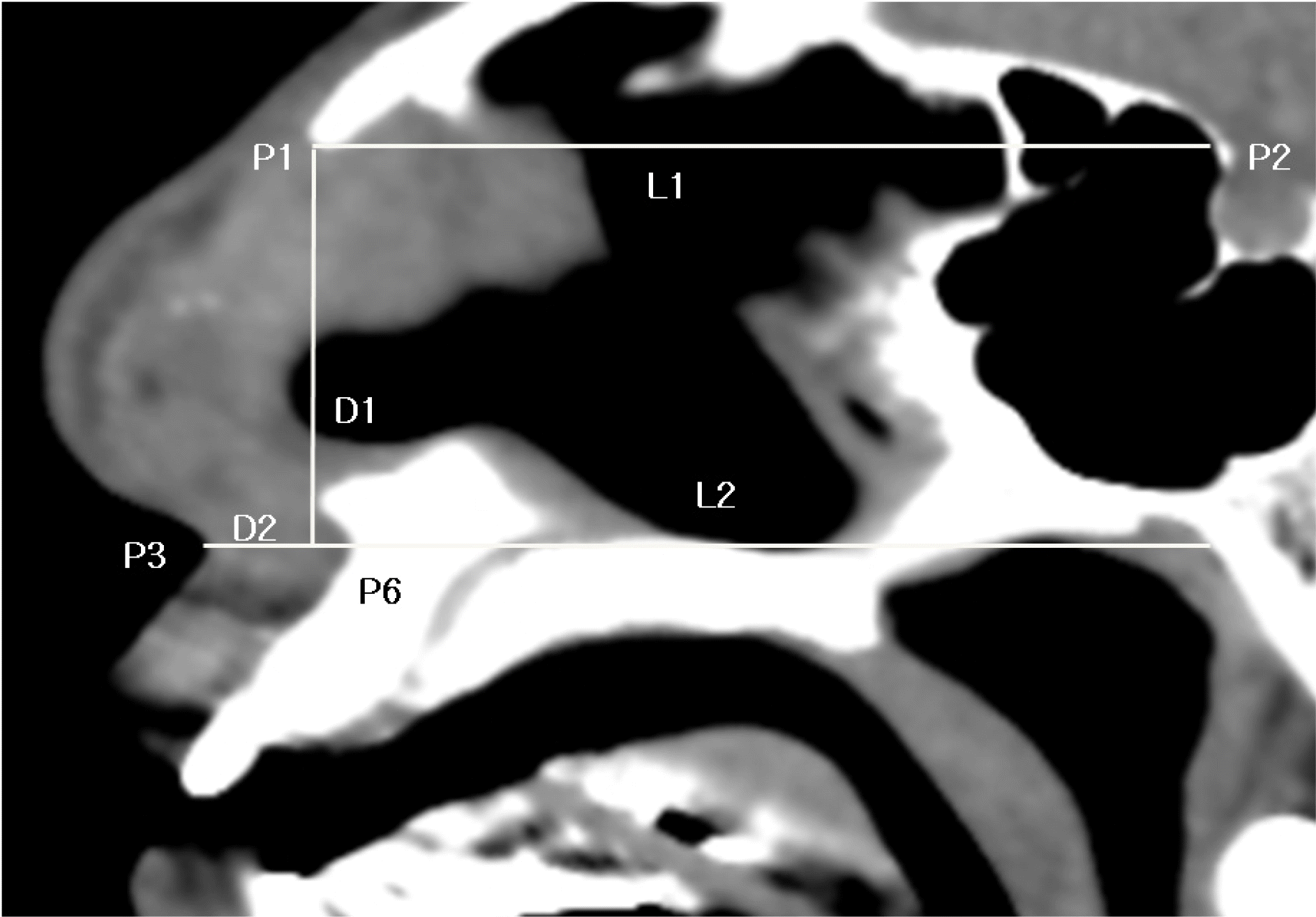
Figure 2.
Plane perpendicular to center sagittal plane and goes through nasal bone tip and the middle point of tuberculum sellae line. L3: bisector of right and left and goes through P1; P4: orbit end of the optic canal; P5: cranium end of the optic canal; D3: distance between P1 and P4; D4: distance between P1 and P5; D5: distance between P4 and L3; D6: distance between P5 and L3; D7: medial canal wall length; D8: width of orbit end of the optic canal; D9: width of cranium end of the optic canal; P1: nasal bone tip; A1: angle between P1P4 and L3; A2: angle between P1P5 and L3.
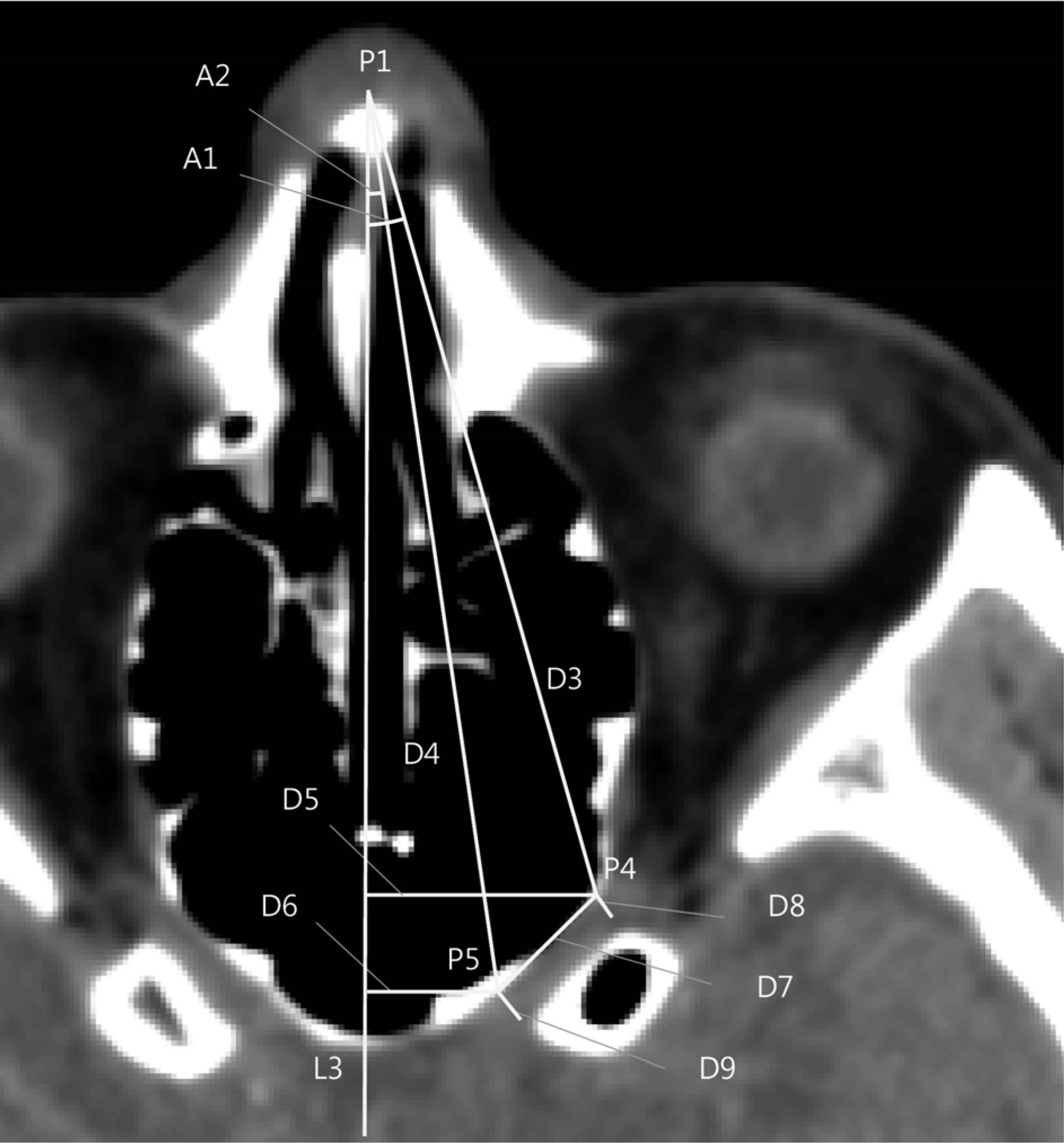
Figure 3.
Plane goes through the root of columella nasi and cranium end of the optic canal and orbit end of the optic canal. P3: root of columella nasi; P4: orbit end of the optic canal; P5: cranium end of the optic canal; D10: distance between P3 and P4; D11: distance between P3 and P5; A3: angle between P3P5 and P3P4.
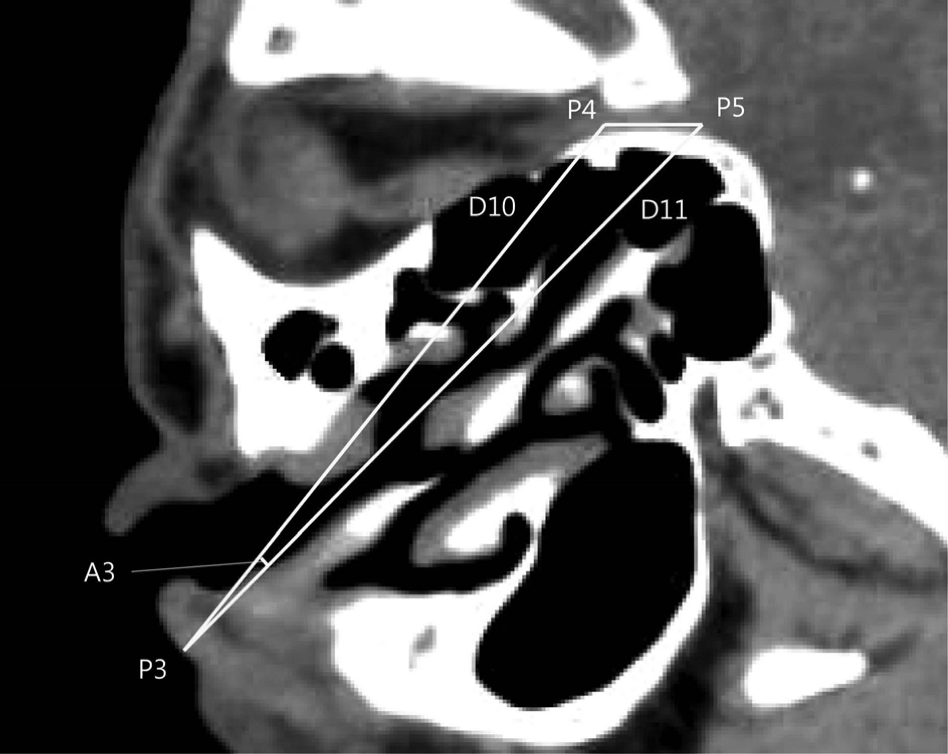
Table 1.
Parameters measured in this study
Table 2.
Patients demographics
| Male | Female | p-value | |
|---|---|---|---|
| Patients | 25 | 25 | |
| Age (years) | 38.6 ± 13.0 | 37.8 ± 12.2 | 0.841* |
Table 3.
Measurement results of D1 and D2
| Male (mm) | Female (mm) | p-value | |
|---|---|---|---|
| D1 | 32.05 ± 3.03 | 29.97 ± 1.64 | 0.04* |
| D2 | 11.35 ± 2.85 | 10.45 ± 2.60 | 0.251* |
Table 4.
Measurement results of D3-D11, A1-A3
| OD | p-value | OS | p-value | |||
|---|---|---|---|---|---|---|
| Male | Female | Male | Female | |||
| D3 (mm) | 65.18 ± 2.65 | 60.25 ± 2.73 | 0.001* | 64.80 ± 2.64 | 59.86 ± 3.00 | 0.001* |
| D4 (mm) | 71.84 ± 2.94 | 66.41 ± 3.17 | 0.001* | 71.59 ± 3.05 | 66.00 ± 3.18 | 0.001* |
| D5 (mm) | 15.01 ± 1.49 | 13.79 ± 1.51 | 0.006* | 15.38 ± 1.90 | 13.57 ± 1.40 | 0.001* |
| D6 (mm) | 7.69 ± 1.34 | 6.98 ± 1.55 | 0.091* | 7.48 ± 2.01 | 6.56 ± 1.43 | 0.070* |
| D7 (mm) | 10.94 ± 1.32 | 10.23 ± 1.24 | 0.056* | 11.07 ± 1.37 | 10.30 ± 1.25 | 0.043* |
| D8 (mm) | 2.48 ± 0.47 | 2.38 ± 0.54 | 0.501* | 2.48 ± 0.42 | 2.31 ± 0.52 | 0.219* |
| D9 (mm) | 4.11 ± 0.62 | 3.58 ± 0.70 | 0.008* | 4.04 ± 0.59 | 3.63 ± 0.78 | 0.041* |
| D10 (mm) | 81.50 ± 3.29 | 75.65 ± 3.08 | 0.001* | 81.76 ± 4.17 | 75.44 ± 3.25 | 0.001* |
| D11 (mm) | 88.52 ± 3.31 | 82.11 ± 3.24 | 0.001* | 88.42 ± 4.16 | 82.05 ± 3.49 | 0.001* |
| A1 (°) | 13.52 ± 1.30 | 13.13 ± 1.45 | 0.326* | 14.06 ± 1.63 | 13.49 ± 1.69 | 0.410† |
| A2 (°) | 5.85 ± 0.91 | 5.93 ± 1.53 | 0.808* | 6.43 ± 1.24 | 6.00 ± 1.34 | 0.227* |
| A3 (°) | 5.71 ± 1.11 | 6.01 ± 0.93 | 0.314* | 5.82 ± 0.95 | 5.69 ± 0.84 | 0.410† |
Values are presented as mean ± SD; Difference between both eyes: p > 0.05 by independent t-test; D3: distance between P1 and P4; D4: distance between P1 and P5; D5: distance between P4 and L3; D6: distance between P5 and L3; D7: medial wall length of optic canal; D8: width of orbital end of optic canal; D9: width of cranial end of optic canal; D10: distance between P3 and P4; D11: distance between P3 and P5; A1: angle between P1P4 and L3; A2: angle between P1P5 and L3; A3: angle between P3P5 and P3P4.
 XML Download