Abstract
Case summary
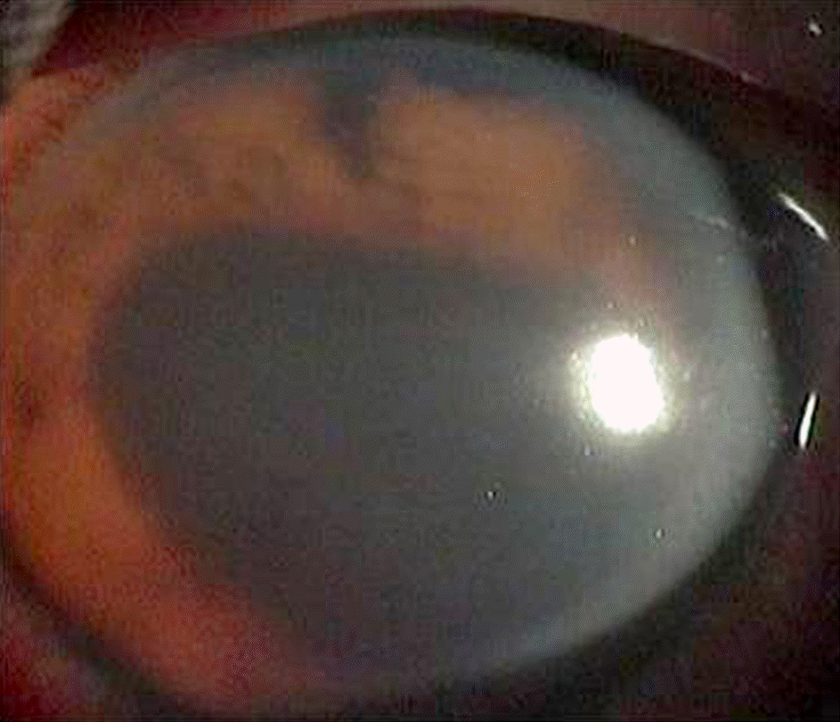
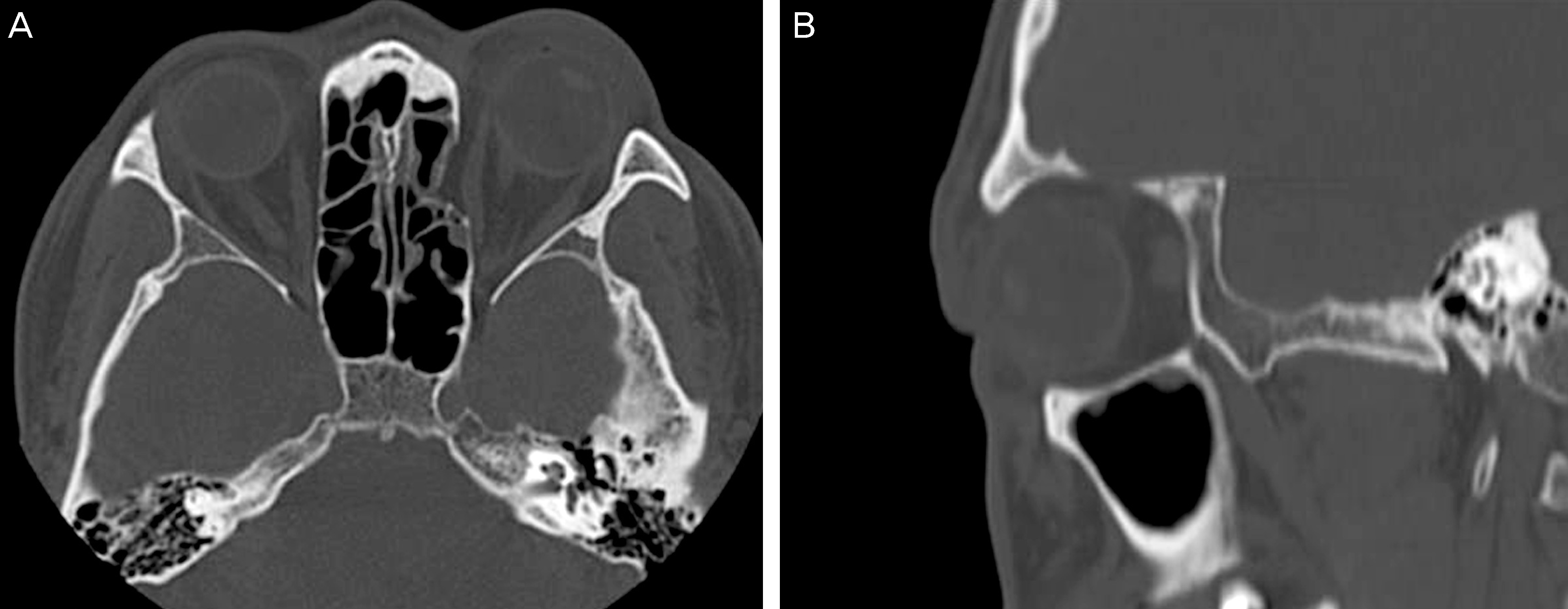
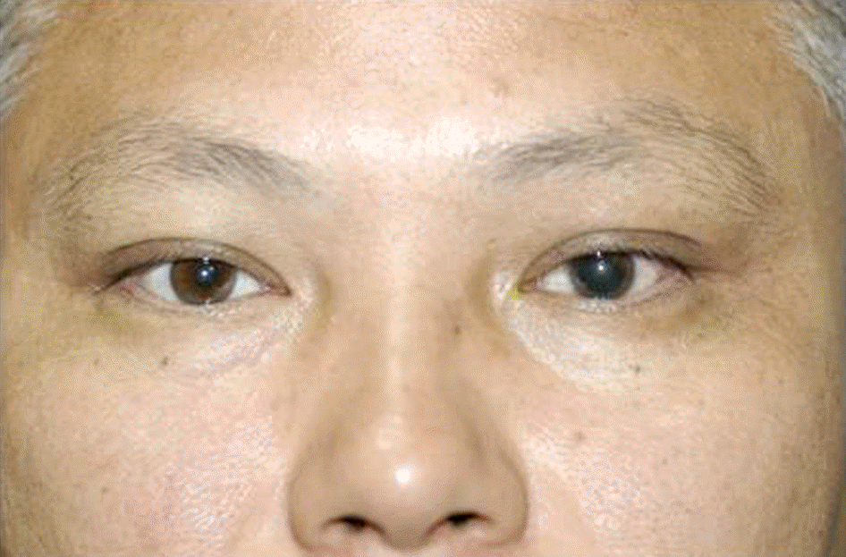
A previously healthy 41-year-old male was evaluated for decreased visual acuity and blepharoptosis in the left eye after ocular trauma. On ophthalmologic examination, visual acuity in the left eye was hand motion, intraocular pressure was 29 mm Hg, hematoma and eyelid edema were minimal. The patient had complete unilateral ptosis with superficial upper eyelid laceration. Additional findings in the left eye included fracture of the medial orbital wall, hyphema, iris sphincter muscle tear, iridodialysis and conjunctival laceration. The other examinations were unremarkable with full ocular motility. Because of iris sphincter muscle tear and iridodialysis, the pupillary reaction could not be evaluated. His left upper eyelid drooped completely and levator function test (LFT) was 0 mm. He was diagnosed with an isolated neurogenic blepharoptosis and received oral prednisolone at a dose of 1 mg/kg per day for 7 days with gradual tapering. One month later, the patient had normal symmetric lid height and completely restored levator function.
Go to : 
References
1. Jang BL. Neuro-ophthalmology. 2nd ed.Seoul: Ilchokak;2004. p. 287–9.
2. Crawford JS. Ptosis as a result of trauma. Can J Opthalmol. 1974; 9:244–8.
3. Boyle NS, Chang EL. Traumatic Blepharoptosis. In : Cohen AJ, Weinberg DA, editors. Evaluation and management of blepharoptosis. Weinberg: Springer Science Business Media;2011. p. 129–40.
4. McCulley TJ, Kersten RC, Yip CC, et al. Isolated unilateral neurogenic blepharoptosis secondary to eyelid trauma. Am J Ophthalmol. 2002; 134:626–7.

6. Ahn HB. Ophthalmic plastic and reconstructive surgery. 2nd ed.Seoul: Naewaeh-haksool;2009. p. 116–21.
7. Callahan MA, Beard C. Beard's Ptosis. 4th ed.Birmingham, AL: Aesculapius publishing co.;1990. p. 67–8.
8. Kersten RC, de Conciliis C, Kulwin DR. Acquired ptosis in the young and middle-aged adult population. Ophthalmology. 1995; 102:924–8.

9. Thean JH, McNab AA. Blepharoptosis in RGP and PMMA hard contact lens wearers. Clin Exp Optom. 2004; 87:11–4.

10. Silkiss RZ, Baylis HI. Management of traumatic ptosis. Adv Ophthalmic Plast Reconstr Surg. 1987; 7:149–55.
11. Memon MY, Paine KW. Direct injury of the oculomotor nerve in craniocerebral trauma. J Neurosurg. 1971; 35:461–4.

12. Bun RJ, Vissink A, Bos RR. Traumatic superior orbital fissure syndrome: report of two cases. J Oral Maxillofac Surg. 1996; 54:758–61.

13. Sugamata A. Orbital apex syndrome associated with fractures of the inferomedial orbital wall. Clin ophthalmol. 2013; 7:475–8.

14. Derakhshan I. Superior branch palsy of the oculomotor nerve with spontaneous recovery. Ann Neurol. 1978; 4:478–9.

15. Jung JW, Chi MJ. Temporary unilateral neurogenic blepharoptosis after orbital medial wall reconstruction: 3 cases. Ophthalmologica. 2008; 222:360–2.

16. EO S, Kim JY, Azari K. Temporary orbital apex syndrome after repair of orbital wall fracture. Plast Reconstr Surg. 2005; 116:85e–9e.

17. Lipkin AF, Woodson GE, Miller RH. Visual loss due to orbital fracture. The role of early reduction. Arch Otolaryngol Head Neck Surg. 1987; 113:81–3.

18. Melcangi RC, Magnaghi V, Galbiati M, Martini L. Formation and effects of neuroactive steroids in the central and peripheral nervous system. Int Rev Neurobiol. 2001; 46:145–76.
Go to : 
 | Figure 1.Left upper eyelid ptosis after trauma. Complete ptosis with minimal eyelid edema were noted. |
 | Figure 2.Photographs show no limitations of extraocular muscles at 9 cardinal gazes were observed at 1 day. Especially preservation of superior rectus muscle functions were noted. |


 PDF
PDF ePub
ePub Citation
 Print
Print


 XML Download