Abstract
Purpose
To report a case of syphilitic scleritis initially misdiagnosed as noninfectious nodular or fungal scleritis.
Case summary
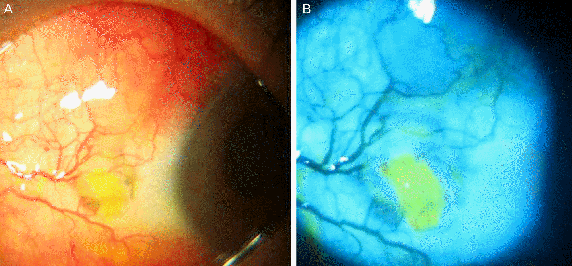
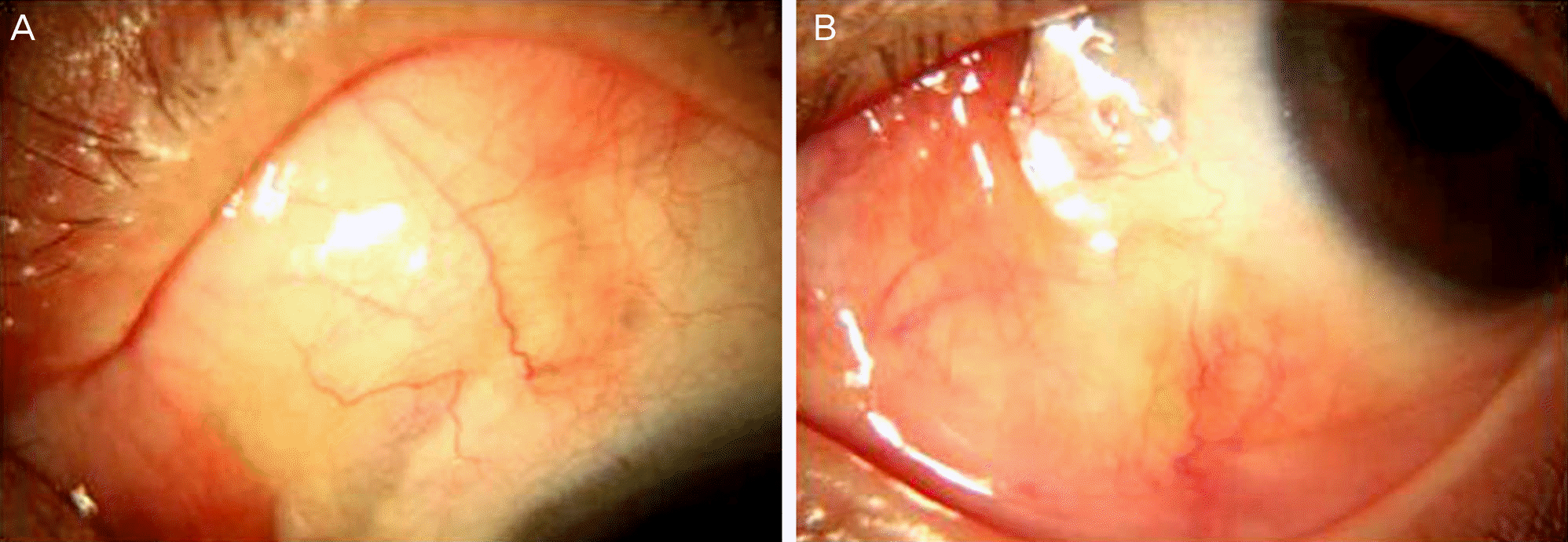
A 63-year-old female, who had severe headaches and ocular pain in her left eye despite treatment with topical and oral NSAIDs for the past 4 months, was transferred from a local clinic. The patient had a history of pterygium excision in the same eye 4 years prior. Upon presentation, she had a scleromalacia with calcified plaque at the nasal conjunctiva. An erythematous nodular elevated lesion was observed in the superonasal sclera. Microbiological smear and cultures were performed to exclude infectious scleritis. Under the suspicion of noninfectious nodular scleritis, the patient was prescribed topical oral steroid and oral NSAIDs. Candida parapsilosis was identified by the microbiological culture. Under the suspicion of fungal scleritis, oral fluconazole and topical amphotericin B were administered, but the lesions did not improve. On the 23rd day of treatment, we discovered the patient had a history of syphilis. The serology test was negative for RPR and FTA-ABS IgM but positive for FTA-ABS IgG. Under the suspicion of syphilitic scleritis, oral doxycycline (200 mg bid) was administered and benzathine penicillin M (2.4 million units) was injected intramuscularly 3 times at 1-week intervals. After the doxycycline and benzathine penicillin therapy, the pain and nodular erythematous lesions were completely resolved.
Conclusions
As shown in this case, syphilitic scleritis should be considered when the patient is resistant to other conventional treatments and shows positive serological tests for syphilis. This is important because syphilitic scleritis is usually aggravated by steroid treatment but can be cured by proper anti-syphilitic chemotherapy.
Go to : 
References
1. Reynolds MG, Alfonso E. Treatment of infectious scleritis and keratoscleritis. Am J ophthalmol. 1991; 112:543–7.

2. O'Donoghue E, Lightman S, Tuft S, Watson P. Surgically induced necrotising sclerokeratitis (SINS)–precipitating factors and response to treatment. Br J Ophthalmol. 1992; 76:17–21.
3. Diaz-valle D, Benitez del Castillo JM, Castillo A, et al. Immunologic and clinical evaluation of postsurgical necrotizing sclerocorneal ulceration. Cornea. 1998; 17:371–5.

4. Kim YK, Kim TY. 4 case of Pseudomonas scleritis after pterygium excision. J Korean Ophthalmol Soc. 1999; 40:2304–12.
6. Rao Na, Marak GE, Hydayat AA. Necrotizing scleritis. A clinico-pathologic study of 41 cases. Ophthalmology. 1985; 92:1542–9.
7. Su CY, Tsai JJ, Chang YC, Lin CP. Immunologic and clinical manifestations of infectious scleritis after pterygium excision. Cornea. 2006; 25:663–6.

9. Cunningham MA, Alexander JK, Matoba AY, et al. Management and outcome of microbial anterior scleritis. Cornea. 2011; 30:1020–3.

11. Foster CS, Tamesis RR. Ocular syphilis. In : Albert DM, Jakobiec FA, editors. Principles and Practice of Ophthalmology. 1st ed.Philadelphia: WB Saunders Co;1994. p. 968–72.
12. Yoon KC, Im SK, Seo MS, Park YG. Neurosyphilitic episcleritis. Acta Ophthalmol Scand. 2005; 83:265–6.

14. Centers for Disease control and prevention. Sexually transmitted diseases treatment guidelines 2002. MMWR Recomm Rep. 2002; 51:1–78.
Go to : 


 PDF
PDF ePub
ePub Citation
 Print
Print


 XML Download