Abstract
Purpose
To report a case of treating a patient with intralesional cidofovir injection who had frequently recurring lacrimal sac squamous papilloma after several excision surgeries.
Case summary
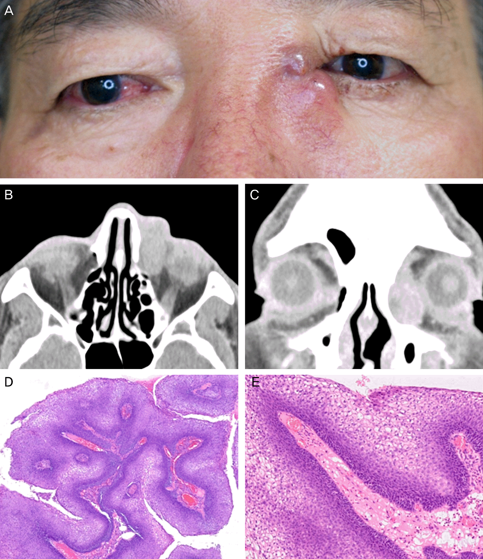
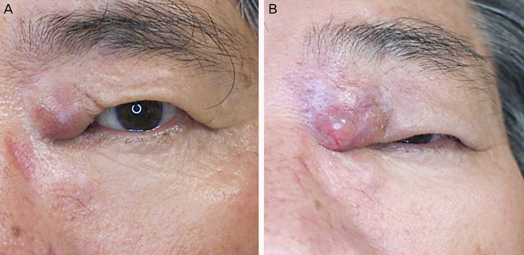
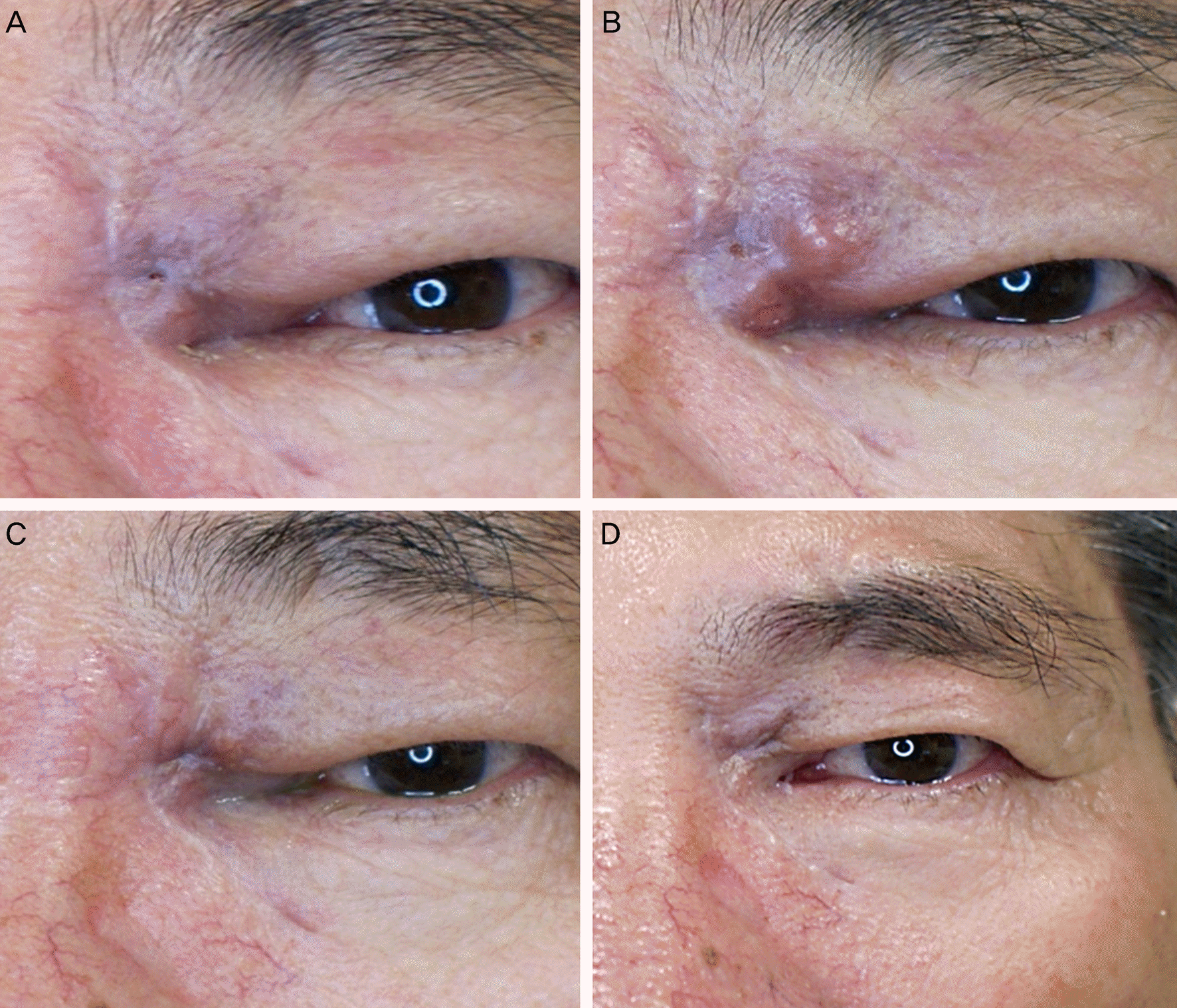
A 59-year-old man who had mass excision surgery at a different clinic nine months previously, visited our clinic to treat a recurring erythematous protruding mass near his left medial canthus that developed two months prior. Orbit CT showed a 15 × 25 mm-sized large mass located on the lacrimal sac adherent to medial orbital wall. An excision biopsy was performed and the histopathologic examination showed typical findings of squamous papilloma. Because the tumor recurred six months after the second surgery, we decided to perform adjuvant therapy using the antiviral agent cidofovir. The patient was treated with a 5 mg/mL intralesional cidofovir injection every three weeks. A transient recurrence presented on the upper lid at the third intralesional cidofovir injection site two months after the surgery, but the recurrent lesion improved after repeated injections. During the 12 months of follow-up, there were no complications and no evidence of recurrence.
Go to : 
References
1. Hung SL, Ma L. Recurrent lacrimal sac papilloma: case report. Chang Gung Med J. 2000; 23:113–7.
2. Madreperla SA, Green WR, Daniel R, Shah KV. Human papil-lomavirus in primary epithelial tumors of the lacrimal sac. Ophthalmology. 1993; 100:569–73.
3. Sjo NC, von Buchwald C, Cassonnet P, et al. Human papillomavirus: cause of epithelial lacrimal sac neoplasia. Acta Ophthalmol Scand. 2007; 85:551–6.

4. Weber RS, Shillitoe EJ, Robbins KT, et al. Prevalence of human papillomavirus in inverted nasal papillomas. Arch Otolaryngol Head Neck Surg. 1988; 114:23–6.

5. Krouse JH. Endoscopic treatment of inverted papilloma: safety and efficacy. Am J Otolaryngol. 2001; 22:87–99.

6. Lawson W, Kaufman MR, Biller HF. Treatment outcomes in the management of inverted papilloma: an analysis of 160 cases. Laryngoscope. 2003; 113:1548–56.

7. Vural E, Suen JY, Hanna E. Intracranial extension of inverted papilloma: An unusual and potentially fatal complication. Head Neck. 1999; 21:703–6.

8. Chaudhry IA, Taiba K, Al-Sadhan Y, Riley FC. Inverted papilloma invading the orbit through the nasolacrimal duct: a case report. Orbit. 2005; 24:135–9.

9. Elner VM, Burnstine MA, Goodman ML, Dortzbach RK. Inverted papillomas that invade the orbit. Arch Ophthalmol. 1995; 113:1178–83.

10. Williams R, Ilsar M, Welham RA. Lacrimal canalicular papillomatosis. Br J Ophthalmol. 1985; 69:464–7.

11. Snoeck R, Wellens W, Desloovere C, et al. Treatment of severe laryngeal papillomatosis with intralesional injections of cidofovir [(S)- 1-(3-hydroxy-2-phosphonylmethoxypropyl) cytosine]. J Med Virol. 1998; 54:219–25.
12. Bielamowicz S, Villagomez V, Stager SV, Wilson WR. Intralesional cidofovir therapy for laryngeal papilloma in an adult cohort. Laryngoscope. 2002; 112:696–9.

Go to : 
 | Figure 1.(A) Preoperative photograph showing left medial orbital erythematous swelling, fixed to the deep orbital planes on palpation. Orbit CT shows 15 × 25 mm-sized round, well-defined mass. Axal (B) and coronal (C) view. (D) Histopathologic finding shows the mass composed of mature squamous epithelium surrounding the central fibrovascular stalks without cellular atypia (Hematoxylin-eosin, × 40). (E) Irregular and severe epithelial hyperplasia is noted (Hematoxylin-eosin, ×400). |
 | Figure 2.(A) Recurred erythematous swelling at the operated site is shown at 7 months after first excision. (B) Preoperative clinical photograph before second operation. |
 | Figure 3.(A) Clinical photograph at 3 weeks after the first intralesional cidofovir injection. (B) Transient recurrence is observed 8 weeks after third intralesional cidofovir injection. Clinical photographs at the forth intralesional cidofovir injection (C), at 8 months after the second operation (D). |


 PDF
PDF ePub
ePub Citation
 Print
Print


 XML Download