Abstract
Purpose
To determine whether rat eyes develop increases in intraocular pressure (IOP) in response to a topically applied corticosteroid and to investigate the relationship between ocular hypertension and apoptosis of retinal ganglion cells.
Methods
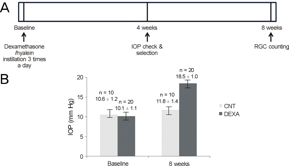
IOP was monitored by rebound tonometry in a group of 10 rats that received topically administered dexamethasone in both eyes (experimental) and in another group of 5 rats that received artificial tears (control) three times daily for 4 weeks after the establishment of baseline IOP values. Only eyes that increased by more than 50% compared with the basal IOP were administered once per day for 5 weeks. After 8 weeks, selective immunofluorescence stain for retinal ganglion cells and terminal deoxynucleotidyl transferase dUTP nick end labeling (TUNEL) stain were conducted.
Results
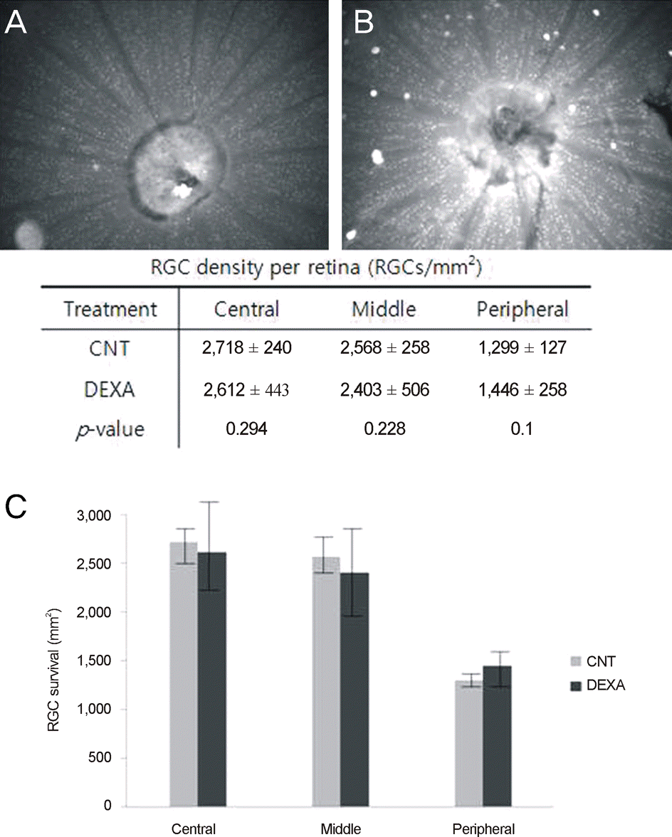
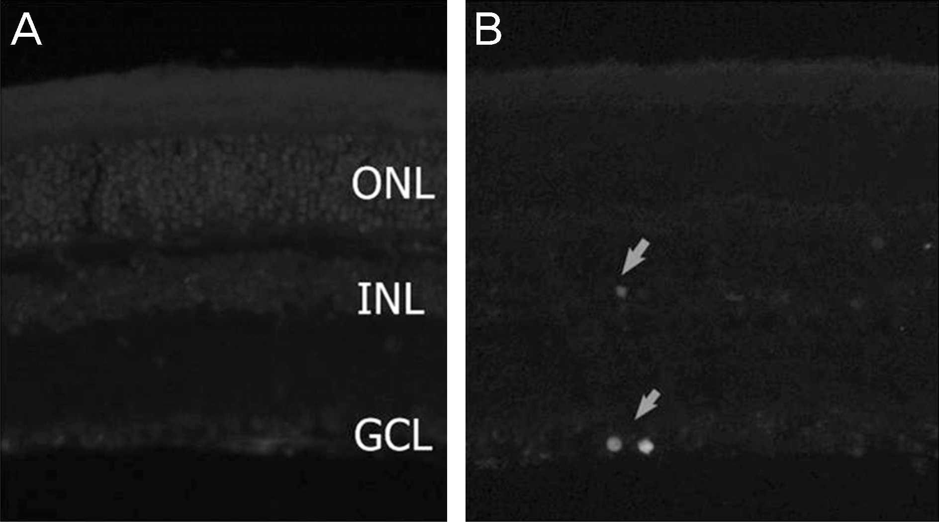
Among 20 experimental eyes, 11 eyes (55%) showed a greater than 50% increase in IOP compared with basal IOP. After 8 weeks, the mean IOPs for the experimental and control groups were 11.8 ± 1.4 mm Hg and 18.5 ± 1.0 mm Hg, respectively (p < 0.01). The counts of central retinal ganglion cells (RGCs) were 2718 ± 240 and 2612 ± 443, respectively (p = 0.294). The results of the TUNEL stain also showed no differences.
Go to : 
References
1. Jonas JB, Budde WM. Diagnosis and pathogenesis of glaucomatous optic neuropathy: morphological aspects. Prog Retinal Eye Res. 2000; 19:1–40.
2. Kass MA, Heuer DK, Higginbotham EJ, et al. The ocular hypertension treatment study—a randomized trial determines that topical ocular hypotensive medication delays or prevents the onset of primary open angle glaucoma. Arch Ophthalmol. 2002; 120:701–13.
3. Kim JH, Park CK. Expression of glutamate receptors in experimental rat model of chronic gluacoma. J Korean Ophthalmol. 2006; 47:468–76.
4. Shimazawa M, Nkamura S, Miwa M, et al. Establishment of the ocular hypertension model using the common marmoset. Exp Eye Res. 2013; 111:1–8.

5. Frankfort BJ, Khan AD, Tse DY, et al. Elevated intraocular pressure causes inner retinal dysfunction before cell loss in a mouse model of experimental glaucoma. Invest Ophthalmol Vis Sci. 2013; 54:762–70.

6. Gerometta R, Podos SM, Candia OA, et al. Steroid-induced ocular hypertension in normal cattle. Arch Ophthalmol. 2004; 122:1492–7.

7. Gerometta R, Podos SM, Danias J, Candia OA. Steroid-induced ocular hypertension in normal sheep. Invest Ophthalmol Vis Sci. 2009; 50:669–73.

8. Bhattacherjee P, Paterson CA, Spellman JM, et al. Pharmacological validation of a feline model of steroid-induced ocular hypertension. Arch Ophthalmol. 1999; 117:361–4.

9. Fingert JH, Clark AF, Craig JE, et al. Evaluation of the myocilin (MYOC) glaucoma gene in monkey and human steroid-induced ocular hypertension. Invest Ophthalmol Vis Sci. 2001; 42:145–52.
10. Knepper PA, Breen M, Weinstein HG, Blacik JL. Intraocular pressure and glycosaminoglycan distribution in the rabbit eye: effect of age and dexamethasone. Exp Eye Res. 1978; 27:567–75.

11. Lorenzetti OJ. Effects of corticosteroids on ocular dynamics in rabbits. J Pharmacol Exp Ther. 1970; 175:763–72.
12. Sawaguchi K, Nakamura Y, Sakai H, Sawaguchi S. Myocilin gene expression in the trabecular meshwork of rats in a steroid-induced ocular hypertension model. Ophthalmic Res. 2005; 37:235–42.

13. Zhan GL, Miranda OC, Bito LZ. Steroid glaucoma: cortico- steroidinduced ocular hypertension in cats. Exp Eye Res. 1992; 54:211–8.
14. Whitlock NA, McKnight B, Corcoran KN, et al. Increased intraocular pressure in mice treated with dexamethasone. Invest Ophthalmol Vis Sci. 2010; 51:6496–503.

15. Jones R 3rd, Rhee Dj. Corticosteroid-induced ocular hypertension and glaucoma: a brief review and update of the literature. Curr opin Ophthalmol. 2006; 17:163–7.
16. Kersey JP, Broadway DC. Corticosteroid-induced glaucoma: a review of the literature. Eye (Lond). 2006; 20:407–16.

17. Armaly MF. Statistical attributes of the steroid hypertensive response in the clinically normal eye, I: the demonstration of three levels of response. Invest Ophthalmol. 1965; 4:187–97.
18. Godel V, feiler-Ofry V, Stein R. Systemic steroid and ocular fluid dynamics. II. Systemic versus topical steroids. Acta Ophthalmol. 1972; 50:664–76.
19. Mindel JS, Tavitian HO, Smith H Jr, Walker EC. Comparative ocular pressure elevation by medrysone, fluorometholone, and dex- amethasone phosphate. Arch Ophthalmol. 1980; 98:1577–8.
20. May CA, Lutjen-Drecoll E. Morphology of the murine optic nerve. Invest Ophthalmol Vis Sci. 2002; 43:2206–12.
21. Aihara M, Lindsey JD, Weinreb RN. Twenty-four-hour pattern of mouse intraocular pressure. Exp Eye Res. 2003; 77:681–6.

22. Lee DW, Kim KY, Shim MS, et al. Coenzyme Q10 ameliorates oxidative stress and prevents mitochondrial alteration in ischemic retinal injury. Apoptosis. 2014; 19:603–14.

23. Jackson R, Waitzman M. Effects of some steroids on aqueous humor dynamics. Exp Eye Res. 1965; 4:112–23.

24. Levene RZ, Rothberger MD, Rosenberg S. Corticosteroid glaucoma in the rabbit. Am J Ophthalmol. 1974; 78:505–10.

25. Podos SM. Animal models of human glaucoma. Trans Am Acad Ophthalmol Otol. 1976; 81:632–5.
26. Touvinen E, Liesmaa M, Eslla R. The influence of corticosteroids on intraocular pressure in rabbits. II. The influence of massive subconjunctival doses of dexamethasone and betamethasone. Acta Ophthalmol. 1966; 44:901–5.
27. Hester DE, Trites PN, Peiffer RL, Petrow V. Steroid-induced ocular hypertension in the rabbit: a model using subconjunctival injections. J Ocul Pharmacol. 1987; 3:185–9.

28. Cole DF. Ocular fluid. In : Davson H, editor. The Eye. 3rd ed.Orlando, Fla: Academic press;1984. p. 269–390.
29. Clark AF, Brotchie D, Read AT, et al. Dexamethasone alters F-actin architecture and promotes cross-linked actin network formation in human trabecular meshwork tissue. Cell Motil Cytoskeleton. 2005; 60:83–95.

30. Clark AF, Wilson K, McCartney MD, et al. Glucocorticoid-induced formation of cross-linked actin networks in cultured human trabecular meshwork cells. Invest Ophthalmol Vis Sci. 1994; 35:281–94.
31. Wilson K, McCartney MD, Miggans ST, Clark AF. Dexamethasone induced ultrastructural changes in cultured human trabecular meshwork cells. Curr Eye Res. 1993; 12:783–93.

32. Yue BY. The extracellular matrix and its modulation in the trabecular meshwork. Surv Ophthalmol. 1996; 40:379–90.
33. Johnson DH, Bradley JM, Acott TS. The effect of dexamethasone on glycosaminoglycans of human trabecular meshwork in perfusion organ culture. Invest Ophthalmol Vis Sci. 1990; 31:2568–71.
34. Matsumoto Y, Johnson DH. Dexamethasone decreases phagocytosis by human trabecular meshwork cells in situ. Invest Ophthalmol Vis Sci. 1997; 38:1902–7.
35. Wordinger RJ, Clark AF. Effects of glucocorticoids on the trabecular meshwork: towards a better understanding of glaucoma. Prog Retin Eye Res. 1999; 18:629–67.

36. Zhang X, Ognibene CM, Clark AF, Yorio T. Dexamethasone inhibition of trabecular meshwork cell phagocytosis and its modulation by glucocorticoid receptor beta. Exp Eye Res. 2007; 84:275–84.
37. Xiong Miao J, Xi Z, et al. Regulatory effect of dexamethasone on aquaporin-1 expression in cultured bovine trabecular meshwork cells. J Huazhong Univ Sci Technolog Med Sci. 2005; 25:735–7.
38. Underwood JL, Murphy CG, Chen J, et al. Glucocorticoids regulate transendothelial fluid flow resistance and formation of intercellular junctions. Am J Physiol. 1999; 277:C330–42.
39. Goodwin JE, Zhang J, Geller DS. A critical role for vascular smooth muscle in acute glucocorticoid-induced hypertension. J Am Soc Nephrol. 2008; 19:1291–9.

40. Mangos GJ, Whitworth JA, Williamson PM, Kelly JJ. Glucocorticoids and the kidney. Nephrology (Carlton). 2003; 8:267–73.
41. Fardet L, Flahault A, Kettaneh A, et al. Corticosteroid-induced clinical adverse events: frequency, risk factors and patient's opinion. Br J Dermatol. 2007; 157:142–8.

42. Kelly JJ, Mangos G, Williamson PM, Whitworth JA. Cortisol and hypertension. Clin Exp Pharmacol Physiol Suppl. 1998; 25:S51–6.

43. Cavalli A, Lattion AL, Hummler E, et al. Decreased blood pressure response in mice deficient of the alpha1b-adrenergic receptor. Proc Natl Acad Sci U S A. 1997; 94:11589–94.
Go to : 
 | Figure 1.Dexamethasone instillation and induction of intraocular pressure (IOP). (A) Diagram for induction of IOP. Dexamethasone instillation began after baseline IOP check three times a day. After 4 weeks, in eyes with 50% elevated IOP than baseline, dexamethasone instillation was done 1 time a day for 4 weeks more. (B) IOP elevation in the mouse eyes at baseline and 8 weeks after dexamethasone instillation. CNT = control group; DEXA = dexamethason instillation group; RGC = retinal ganglion cell. |
 | Figure 2.Retinal ganglion cell (RGC) survival in control eyes and treated with dexamethasone eyes. Dexamethasone was instilled 3 times a day for 4 weeks in dexa group. After that, instilled once a day for 4 more weeks in eyes with elevated intraocular pressure (IOP) 50% than baseline. Retinal whole-mount immunohistochemistry for Brn3a in control group (A) and experimental group (B). (C) Quantitative analysis of RGC loss. Values are mean. There were no significant changes in RGC survival between control and dex- amethasone treated C57BL mice. Significant at p< 0.05. DEXA = dexamethason instillation group. |
 | Figure 3.Increased intraocular pressure (IOP) mediated apoptotic cell death. (A) There was no TUNEL-positive cells in the retina of control mice treated with artificial tears. (B) However, increased IOP retinas showed TUNEL-positive apoptotic cell death in the GCL and INL. Arrows: TUNEL-positive cell. TUNEL = terminal deoxynucleotidyl transferase dUTP nick end labeling; ONL = outer nuclear layer; INL = inner nuclear layer; GCL = ganglion cell layer. |
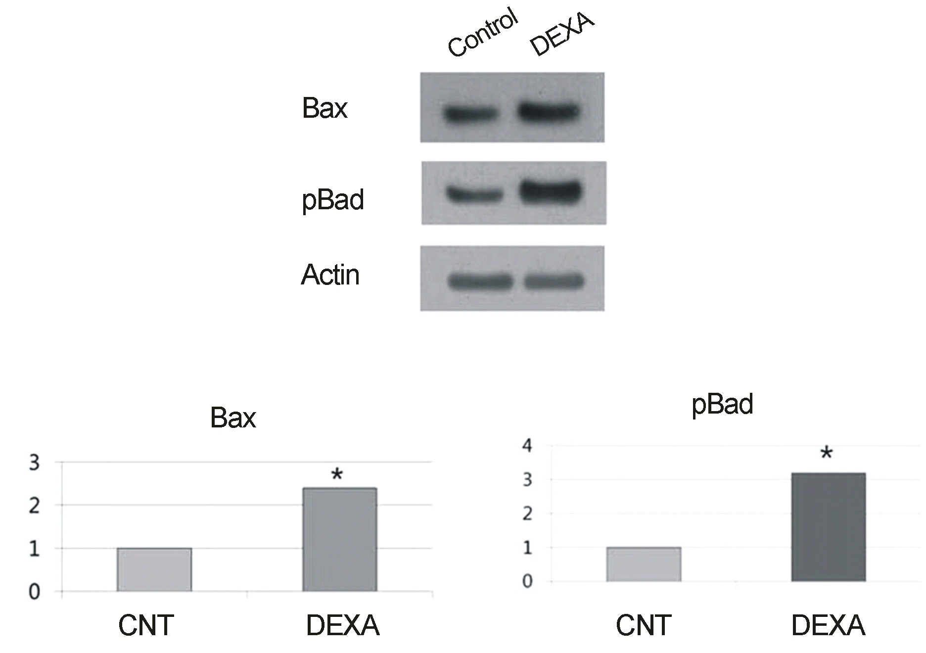
 | Figure 4.Increased intraocular pressure (IOP) induced apoptosis in dexamethasone instilled eyes. Dexamethasone was instilled 3 times a day for 4 weeks in experimental group. After that, instilled 1 time a day for 4 more weeks in eyes with elevated IOP 50% than baseline. Bax and pBad protein expression significantly increased in retina treated with dexamethasone. Values are mean. Significant at *p < 0.05. CNT = control group; DEXA = dexamethason instillation group. |


 PDF
PDF ePub
ePub Citation
 Print
Print


 XML Download