

 PDF
PDF ePub
ePub Citation
 Print
Print


Abstract
Purpose
Eruptive vellus hair cysts (EVHC) are benign lesions that affect the pediatric population and are rarely seen con-genitally or in young adults. EVHCs are small, cystic papules that usually occur on the chest and proximal extremities. EVHCs of the eyelids have been reported infrequently. We experienced a case of solitary EVHC that developed on the eyelid in a middle-aged male. Herein, we present our case with a brief review of the literature.
Case summary
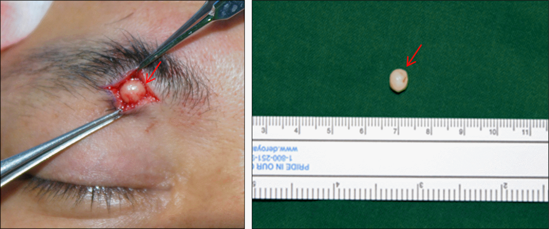
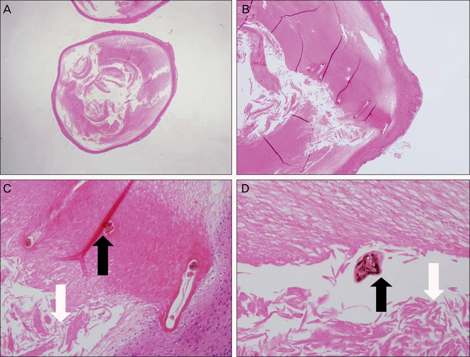
A 44-year-old male presented with a history of an asymptomatic, palpable mass in the right upper eyelid that had been slowly growing for 2 years. Physical examination revealed non-tender, firm and round mass in right upper eyelid. Computed tomography scan of the orbit showed a 7.0 × 9.0 × 9.5 mm-sized focal bulging contour in the right upper eyelid. The patient underwent sub-brow incision and excisional biopsy of the eyelid mass. Histopathological examination revealed a 7.0 × 6.0 × 4.0 mm-sized cystic structure lined by squamous epithelium containing laminated keratinous materials and multiple vellus hair shafts. There was no local recurrence during the postoperative follow-up.
Go to : 
References
1. Esterly NB, Fretzin DF, Pinkus H.Eruptive vellus hair cysts. Arch Dermatol. 1977; 113:500–3.

2. Choi JH, Chun JS, Seol JE. . A case of eruptive vellus hair cyst involving the face, trunk, and extremities. Korean J Dermatol. 2009; 47:1154–7.
3. Hong SD, Frieden IJ.Diagnosing eruptive vellus hair cysts. Pediatr Dermatol. 2001; 18:258–9.
4. Lee HS, Lee HK, Park K, Son SJ.A case of eruptive vellus hair cyst treated by mini-incision. Korean J Dermatol. 2007; 45:410–2.
5. Kumakiri M, Takashima I, Iju M. . Eruptive vellus hair cysts–a facial variant. J Am Acad Dermatol. 1982; 7:461–7.
6. Jun JH, Yun SJ, Kim SJ. . Two cases of eruptive vellus hair cysts developed on the atypical sites. Korean J Dermatol. 2004; 42:74–6.
7. Jung KD, Cho HJ, Kim PS. . A case of the facial variant of eruptive vellus hair cyst. Korean J Dermatol. 2009; 47:498–500.
8. Reep MD, Robson KJ.Eruptive vellus hair cysts presenting as multiple periorbital papules in a 13-year-old boy. Pediatr Dermatol. 2002; 19:26–7.
9. Mieno H, Fujimoto N, Tajima S.Eruptive vellus hair cyst in pa-tients with chronic renal failure. Dermatology. 2004; 208:67–9.
10. Park JH, Her Y, Chun BM. . A case of eruptive vellus hair cysts that developed on the labium major. Ann Dermatol. 2009; 21:294–6.
11. Cho HM, Kim SN.A clinical and histopathological study of 324 cases of epidermal cyst. Korean J Dermatol. 2007; 45:242–8.
12. Hammerton MD, Shrank AB.Superficial sebaceous hyperplasia of the areolae. Br J Dermatol. 1993; 129:649–50.
13. Bovenmyer DA.Eruptive vellus hair cysts. Arch Dermatol. 1979; 115:338–9.
14. Sina B, Burnett JW.Eruptive vellus hair cysts. Cutis. 1984; 33:503–4.
15. Coras B, Hohenleutner U, Landthaler M, Hohenleutner S.Early re-currence of eruptive vellus hair cysts after Er:YAG laser therapy: case report and review of the literature. Dermatol Surg. 2005; 31:1741–4.
Go to :
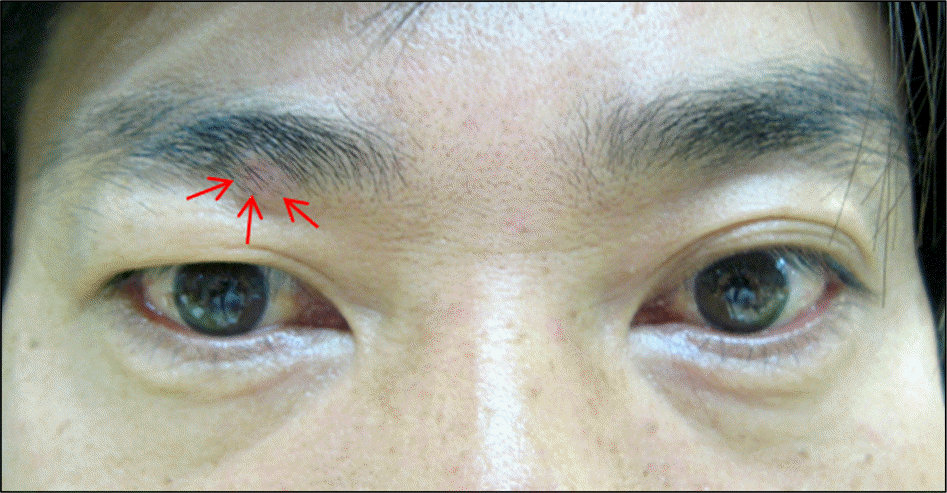 | Figure 1.External appearance of the patient. He presented solitary red-colored mass on his right upper eyelid (arrow). |
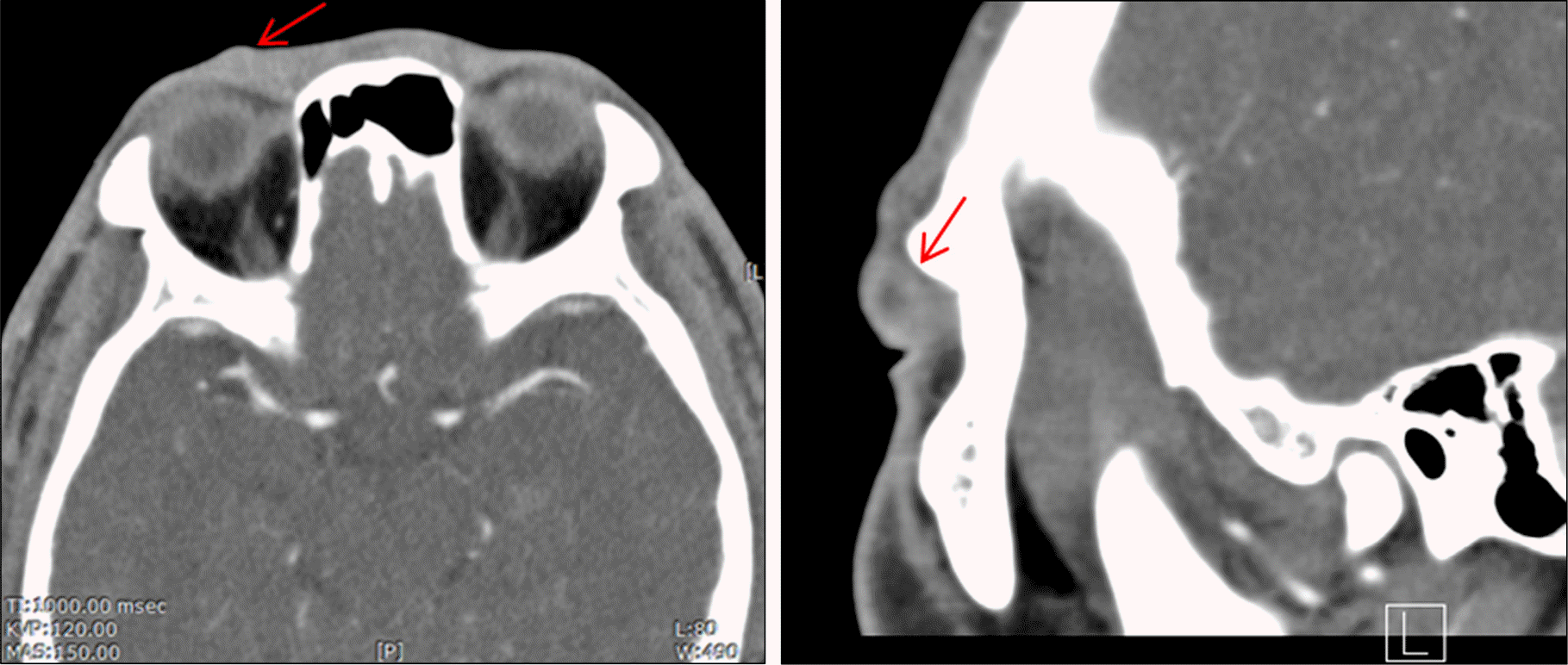 | Figure 2.Computed tomography scan showed a hypo-dense, non-enhancing, focal round mass measuring 7.0 × 9.0 ×9.5 mm on the right upper eyelid (arrow). |
 XML Download