Abstract
Purpose
This study was conducted to measure macular ganglion cell-inner plexiform layer (mGC-IPL) thickness in patients with a history of unilateral single attack of acute primary angle closure (APAC) and to compare it with that of unaffected fellow eyes 8 weeks after resolution using spectrum domain optical coherence tomography (SD-OCT).
Methods
Medical records of 24 patients with history of first episode of unilateral APAC were reviewed retrospectively. Eight weeks after APAC, mGC-IPL thickness and peripapillary retinal nerve fiber layer thickness were measured with SD-OCT and analyzed in eyes affected by APAC (group 1) and fellow eyes (group 2).
Results
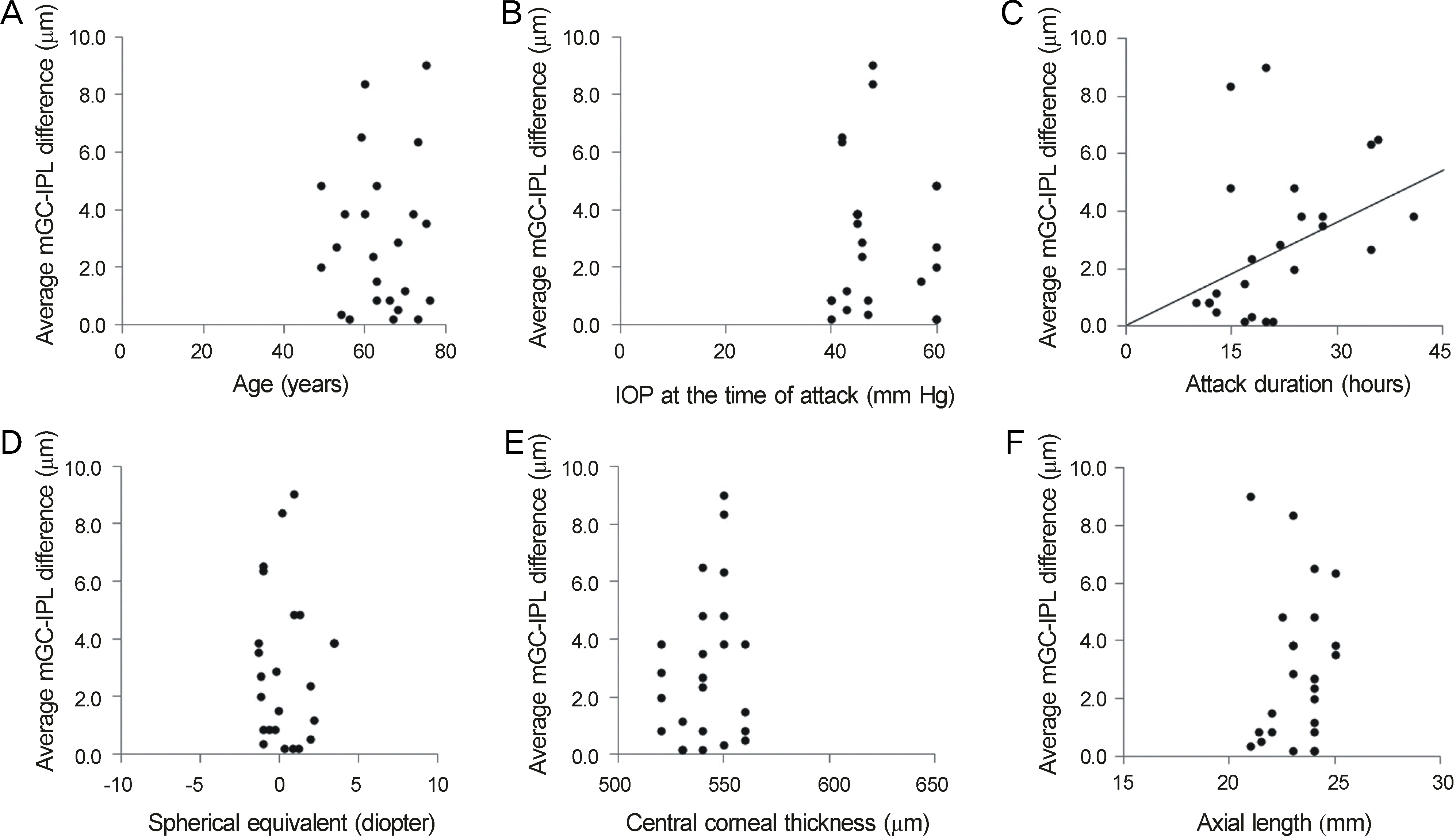
There were no significant differences between the groups with regard to best corrected visual acuity, spherical equivalent, central corneal thickness, or axial length (p > 0.05). There were no significant differences in mGC-IPL thickness in the superotemporal, superior, or superonasal sectors (p > 0.05). However, average, inferonasal, inferior, and inferotemporal sectors of group 1 were significantly thinner than those of group 2 (p = 0.002, 0.002, 0.001, 0.001, respectively). In addition, average mGC-IPL difference between affected eyes and fellow eyes showed a statistically significant correlation with attack duration (correlation coefficient = 0.249, p = 0.019).
Go to : 
References
1. Erie JC, Hodge DO, Gray DT. The incidence of primary angle-closure glaucoma in Olmsted County, Minnesota. Arch Ophthalmol. 1997; 115:177–81.

2. Tan AM, Loon SC, Chew PT. Outcomes following acute primary angle closure in an Asian population. Clin Experiment Ophthalmol. 2009; 37:467–72.

3. McNaught EI, Rennie A, McClure E, Chisholm IA. Pattern of visual damage after acute angle-closure glaucoma. Trans Ophthalmol Soc UK. 1974; 94:406–15.
4. Aung T, Looi AL, Chew PT. The visual field following acute primary angle closure. Acta Ophthalmol Scand. 2001; 79:298–300.

5. Bonomi L, Marraffa M, Marchini G, Canali N. Perimetric defects after a single acute angle-closure glaucoma attack. Graefes Arch Clin Exp Ophthalmol. 1999; 237:908–14.

6. Chew SS, Vasudevan S, Patel HY, et al. Acute primary angle closure attack does not cause an increased cup-to-disc ratio. Ophthalmology. 2011; 118:254–9.

7. Shen SY, Baskaran M, Fong AC, et al. Changes in the optic disc after acute primary angle closure. Ophthalmology. 2006; 113:924–9.

8. Aung T, Husain R, Gazzard G, et al. Changes in retinal nerve fiber layer thickness after acute primary angle closure. Ophthalmology. 2004; 111:1475–9.

9. Wong IY, Yuen NS, Chan CW. Retinal nerve fiber layer thickness after a single attack of primary acute angle-closure glaucoma measured with optical coherence tomography. Ophthalmic Surg Lasers Imaging. 2010; 41:96–9.

10. Mansoori T, Viswanath K, Balakrishna N. Quantification of retinal nerve fiber layer thickness after unilateral acute primary angle closure in Asian Indian eyes. J Glaucoma. 2013; 22:26–30.

11. Mwanza JC, Durbin MK, Budenz DL, et al. Profile and predictors of normal ganglion cell-inner plexiform layer thickness measured with frequency-domain optical coherence tomography. Invest Ophthalmol Vis Sci. 2011; 52:7872–9.

12. Kuehn MH, Fingert JH, Kwon YH. Retinal ganglion cell death in glaucoma: mechanisms and neuroprotective strategies. Ophthalmol Clin North Am. 2005; 18:383–95. vi.

13. Schulz M, Raju T, Ralston G, Bennett MR. A retinal ganglion cell neurotrophic factor purified from the superior colliculus. J Neurochem. 1990; 55:832–41.

14. Quigley H, Anderson DR. The dynamics and location of axonal transport blockade by acute intraocular pressure elevation in primate optic nerve. Invest Ophthalmol Vis Sci. 1976; 15:606–16.
15. Ko ML, Hu DN, Ritch R, et al. Patterns of retinal ganglion cell survival after brain-derived neurotrophic factor administration in hypertensive eyes of rats. Neurosci Lett. 2001; 305:139–42.

16. Flammer J, Orgul S, Costa VP, et al. The impact of ocular blood flow in glaucoma. Prog Retin Eye Res. 2002; 21:359–93.

17. Martin KR, Levkovitch-Verbin H, Valenta D, et al. Retinal glutamate transporter changes in experimental glaucoma and after optic nerve transaction in the rat. Invest Ophthalmol Vis Sci. 2002; 43:2236–43.
18. Medeiros FA, Zangwill LM, Bowd C, et al. Evaluation of retinal nerve fiber layer, optic nerve head, and macular thickness measurements for glaucoma detection using optical coherence tomography. Am J Ophthalmol. 2005; 139:44–55.

19. Greenfield DS, Bagga H, Knighton RW. Macular thickness changes in glaucomatous optic neuropathy detected using optical coherence tomography. Arch Ophthalmol. 2003; 121:41–6.

20. Wollstein G, Schuman JS, Price LL, et al. Optical coherence tomography (OCT) macular and peripapillary retinal nerve fiber layer measurements and automated visual fields. Am J Ophthalmol. 2004; 138:218–25.

21. Oj ima T, Tanabe T, Hangai M, et al. Measurement of retinal nerve fiber layer thickness and macular volume for glaucoma detection using optical coherence tomography. Jpn J Ophthalmol. 2007; 51:197–203.

22. Lee YJ. Analysis of factors associated with variability in measures obtained by spectral domain optical coherence tomography. J Korean Ophthalmol Soc. 2012; 53:639–46.

23. Moon SW, Kim ES, Kim YG, et al. The comparison of macular thickness measurements and repeatabilities between time domain and spectral domain OCT. J Korean Ophthalmol Soc. 2009; 50:1050–9.

24. Pierro L, Gagliardi M, Iuliano L, et al. Retinal nerve fiber layer thickness reproducibility using seven different OCT instruments. Invest Ophthalmol Vis Sci. 2012; 53:5912–20.

25. Yang Q, Reisman CA, Wang Z, et al. Automated layer segmentation of macular OCT images using dual-scale gradient information. Opt Express. 2010; 18:21293–307.

26. Lee HS, Park JW, Park SW. Factors affecting refractive outcome after cataract surgery in patients with a history of acute primary angle closure. Jpn J Ophthalmol. 2014; 58:33–9.

27. Mwanza JC, Durbin MK, Budenz DL, et al. Profile and predictors of normal ganglion cell-inner plexiform layer thickness measured with frequency-domain optical coherence tomography. Invest Ophthalmol Vis Sci. 2011; 52:7872–9.

28. Mwanza JC, Oakley JD, Budenz DL, et al. Macular ganglion cell-inner plexiform layer: automated detection and thickness reproducibility with spectral domain-optical coherence tomography in glaucoma. Invest Ophthalmol Vis Sci. 2011; 52:8323–9.

29. Douglas GR, Drance SM, Schulzer M. The visual field and nerve head in angle-closure glaucoma. A comparison of the effects of acute and chronic angle closure. Arch Ophthalmol. 1975; 93:409–11.
30. Poostchi A, Wong T, Chan KC, et al. Optic disc diameter increases during acute elevations of intraocular pressure. Invest Ophthalmol Vis Sci. 2010; 51:2313–6.

31. Strouthidis NG, Fortune B, Yang H, et al. Longitudinal change detected by spectral domain optical coherence tomography in the optic nerve head and peripapillary retina in experimental glaucoma. Invest Ophthalmol Vis Sci. 2011; 52:1206–19.

32. Tsai JC. Optical coherence tomography measurement of retinal nerve fiber layer after acute primary angle closure with normal visual field. Am J Ophthalmol. 2006; 141:970–2.

33. Hood DC, Raza AS, de Moraes CG, et al. The nature of macular damage in glaucoma as revealed by averaging optical coherence tomography data. Transl Vis Sci Technol. 2012; 1:3.

34. Aung T, Nolan WP, Machin D, et al. Anterior chamber depth and the risk of primary angle closure in 2 East Asian populations. Arch Ophthalmol. 2005; 123:527–32.

35. Leung CK, Palmiero PM, Weinreb RN, et al. Comparisons of anterior segment biometry between Chinese and Caucasians using anterior segment optical coherence tomography. Br J Ophthalmol. 2010; 94:1184–9.

Go to : 
 | Figure 1.Scatter plot of average mGC-IPL difference versus age, IOP at the time of attack, attack duration, spherical equivalent, central corneal thickness and axial length. (A) Correlation between age and average mGC-IPL difference. (B) Correlation between IOP at the time of attack and average mGC-IPL difference. (C) Correlation between attack duration and average mGC-IPL difference. (D) Correlation between spherical equivalent and average mGC-IPL difference. (E) Correlation between central corneal thickness and average mGC-IPL difference. (F) Correlation between axial length and average mGC-IPL difference. MGC-IPL = macular ganglion cell-inner plexiform layer; IOP = intraocular pressure. |
Table 1.
Demographics and intraocular pressure of patients
Table 2.
Clinical characteristics of eyes with APAC (group 1) and fellow eyes (group 2)
Table 3.
Comparison of pRNFL parameter between eyes with APAC (group 1) and fellow eyes (group 2) measured by Cirrus HD-OCT
Table 4.
Comparison of mGC-IPL parameter between eyes with APAC (group 1) and fellow eyes (group 2) measured by Cirrus HD-OCT


 PDF
PDF ePub
ePub Citation
 Print
Print


 XML Download