

 PDF
PDF ePub
ePub Citation
 Print
Print


Abstract
Purpose
To compare the pain scale and time necessary for panretinal photocoagulation (PRP) between Navilas® (OD-OS, Teltow, Germany) and conventional laser in diabetic retinopathy.
Methods
Fifteen patients who required PRP for diabetic retinopathy were enrolled in the present study. PRP was performed using Navilas® (5 × 5 array patterned system) in the superior, nasal and inferior areas, and using conventional laser at the temporal area 1 week later. Total time of laser application and number of laser shots were counted for calculating required time per 100 spots of each laser system. Immediately after the laser photocoagulation, patients were asked to quantify their pain on a visual analog pain scale (0 = no pain; 10 = worst pain).
Go to : 
References
1. Kemt M, Cheuteu RE, Cserhati S, et al. Pain and accuracy of focal laser treatment for diabetic macular edema using a retinal navigated laser (Navilas). Clin Ophthalmol. 2012; 6:289–96.
2. Nakamura Y, Mitamura Y, Ogata K, et al. Functional and morphological changes of macula after subthreshold micropulse diode laser photocoagulation for diabetic macular oedema. Eye (Lond). 2010; 24:784–8.

3. Bolz M, Kriechbaum K, Simader C, et al. In vivo retinal morphology after grid laser treatment in diabetic macular edema. Ophthalmology. 2010; 117:538–44.
4. Chhablani J, Kozak I, Barteselli G, El-Emam S. A novel navigated laser system brings new efficacy to the treatment of retinovascular disorders. Oman J Ophthalmol. 2013; 6:18–22.
5. Chalam KV, Murthy RK, Brar V, et al. Evaluation of a novel, non contact, automated focal laser with integrated (NAVILAS) fluorescein angiography for diabetic macular edema. Middle East Afr J Ophthalmol. 2012; 19:158–62.
6. Kozak I, Oster SF, Cortes MA, et al. Clinical evaluation and treatment accuracy in diabetic macular edema using navigated laser photocoagulator NAVILAS. Ophthalmology. 2011; 118:1119–24.
7. Neubauer AS, Langer J, Liegl R, et al. Navigated macular laser decreases retreatment rate for diabetic macular edema: a comparison with conventional macular laser. Clin Ophthalmol. 2013; 7:121–8.
8. Ober MD, Kernt M, Cortes MA, Kozak I. Time required for navigated macular laser photocoagulation treatment with the Navilas. Graefes Arch Clin Exp Ophthalmol. 2013; 251:1049–53.
9. MeyerSchwickerath G. Light coagulation; a method for treatment and prevention of the retinal detachment. Albrecht Von Graefes Arch Ophthalmol. 1954; 156:234.
10. Photocoagulation treatment of proliferative diabetic retinopathy. Clinical application of Diabetic Retinopathy Study (DRS) findings, DRS Report Number 8. The Diabetic Retinopathy Study Research Group. Ophthalmology. 1981; 88:583–600.
11. Treatment techniques and clinical guidelines for photocoagulation of diabetic macular edema. Early Treatment Diabetic Retinopathy Study Report Number 2. Early Treatment Diabetic Retinopathy Study Research Group. Ophthalmology. 1987; 94:761–74.
12. Pahor D. Visual field loss after argon laser panretinal photocoagulation in diabetic retinopathy: full- versus mild-scatter coagulation. Int Ophthalmol. 1998; 22:313–9.
13. Lewis H, Schachat AP, Haimann MH, et al. Choroidal neovascularization after laser photocoagulation for diabetic macular edema. Ophthalmology. 1990; 97:503–10.
14. Yuki T, Kimura Y, Nanbu S, et al. Ciliary body and choroidal detachment after laser photocoagulation for diabetic retinopathy. A high-frequency ultrasound study. Ophthalmology. 1997; 104:1259–64.
15. Kernt M, Cheuteu R, Vounotrypidis E, et al. Focal and panretinal photocoagulation with a navigated laser (NAVILAS®). Acta Ophthalmol. 2011; 89:e662–4.
16. Kozak I, Kim JS, Oster SF, et al. Focal navigated laser photo-coagulation in retinovascular disease: clinical results in initial case series. Retina. 2012; 32:930–5.
17. Yang JW, Lee YC. Comparison of the effects of patterned and conventional panretinal photocoagulation on diabetic retinopathy. J Korean Ophthalmol Soc. 2010; 51:1590–7.
Go to :
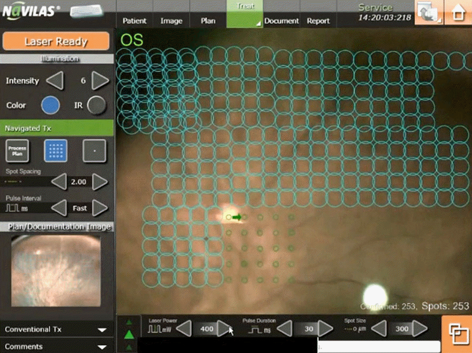 | Figure 1.Photograph of the monitor of the Navilas®. Blue circles show treated spots. Green small circles show the aimed spots (5 × 5 array pattern) (Image courtesy of WWW.OD-OS.com). |
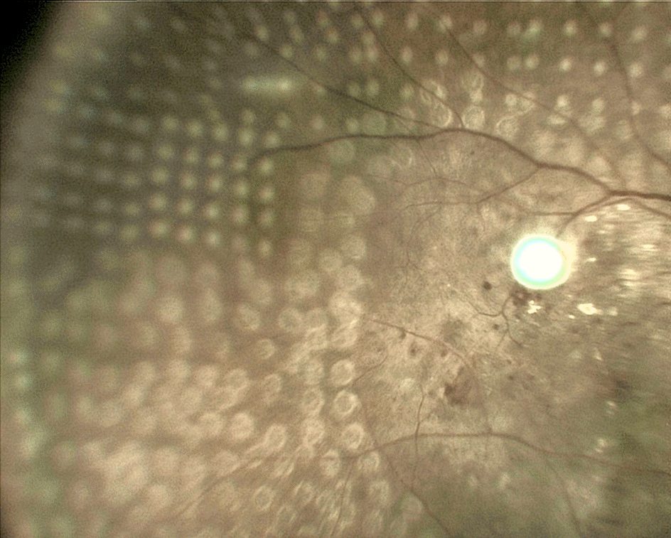 | Figure 2.Color fundus photograph after photocoagulation. The upper half shows burn scars with Navilas®, the lower half shows burn scars with conventional laser. |
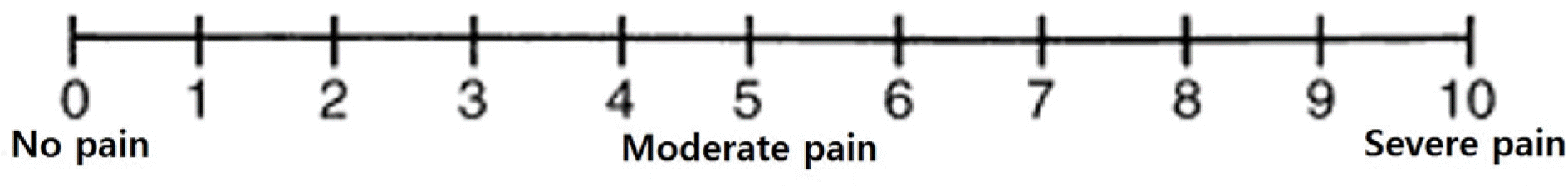
Table 1.
Comparisons of laser parameters, required time and pain scale in both groups
| Navilas® | Conventional laser | p-value* | |
|---|---|---|---|
| Laser power (mW) | 373.3 ± 38.1 | 209.3 ± 20.5 | <0.001 |
| Laser exposure time (sec) | 0.03 | 0.1 | <0.001 |
| Time for treatment (sec) | 562.2 ± 39.0 | 668.6 ± 51.5 | <0.001 |
| Number of laser spot | 2055.2 ± 288.7 | 658.0 ± 65.3 | <0.001 |
| Time per 100 laser spot (sec) | 27.7 ± 3.2 | 102.0 ± 6.6 | <0.001 |
| Visual analog pain scale | 3.3 ± 1.2 | 6.9 ± 1.1 | <0.001 |
 XML Download