Abstract
Purpose
We evaluated cultured specimens from silicone tubes removed from patients with congenital nasolacrimal duct obstruction and determined the antibiotic sensitivities of the specimens.
Methods
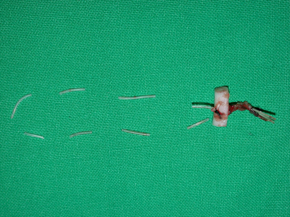
This study included 26 eyes of 22 patients who had received endonasal silicone tube intubation for congenital nasolacrimal duct obstruction. The removed silicone tubes were divided into canaliculus, lacrimal sac, nasolacrimal duct and nasal cavity parts according to insertion state. Then, bacteria and fungus cultures were performed and their antibiotic sensitivity was tested.
Results
Bacteria culture rate was 80.8% in the canaliculus and the lacrimal sac, and 88.5% in the lacrimal duct, and the nasal cavity, which was not significantly different according to insertion site. Fungus culture rate was significantly higher in the nasal cavity than in the nasolacrimal duct and in the nasolacrimal duct than in the lacrimal sac and the canaliculus (p-value < 0.05). The species of cultured Gram-positive bacteria were in the following order: Staphylococcus aureus, Streptococcus pneumonia and coagulase negative Staphylococcus. Common species of cultured Gram-negative bacteria were Pseudomonas and Serratia marcescens. All six species of cultured fungi were Candida. Among 12 Staphylococcus aureus cultured, eight species showed resistance to methicillin (MRSA). In all patients, the symptoms and the signs of nasolacrimal duct obstruction improved after the tube removal.
Conclusions
Bacterial and fungal infection of the silicone tube in patients with congenital nasolacrimal duct obstruction does not appear to affect directly the outcome of silicone tube intubation. Further studies of bacterium and fungi in the nasolacrimal duct before silicone tube intubation are needed for determining the infection causing nasolacrimal duct obstruction.
Go to : 
References
1. Cho KW, Lee SY, Kim SJ. Treatment of congenital nasolacrimal duct obstruction using silicone intubation set. J Korean Ophthalmol Soc. 1995; 36:553–8.
3. Nelson LR, Calhoun JH, Menduke H. Medical management of congenital nasolacrimal duct obstruction. Ophthalmology. 1985; 92:1187–90.

4. Baker JD. Treatment of congenital nasolacrimal system obstruction. J Pediatr Ophthalmol Strabismus. 1985; 22:34–6.

5. Usha K, Smitha S, Shah N, et al. Spectrum and the susceptibilities of microbial isolates in cases of congenital nasolacrimal duct obstruction. J AAPOS. 2006; 10:469–72.

6. Lee JJ, Ahn JH, Kim JL, Yang JW. The clinical outcome of endoscopic silicone tube intubation for congenital nasolacrimal duct obstruction. J Korean Ophthalmol Soc. 2012; 7:929–33.

7. Vanderveen DK, Jones DT, Tan H, Petersen RA. Endoscopic dacryocystorhinostomy in children. J AAPOS. 2001; 5:143–7.

8. Shin HM, Lew H, Yun YS. Fungus at removed silicone tubes in nasolacrimal duct obstruction patients. J Korean Ophthalmol Soc. 2004; 12:1967–72.
9. Kuchar A, Lukas J, Steinkogler FJ. Bacteriology and antibiotic therapy in congenital nasolacrimal duct obstruction. Acta Ophthalmol Scand. 2000; 78:694–8.

10. Ruby AJ, Lissner GS, O'Grady R. Surface reaction on silicone tubes used in the treatment of nasolacrimal drainage system obstruction. Ophthalmic Sur. 1991; 22:745–8.

11. Shin HM, Lew HL, Lee JM. Cytologic study of removed silicone tube in nasolacrimal duct obstruction patients with the liquid-based thin layer preparation techinique. J Korean Ophthalmol Soc. 2004; 45:707–13.
Go to : 
 | Figure 1.Detailed portion of removed silicone tube (canaliculus, lacrimal sac, nasolacrimal duct, nasal cavity). |
Table 1.
Incidence of the various microorganisms as found in the silicone tube removed from 26 eyes
| Bacteria culture (%) | 96.2 | Gram (+) | 50 |
| Gram (-) | 26.9 | ||
| Both Gram (+), (-) | 19.2 | ||
| Fungus culture (%) | 23.1 |
Table 2.
Incidence of the various microorganisms as found in the silicone tube removed from 26 eyes
| % of isolates (n = 26) | |
|---|---|
| S. aureus | 46.2 (11) |
| S. pneumoniae | 11.5 (3) |
| CNS | 3.8 (1) |
| P. aeruginosa | 15.4 (4) |
| S. marcescens | 15.4 (4) |
| Gram (-) bacilli | 15.4 (4) |
| Candida | 23.1 (6) |
Table 3.
Correlation between microbiology and location of silicone tube in 26 eyes
| Canaliculus(%) | Lacrimal sac(%) | Nasolacrimal duct(%) | Nasal cavity(%) | |
|---|---|---|---|---|
| S. aureus | 42.3 | 42.3 | 42.3 | 42.3 |
| S. pneumoniae | 11.5 | 11.5 | 11.5 | 11.5 |
| CNS | 3.8 | 0 | 3.8 | 0 |
| P. aeruginosa | 11.5 | 11.5 | 15.4 | 15.4 |
| S. marcescens | 15.4 | 15.4 | 15.4 | 15.4 |
| Gram (-) bacilli | 15.4 | 15.4 | 15.4 | 15.4 |
| Candida* | 0 | 0 | 3.8 | 19.2 |
Table 4.
Antibiotic susceptibility of S. aureus of 12 eyes
| Clindamycin (n = 12) | Ciprofloxacin (n = 12) | Erythromycin (n = 12) | Fusidic acid (n = 12) | Gentamicin (n = 12) | Oxacilin* (n = 12) | Penicillin* (n = 12) | Tetracycline (n = 12) | Vancomycin (n = 12) | |
|---|---|---|---|---|---|---|---|---|---|
| Susceptible | 6 | 12 | 5 | 12 | 11 | 4 | 1 | 10 | 12 |
| Intermediate | 0 | 0 | 0 | 0 | 1 | 0 | 0 | 0 | 0 |
| Resistant | 6 | 0 | 7 | 0 | 0 | 8 | 11 | 2 | 0 |


 PDF
PDF ePub
ePub Citation
 Print
Print


 XML Download